When there are different symptoms and signs of certain diseases, video thoracoscopy is a medical procedure that can be useful to help doctors in Turkey come up with a final diagnosis.
Video thoracoscopy is often used to examine changes in the center of the chest (mediastinum), which runs between the lungs from the lower neck to the diaphragm and from the sternum to the spine. Tissues outside the mediastinum but within the pleural cavity and chest cavity may also be evaluated. Treatment measures for the disease in question are often taken during a video thoracoscopy procedure.

Reasons why a video thoracoscopy may be required
Doctors use diagnostic video thoracoscopy when there are or are suspected to be changes in various organs in the chest, including the lungs, diaphragm, thymus gland, part of the immune system, esophagus, and lymph node disease. This can include the lungs, diaphragm, thymus, part of the immune system, esophagus, lymph node disease, and other changes in the mediastinum (center of the chest), such as diagnosis and sampling of abscesses and masses within the lungs and chest cavity. Video thoracoscopy may also be performed in case of fluid accumulation (effusion) in the pleura or pericardium.
Pleural effusions can be watery, purulent, or bloody (hemothorax) and can sometimes contain tumor cells. Video thoracoscopy can be used in the treatment of lung cancer, hyperhidrosis, tracheal disorders, pectus excavatum, diaphragmatic hernias, and all chest wall diseases.
Symptoms
Symptoms depend on the specific disease that the diagnostic thoracoscopy reveals. There is often shortness of breath and pain on examination.
Diagnostics
It is important that the patient is questioned about previous complaints and illnesses. A physical examination can also be helpful, where the lungs, in particular, are listened to and auscultated. Relevant findings can often be represented using imaging methods, such as X-ray examination, ultrasound examination, or computerized tomography (CT). In addition, a blood test is performed where attention is paid, for example, in empyema (purulent collection), to signs of inflammation.
Differential diagnosis
Potential diseases must be distinguished from each other, which is why diagnostic thoracoscopy is performed.
Treatment
Preventive treatment
There are various scenarios and options for non-surgical internal treatment, including administering medications or undergoing general measures performed by a specialist.
Surgery
Video thoracoscopy is performed under general anesthesia. Ventilation is provided through the lungs on the healthy side.
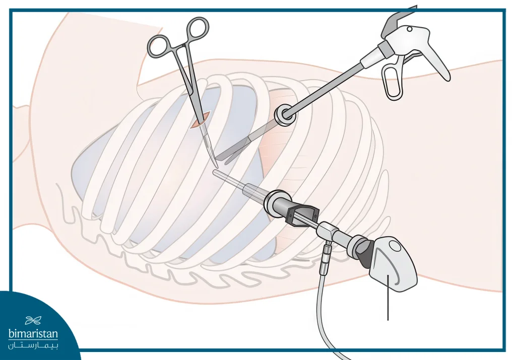
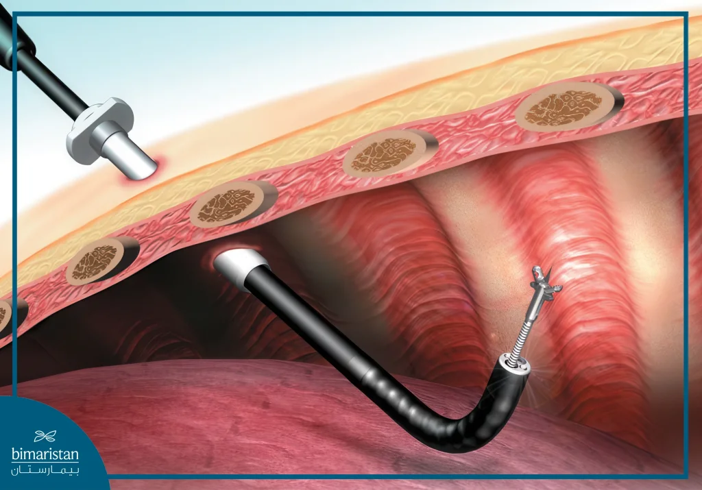
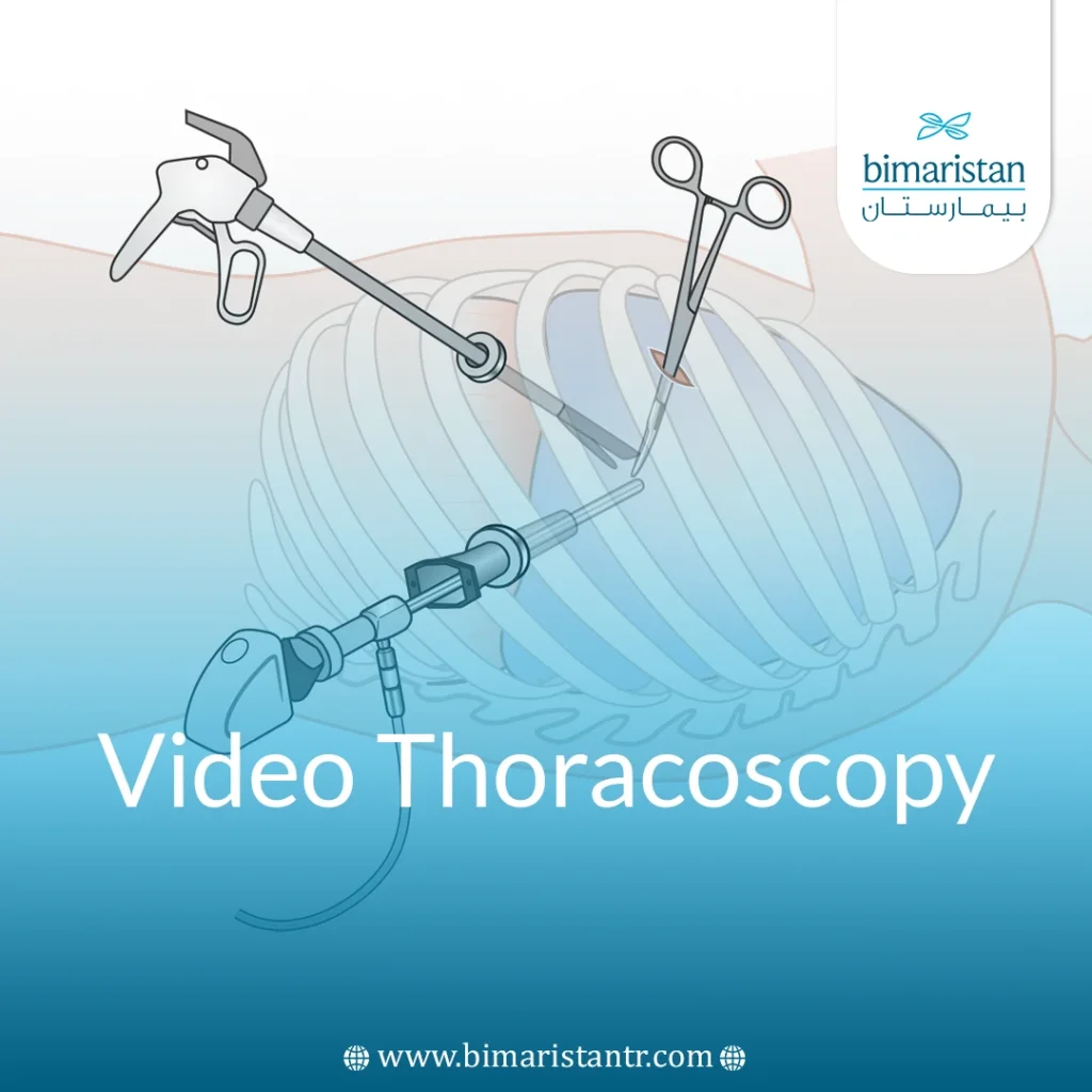
Video thoracoscopy is a medical procedure that involves making small incisions in the chest cavity in the space between the ribs, through which a tiny optical device called a thoracoscope, equipped with a special video camera, surgical instruments, and thoracoscopic instruments, can be inserted.
The surgeon can follow the image from the small camera on the monitor, evaluate the organs and tissues, and, if necessary, perform procedures and biopsy. Depending on the results, different procedures can be performed through the chest wall.

In video thoracoscopy, a special device is used to aspirate the pleural effusion. This removes the restrictions on the lungs. If there is a purulent effusion (pleural empyema), a tube is inserted to drain the secretion and rinsed several times a day with antibiotics and chest antiseptic fluid.
If adhesions and tissue roughness (crusts) are present in the pleura, often caused by inflammation or effusion, video thoracoscopy can be used to loosen or remove them.
Sometimes the roughness can also be removed directly from the lung tissue by video thoracoscopy; in other cases, medical intervention with traditional open surgery is necessary for treatment.
In video thoracoscopy, in the case of pericardial effusion, the solid pericardial tissue is opened by making a 2 × 2 cm incision. This allows the fluid to drain into the pleural space, so that the excessive pressure that threatens the heart is prevented or relieved. Due to the small amount, the fluid can usually be easily absorbed by the pleura.
When a malignant tumor is suspected, video thoracoscopy enables an endoscopist to remove larger portions of the pleura and then subject them to a tissue examination (biopsy). If the suspicion of cancer is confirmed, the remaining part of the pleura must also be removed laparoscopically and/or, in the vast majority of cases, open chest surgery is necessary for this purpose. As a result, respiratory function is unaffected or only slightly impaired.
In video thoracoscopy, if the pleural effusion is found to be the cause of a malignant tumor and the cause of the spread of cancer cells in the pleura (pleural cancer), the entire pleural cavity is considered an affected area. For this purpose, talcum powder is inserted into the cavity. This can also help with pleural mesothelioma, which starts from the pleura. This substance prevents the fluid from collecting again. After using this method, respiratory function is also unrestricted or barely restricted.
In the case of tumors or enlargements, the thymus often has to be removed. The thymus is an organ in which some immune cells develop in childhood and adolescence, but turn into fatty tissue in adulthood. And video thoracoscopy makes sense here because fewer scars are created. However, due to the position and complex vascular supply, the operation can be difficult, so it is often necessary to convert it to an open operation. The removal of the thymus gland does not result in any defects or diseases.
Following proper procedures in video thoracoscopy, one or two drainage tubes are usually inserted into the patient’s chest cavity in order to absorb the wound fluid and thus enable the lungs to expand to normal size. After several days, the tubes can usually be withdrawn back through the chest wall.
Possible additional procedures for the process
When there are unexpected findings or complications, it may be necessary for the doctor to expand the procedure or modify the method. It may be necessary to switch from thoracoscopy to open surgery.
Complications of video thoracoscopy
Surrounding anatomical structures or organs can be injured during video thoracoscopy.
This can lead to primary bleeding and secondary bleeding, and even also nerve damage, which can often lead to temporary sensitivity disorders or paralysis.
Injury to the phrenic nerve can lead to breathing difficulties, injury to the visceral nerve (vagus nerve) can lead to irregular heartbeats, and weakness of the vocal cord nerve can cause voice disorders and possibly shortness of breath.
Furthermore, inflammation, wound healing disorders, and visible scars with possible functional or aesthetic effects can occur.
Similarly, allergic reactions of varying degrees of severity cannot be ruled out.
Interventions in the thymus can lead to a type of muscle weakness (myasthenia gravis).
Sutures can also become loose in the esophagus, causing esophageal, gastroesophageal, and thoracic issues.
Video thoracoscopy instructions for patients
Before the procedure
Medications that negatively affect blood clotting, such as so-called marcomars or aspirin, should often be discontinued or cut back in consultation with your cardiologist.
Smoking can lead to wound healing disorders and disabilities after the operation, so the patient should stop before the laparoscopic operation if possible.
Before the procedure
Special breathing exercises, physiotherapy, and chest swimming are very useful after the operation to maintain health and minimize issues after such surgeries.
If you notice any abnormalities or just pains that may indicate complications, don’t hesitate to contact your doctor.
Regular check-ups should be done after the removal of the malignant findings.
Looking for the best video thoracoscopy clinics in Turkey?
At Bimaristan, we guide you to top specialists and hospitals offering advanced video thoracoscopy procedures.
Contact us today for expert care and trusted support.
Sources:

