Chest wall tumor treatment is becoming increasingly important as chest tumors now account for 5% of all thoracic tumors globally, according to recent statistics. Early detection plays a critical role in improving treatment outcomes and significantly boosts the chances of recovery.
Chest wall tumor treatment options range from precise surgical procedures to advanced therapeutic techniques, with success rates reaching up to 90% in early-diagnosed cases. Therefore, early diagnosis and expert care by experienced medical professionals are key elements in achieving complete recovery.
What are chest wall tumors or lung wall tumors?
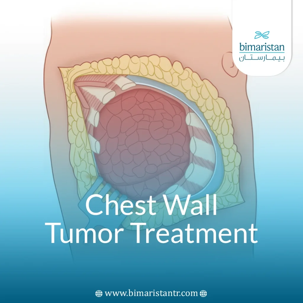
Chest wall tumors are an abnormal proliferation of cells in the chest wall (the structure surrounding the heart, lungs, and liver) that arise at the expense of the bone structure, muscle tissue, or soft tissue forming the chest wall, whether in the front or back of the rib cage. Chest wall tumors are more common in adults over the age of 40 and are divided into benign and malignant tumors, as well as primary and secondary tumors.
Benign chest wall tumors are more common in children and adolescents (such as chondroma, fibroma, or lipoma of the chest wall), while malignant chest wall tumors are more common in adults due to chronic exposure to carcinogens and cellular abnormalities in cell growth and proliferation with age.
Types of rib cage tumors
I: benign tumors
1. Benign bone tumors
- Osteoma
- Osteochondroma: This is the most common benign tumor of the ribs
- Fibrous dysplasia
- Unicameral bone cyst
2. Benign cartilage tumors
- Chondroma: May be difficult to differentiate from chondrosarcoma by histopathology in small biopsies.
3. Benign soft tissue tumors
- Lipoma
- Fibroma
- Hemangioma
- Benign peripheral nerve tumor, such as neurofibroma or schwannoma
II: Malignant tumors
1. Malignant primary tumors
A. Malignant bone tumors
- Osteosarcoma
- Chondrosarcoma: The most common primary malignant tumor of the rib cage
- Ewing sarcoma: Affects young people
B. Malignant soft tissue tumors
- Fibrosarcoma
- Rhabdomyosarcoma
- Undifferentiated sarcoma
2. Secondary tumors (metastases to the chest wall)
- Breast cancer: The most common type of cancer that travels to the rib cage
- Lung cancer
- Kidney cancer
- Thyroid cancer
- Melanoma cancer
Important notes:
- Benign tumors are usually slow-growing, painless, and may be discovered accidentally.
- Malignant tumors tend to grow rapidly, can be painful, and cause destruction of bone or surrounding tissue.

Can chest wall tumors be metastatic from other organs?
Chest tumors can be metastatic from other organs, in which case they are called “secondary chest wall tumors.” Metastases come directly from neighboring tissues, or may come from distant organs such as the prostate, kidneys, and thyroid via the bloodstream and lymphatic system.
Secondary tumors of the chest wall are more common than primary cancers, accounting for approximately 80%, and are the most common:
- Breast cancer
- Lung cancer
- Kidney cancer
- Thyroid cancer
- Lymphomas
It is transmitted either by direct contact, as in lung and breast cancer, or through the bloodstream, as in lung and kidney cancer, or through the lymph nodes and peritoneum.
How to accurately diagnose chest wall tumors
The patient shows a number of symptoms and signs that are visible, which the doctor can rely on to establish an initial diagnosis of the disease, but these symptoms are not mainly diagnostic for chest wall tumors, due to the similarity of the symptoms of chest wall tumors with systemic tumors that can appear in the chest cavity or any organ of the body, and the symptoms of thoracic cage tumors:
- Localized pain in the chest or back that increases with movement and deep breathing, where the pain does not go away with painkillers and lasts for a long time
- The appearance of a palpable mass in the chest wall, which may be mobile or solid.
- Unexplained weight loss in malignant tumors
- In rare cases, the tumor may press on the esophagus, causing difficulty swallowing.
- The tumor may compress large veins, causing swelling in the face or extremities (rare)
- General fatigue and weakness
Although these symptoms are apparent and clear, they are not diagnostic for chest wall tumors specifically, so a biopsy or tissue sample is taken from the patient to confirm the diagnosis and determine the nature of the tumor, but it requires an auxiliary imaging technique (such as CT), in addition to not giving enough tissue for accurate diagnosis, so when a combination of these symptoms are present together, the doctor asks the patient to undergo imaging tests to confirm the diagnosis, including imaging tests:
- X-ray imaging: It is useful in bone tumors such as chondrosarcoma.
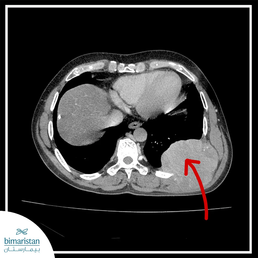
- Computed tomography (CT): Determines the size of the tumor and its relationship with neighboring organs, as well as detects bone and thoracic metastases.
- Magnetic resonance imaging (MRI): It is useful in evaluating soft tissues such as muscles and nerves and shows fine details in the tumor.
- PET-CT: Used to distinguish between benign and malignant tumors and detect distant metastases. It also helps determine the method of biopsy.
These imaging methods are fully diagnostic for chest tumors, as they give a definite idea of the dimensions of the mass, show whether these tumors are benign or malignant, and reveal the extent of the tumor in the case of malignant chest tumors.
Biopsy diagnostic methods
The type of chest wall cancer is predicted by its shape on CT scan, where it is located in the chest, and the age of the affected patient.
A biopsy is taken to confirm the type of cancer. If the size of the mass is smaller than 4 cm and is not invasive to other tissues, an excisional biopsy is preferred. If the mass is large, invasive to neighboring tissues, or there are metastases in other places, then a core needle biopsy (Core needle biopsy or Tru-cut) or through a small incision under local anesthesia is preferred.
Chest wall tumor treatment: Multiple options
Chest wall tumor treatment methods vary between surgical treatment, radiation, and chemotherapy. Surgical treatment with resection is considered the primary approach for most chest wall tumors, especially when the tumors are primary (not metastases from other organs). In cases of benign tumors, they can often be completely removed while preserving the surrounding healthy tissues without causing damage.
However, malignant tumors require a different approach. In cases of small malignant tumors, the tumor must be removed along with a margin of healthy tissue to reduce the risk of recurrence. For larger malignant tumors, surgeons often perform a wide resection, which may involve removing the tumor along with surrounding muscles and bones. Afterward, reconstructive surgery may be necessary to restore the chest wall structure and allow the patient to continue living a normal life.
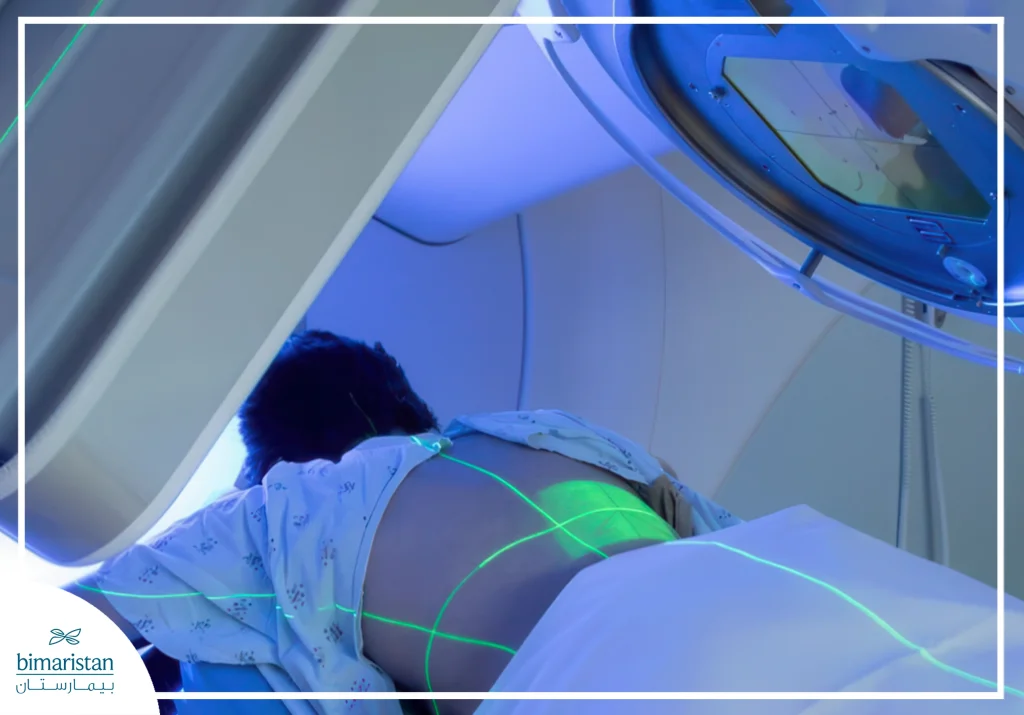
In some cases, it may not be possible to remove the tumor, so radiation therapy or chemotherapy is used to remove this tumor, and the use of each treatment varies according to the case, as radiation therapy is used to relieve pain in advanced cases, and may be used as an adjuvant treatment with surgical treatment, reducing the size of the tumor before surgery, or destroying the remaining cancer cells after surgery.
Chemotherapy is used for malignant tumors that spread quickly, as well as to control metastases when the tumor has spread, and may also be used as an adjunctive treatment to surgery to destroy cancer cells that remain after surgery.

Surgery alone vs. surgery with adjuvant therapy
Some cases of chest tumors require synergy between surgery and adjuvant therapy, such as advanced malignant tumors, but in cases of benign tumors, adjuvant therapy is not required, and results over the years show that surgery alone may be better than surgery with adjuvant therapy due to fewer complications and faster recovery compared to surgery with adjuvant therapy.
| Surgery with adjuvant therapy | Surgery alone | Criterion |
| Higher in advanced malignant tumors | Elevated in benign tumors | Success rate |
| A little more | less | Complications |
| Relatively longer | Relatively faster | Recovery time |
| Continuous monitoring | Simple patrol | The need for follow-up |
Resection of chest wall tumors: Surgical steps and reconstruction techniques
Thoracic tumor resection is performed under general anesthesia, and endotracheal intubation is performed to preserve the patient’s respiratory function during the surgery. The surgeon locates the tumor through pre-operative imaging and then makes an incision over the tumor area, taking care to preserve the surrounding nerves and blood vessels. In the case of benign chest tumors, the surgeon removes only the tumor and preserves healthy tissue, while in the case of malignant tumors, the surgeon removes the tumor and a portion of the surrounding tissue (usually between 2-5 cm), and some ribs may be removed if the malignant tumor has reached them.
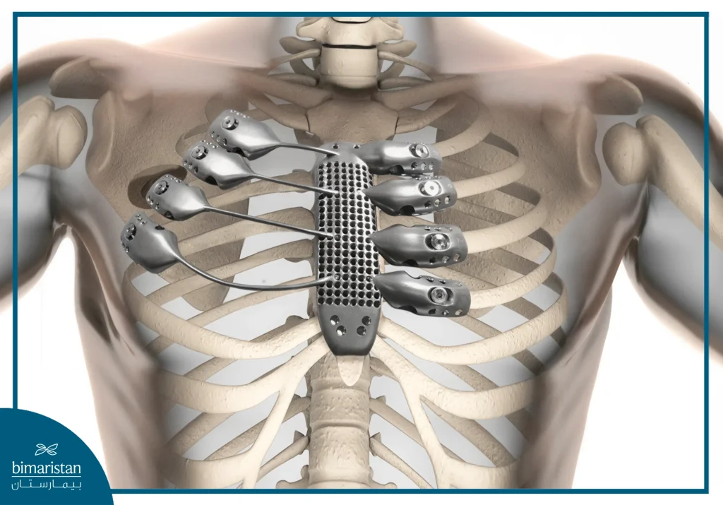
When the surgeon removes a large number of ribs, the chest may become unstable, so in order to stabilize the rib cage, the surgeon uses artificial stents to stabilize the rib cage and prevent it from moving. In addition, grafts using mesh or muscle grafts may be used in certain cases, such as large chest wall defects (greater than 5 cm) after tumor resection and the need to enhance blood perfusion, and these mesh and muscle grafts are also useful to reduce the risk of infection after resection.

What effect does surgery have on breathing and movement?
Chest wall surgeries may affect breathing and movement temporarily or permanently, but depending on whether the surgeon has removed a large area of ribs or muscle, the most important effects on breathing and movement after the operation:
- Immediate postoperative effects: Painful breathing that limits deep breathing, pneumothorax may occur if the pleura is punctured during surgery, and mobility, the patient may experience temporary muscle weakness and stiffness in the joints, making them unable to raise their arms for a few weeks.
- Long-term effects: Surgery for large thoracic tumors weakens the support of the rib cage, altering the natural breathing mechanism and increasing reliance on the diaphragm, and in rare cases, can cause chronic respiratory failure, especially when more than four ribs are removed.
The temporary effect usually disappears after 4-8 weeks and is the most common; on the contrary, the permanent effect is irreversible but very rare, and occurs only in major resections without proper restoration afterwards, as restoration with mesh or grafts eliminates complications by 50%, reducing the risk of permanent effect.
Rehabilitation after surgery
After thoracic tumor surgery, the patient undergoes a rehabilitation program that includes exercises and physical therapy. These exercises help the patient to return to his/her normal life fully, and include rehabilitation programs:
- Deep Breathing Exercises
- Progressive physical therapy for shoulder and upper body movement
- Muscle strengthening exercises, especially the back and chest muscles
The rehabilitation program takes about 2-3 months, and the duration varies from person to person, and during this period some warning signs may appear, such as increased shortness of breath, the appearance of severe pain that does not improve with painkillers, or the appearance of swelling or redness in the area, and when these signs appear, the patient should see his doctor as soon as possible.
Regular Restoration vs. Prosthetic Reconstruction
Some thoracic tumor surgeries may require reconstruction afterwards to preserve the normal function of the rib cage and surrounding muscles, and to prevent complications as much as possible. There are two types of reconstruction: natural reconstruction (muscles or ribs) and artificial reconstruction (titanium mesh or grafts), each of which has different specifications:
| Prosthetic Reconstruction | Regular Restoration | Type |
| Very good | Excellent | Body Compatibility |
| Slightly possible | Very rare | Risk of rejection |
| More complex cases | Simpler cases | Type of case |
| Greater likelihood of occurrence | Less likely to occur | The occurrence of sepsis |
| Top | Relatively less | Cost |
| shorter | Longer | Surgical time |
When is reconstruction necessary after resection?
Surgical reconstruction is necessary after resection of thoracic tumors in specific cases, as the surgeon decides the need for it during the operation based on the size and location of the resulting defect. Reconstruction is mandatory when three or more ribs are removed, when there are large chest wall defects (usually greater than 5 cm), or when parts of the sternum are removed, while in other cases such as partial rib resection or risk of infection, reconstruction is recommended but not mandatory, and the choice of reconstruction method (artificial mesh or muscle grafts) depends on the patient’s condition and the characteristics of the surgical defect.
Recovery rate and outlook
The success of chest wall tumor treatment depends on the type of tumor and the stage of diagnosis, as the cure rate for benign tumors after resection of chest tumors reaches 100%, while it ranges between 40% and 80% for malignant tumors, depending on their type and extent. Surgery plays the primary role in treatment, especially for complete resection with a surgical safety margin, while radiation and chemotherapy are used as adjuvant agents in malignant cases. The outlook remains positive with early detection and appropriate treatment, although relapse may occur in some cases.
| Recovery rate after complete resection | Tumor type |
| 60-80% | Chondrosarcoma |
| 40-60% | Osteosarcoma |
| 30-50% | Soft tissue sarcoma |
| Varies depending on the original tumor, usually low unless it is localized and resectable | Secondary tumors (metastases) |
In conclusion, Bimaristan Medical Center in Turkey offers advanced and precise chest wall tumor treatment, combining medical expertise and modern technologies at competitive costs compared to Europe and America. We guarantee you a safe journey from diagnosis to recovery, because your health deserves the best. Choose medical excellence in the heart of Turkey.
Sources:
- National Cancer Institute. (n.d.). Soft tissue sarcoma. U.S. Department of Health and Human Services.
- American Cancer Society. (n.d.). Soft tissue sarcoma.

