Cerebral collapse (acute cerebral hemorrhage) is a serious neurological emergency caused by the rupture of cerebral blood vessels, leading to blood accumulation and disruption of neurological functions. Conventional surgery has always been the only treatment option, but it carries a high risk of complications. Relying on advanced technologies such as embolic catheterization and thermal frequency under precise imaging guidance, brain bleed interventional radiology treatment provides an optimal solution by precisely stopping the bleeding without opening the skull, making it a pillar in the treatment of cerebral hemorrhage without surgery, while minimizing complications and improving recovery.
What is a cerebral hemorrhage?
Cerebral collapse or cerebral hemorrhage is the second most common cause of stroke, caused by the sudden rupture of a blood vessel inside the skull, which leads to blood leaking into the brain tissue or the spaces surrounding it, the blood collects in the skull and brain and forms a pool that puts pressure on the brain, preventing oxygen and nutrients from reaching the brain tissues and cells.
Cerebral hemorrhage is a life-threatening medical emergency in which brain cells die within three to four minutes if they don’t get enough oxygen. Brain hemorrhage often occurs after a fall or traumatic injury, and it often occurs in people with uncontrolled high blood pressure.
Causes of brain collapse
- Head injuries: Car accidents or falls that rupture superficial vessels
- An aneurysm: A cystic bulge in an artery that bursts under pressure
- Buildup of fatty deposits in the arteries (atherosclerosis)
- blood clot
- Congenital vascular malformations: Abnormal intertwining of arteries and veins (AVM)
- Leakage from abnormal connections between arteries and veins (arteriovenous malformation (AVM) or arteriovenous malformation (AVM))
- Cerebral Amyloid Angiopathy
- Brain tumor
Symptoms of cerebral hemorrhage
Although the symptoms of cerebral hemorrhage in adults are many and varied, in most cases, at least one of these symptoms will be present:
- Sudden, severe headache
- Muscle weakness, tingling or numbness, and partial or complete paralysis of movement in one hemisphere (right or left)
- Vomiting and nausea
- Vision changes (blurry or reduced vision, or even blindness)
- Dizziness and confusion
- Difficulty swallowing and chewing food
- Impaired ability to balance (cerebellar hemorrhage)
- Aphasia or slurred speech
- Difficulty reading or writing
- Impaired sense of taste
- Irregular heartbeat and trouble breathing (brainstem hemorrhage)
- Changes in consciousness (drowsiness and coma may occur)
- Epileptic seizures
What are the risk factors for brain hemorrhage?
Brain hemorrhages can affect anyone at any age, from newborns to adults, and are more common in adults older than 65. You may be at higher risk of a brain hemorrhage if you have the following:
- High blood pressure (the most important cause)
- Drug addiction
- Smoking
- Bleeding conditions or conditions that require treatment with blood thinners (anticoagulants)
- Conditions associated with pregnancy and childbirth (preeclampsia, postpartum vasculopathy, or neonatal intraventricular hemorrhage)
- Conditions that affect how blood vessel walls form.
Types of cerebral hemorrhage:
- Inside the skull but outside the brain tissue:
- Epidural hemorrhage: This bleeding occurs between the skull bone and the outer membrane layer, the dura mater.
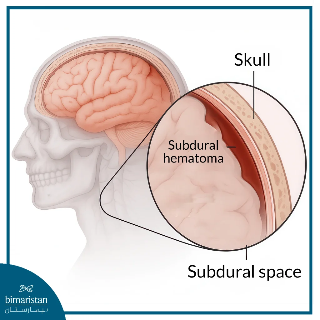
- Subdural hemorrhage: This bleeding occurs between the dura mater and the arachnoid membrane, often resulting from head trauma.
- Subarachnoid hemorrhage: This bleeding occurs between the arachnoid membrane and the arachnoid mater, often due to an aneurysm.
- Intracerebral hemorrhage (ICH): This is the most serious type, occurring deep within the brain tissue.
- Intracerebral hemorrhage: This bleeding occurs in the lobes of the brain, brainstem, and cerebellum, and is bleeding anywhere within the brain tissue itself.
- Intraventricular hemorrhage: This bleeding occurs in the ventricles of the brain, which are specific areas (cavities) of the brain where cerebrospinal fluid (the fluid that protects the brain and spinal cord) is secreted.

Why treat brain collapse with brain bleed interventional radiology treatment?
Until the last decade, craniotomy surgery was the only solution, but it causes serious physical symptoms:
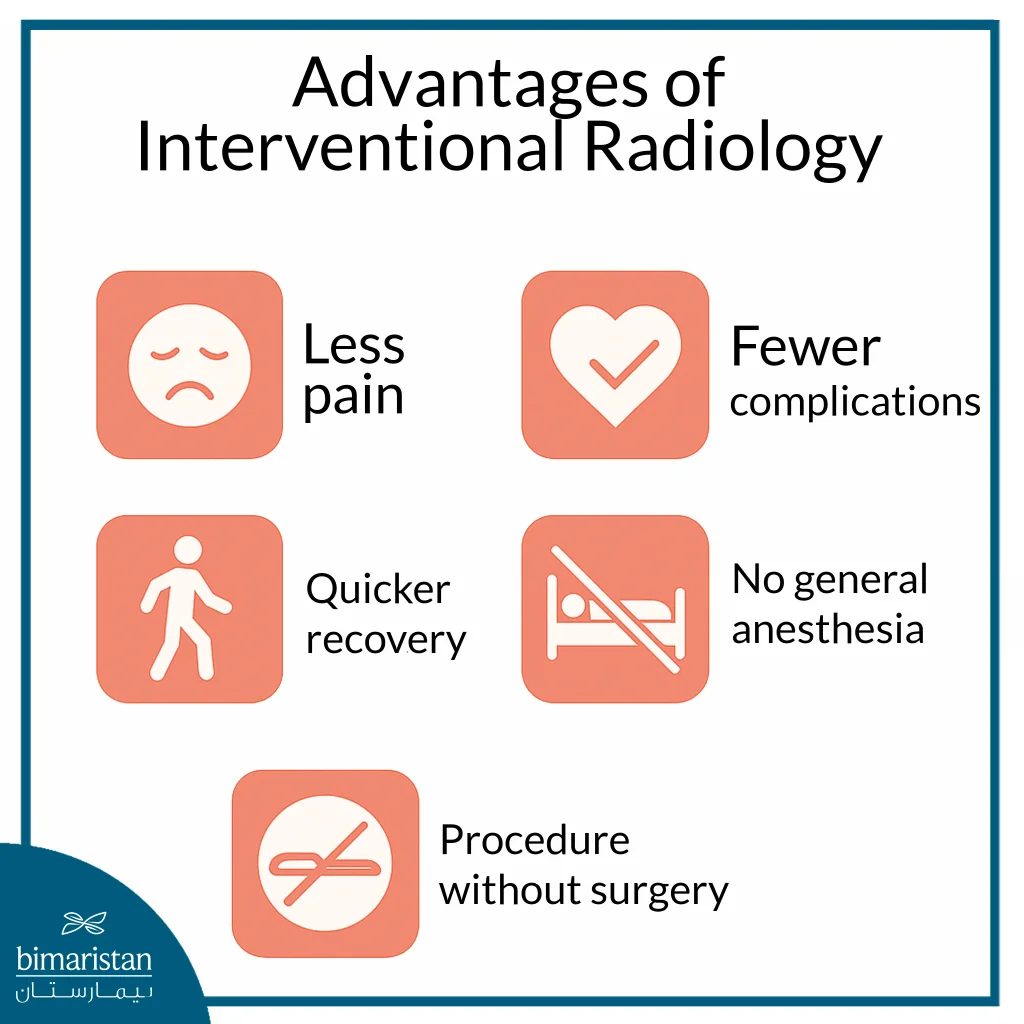
- Less pain: Pain after the procedure is usually less severe.
- No general anesthesia: Performed under local anesthesia only, reducing heart and lung risks.
- Lack of complications: Avoiding opening the skull eliminates the risk of damage to healthy tissue and infection.
- Greater accuracy: Imaging guidance (such as digital subtraction angiography (DSA) allows for targeting bleeding with micrometer precision.
- Faster recovery: Due to the less invasive nature, the recovery period is much shorter compared to open surgery.
- Shorter hospital stay: Many patients can go home the same day or soon after, compared to 7-10 days for open surgery.
- Suitable for high-risk cases: It may be the only viable option for patients who cannot tolerate open surgery due to their overall health condition.
- No significant blood loss
- Fewer infections or clots

How is brain collapse treated with brain bleed interventional radiology treatment?
Interventional radiology is performed in an interventional radiology room equipped with advanced imaging techniques, in just 60-120 minutes, here are the steps of interventional radiology treatment for brain collapse:
- Anesthesia: In most cases, local anesthesia is used in the groin or arm (the catheter insertion site). General anesthesia may be used in some complex cases or for children.
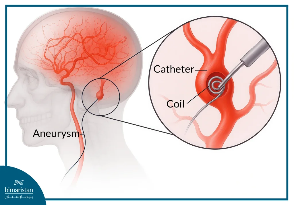
- Catheter Insertion and Guidance: The surgeon makes a small incision in the groin or arm. Using X-rays, the surgeon guides a catheter, thinner than a hair, precisely into the blood vessels in the brain. A contrast material (dye) is then injected to highlight the blood vessels and identify the location of the bleeding or aneurysm, while the physician monitors its path on a live DSA monitor.
- Treatment: Small metal coils are used to seal the aneurysm and prevent blood flow to it in the case of an aneurysm. In the case of bleeding or vascular malformations, adhesives or foam are injected to seal off the affected vessels. In the case of clots, the clot is removed through the catheter or dissolved in special cases.
- Catheter Removal: After confirming the success of the procedure, the catheter is removed, and pressure is applied to the insertion site for 10 minutes to prevent bleeding.
- Resuscitation: The patient is monitored for several hours to ensure their condition is stable.

What are the candidates for brain bleed interventional radiology treatment?
Brain bleed interventional radiology treatment is an option for many cases, especially those that are stable, such as:
- Patients with small aneurysms (less than 5 cm in diameter): The aneurysm’s structure allows for precise insertion of platinum coils without compressing surrounding tissue.
- Arteriovenous malformations (AVMs) in areas such as the visual cortex and brainstem: In these cases, interventional radiology is superior to open surgery in avoiding permanent neurological damage.
- Patients with chronic diseases:
- Heart diseases: such as heart failure and atrial fibrillation.
- Lung diseases: such as COPD and pulmonary fibrosis.
- Unstable diabetes: avoiding the risks of general anesthesia.
- Hunt & Hess grade 1-3 subarachnoid hemorrhage: These are relatively stable cases with partial retention of consciousness.
Differences between young and old for interventional radiology treatment of cerebral infarction
- Older adults (>65 years):
- First choice for those with poor tolerance to surgery.
- Success rates of up to 85% (compared to 55% with surgery).
- Slightly increased risk of kidney stress due to contrast dye.
- Younger adults (<65 years):
- 40% faster recovery.
- Success rates of up to 95% with metal coils.
Critical emergencies
In some cases, urgent intervention is required through interventional radiology treatment of brain collapse:
- Rapidly expanding bleeding: A temporary balloon occlusion is used to stop blood flow within 90 minutes, followed by interventional radiology treatment.
- Loss of consciousness with brainstem bleeding: Detected by emergency CT angiography.
- Severe clotting disorders: Clotting factors, such as vitamin K, are injected during the procedure.
What are the complications of brain bleed interventional radiology treatment?
Complications after brain bleed interventional radiology treatment are not common but may occur, the most common complications are listed below:
- Thrombosis: The catheter interacts with the vessel wall. This is treated with thrombolytics (rt-PA). Heparin is usually injected during the procedure to prevent this.
- Vasospasm: This causes sudden headache and nausea. This is treated with intravenous nitroglycerin.
- Allergy to contrast dye: Symptoms include a rash and difficulty breathing. This can be prevented with allergy testing and prophylactic corticosteroids.
- Vessel perforation: This occurs in patients with advanced atherosclerosis and is immediately closed with platinum coils.
- Failure to completely close a bleeding hemorrhage: This occurs especially in large arteriovenous malformations (AVMs >3 cm).
- Transient ischemic stroke (TIA): A temporary vascular blockage that resolves spontaneously within 24 hours.
Recovery after brain bleed interventional radiology treatment
During the recovery phases after the brain bleed interventional radiology treatment in the beginning and during the first 24 hours, vital signs are monitored every two hours and a CT scan of the brain, after that the patient is instructed to practice breathing exercises to prevent pulmonary complications and perform a neurological evaluation, after a period of about 6 weeks, physical therapy is started to restore motor functions and the patient attends speech sessions if the speech center is affected through 3 sessions per week.
Clots should be prevented by injecting heparin under the skin by the nurse or medical team. The use of compression stockings helps, and rehabilitation therapy should be started according to the results after brain bleed interventional radiology treatment of the cerebral collapse.
- For paraplegia: Neuroelectrical stimulation and flexible exercises 3 times a day.
- For speech difficulties: Speech sessions using brain imaging (fMRI).
- For imbalance: Balance rehabilitation exercises.
Is brain bleed interventional radiology treatment effective?
The results of brain bleed interventional radiology treatment show remarkable effectiveness, often surpassing traditional surgical methods, especially in dealing with complex and delicate cases within the brain. According to recent studies, success rates in treating brain aneurysms are around 91% when metal coils are used, rising to around 94% when special stents (Flow Diverters) are used. In cases of arteriovenous malformations (AVM), the success rate is 85% using medical glue, while interventional radiology techniques have shown 89% effectiveness in cases of subarachnoid hemorrhage.
When compared to open surgery, the superiority of interventional radiology is clear. The mortality rate is only about 5%, compared to 18% in traditional surgery, and infection complications are less than 1%, compared to about 12%. Patients also enjoy a shorter recovery period, with a hospital stay of only 2 to 3 days, compared to 10 to 14 days for surgery, and many patients are able to return to work in 4 to 6 weeks, instead of waiting 3 to 6 months as in major surgeries.
For long-term follow-up, a magnetic resonance angiography (MRA) is recommended 6 weeks after treatment, followed by a periodic digital subtraction angiography (DSA) annually for five years. It should be noted that about 30% of cases with significant abnormalities may require additional interventional sessions later on, highlighting the importance of continuous follow-up to ensure sustainable results.
Cost of brain bleed interventional radiology treatment
The cost of brain bleed interventional radiology treatmentin Turkey ranges between $ 10,000 and $ 15,000 USD, which is significantly lower compared to Europe and America. This table shows the prices of brain bleed interventional radiology treatment in Turkey and some countries:
| State | Cost (USD) |
| Turkey | 10,000 – 15,000 |
| United States of America | 30,000 – 95,000 |
| Saudi Arabia | 20,000 – 30,000 |
| Germany | 35,000 – 50,000 |
Additional cost factors for brain bleed interventional radiology treatment
- Bleeding location: Brain stem hemorrhages are about +40% more expensive, while temporal lobe hemorrhages are about +20% more expensive.
- Doctor’s experience: The more experience a cardiologist has, the higher the cost due to higher efficiency.
- Technologies used: The Flow Diverter costs around $8,000, and the Navigation Catheter costs around $3,000.
- Hospital and equipment: The cost varies depending on the medical center’s equipment and international accreditation.
- The patient’s health status: The presence of chronic diseases or complex cardiac conditions may require additional tests and intensive care.
- Accompanying services: Such as accommodation, pre-operative tests, post-operative follow-up, and translation services.
Finally, brain bleed interventional radiology treatment is no longer a medical luxury but has become the global gold standard for non-invasive treatment of cerebral hemorrhage, combining precision, effectiveness, and minimal intervention. It significantly reduces risks and accelerates recovery. This treatment is especially valuable in cases caused by aneurysms or arteriovenous malformations, allowing access to deep brain tissues without open surgery.
Bimaristan Medical Center in Turkey is a pioneer in applying neuro-interventional radiology techniques, thanks to its specialized staff and advanced equipment that meet international standards. The center offers patients comprehensive care including precise diagnosis, individualized treatment plans, and continuous follow-up after the procedure, making it a trusted destination for advanced brain bleed interventional radiology treatment.
Sources:
- Mitchell, P. J., & Chong, W. (2017, November 13). Interventional radiological treatment of intracranial (brain) aneurysms. InsideRadiology
- RadiologyInfo.org. (2024, August 5). Brain aneurysm embolization. RadiologyInfo

