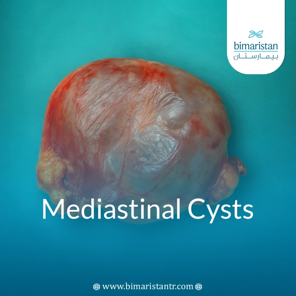
Mediastinal cysts are abnormal fluid-filled cavities located in the mediastinum, the anatomical space between the lungs that houses the heart, major vessels, trachea, and esophagus. These cysts are considered non-cancerous tumors and are often discovered incidentally during chest imaging.
They can be congenital (present at birth) or acquired as a result of infections or surgical complications. Although most cysts are asymptomatic, some may compress neighboring organs, leading to shortness of breath, coughing, and chest pain. An accurate diagnosis is necessary to determine the type of cyst and plan appropriate treatment to avoid complications.
What are mediastinal cysts?
Mediastinal cysts are fluid-filled cavities that arise within the mediastinum and are considered non-cancerous formations, as they often arise for congenital reasons due to a defect in the development of fetal tissues, such as bronchial cysts or cysts of the gastrointestinal tract, or, rarely, acquired due to infections or surgeries.
Difference between cysts and tumors
Cysts are fluid-filled cavities, while tumors are solid and grow abnormally from cells, and unlike some tumors, cysts do not grow rapidly and do not spread.
Are mediastinal cysts dangerous?
Mediastinal cysts are often considered benign and non-cancerous, they do not tend to become malignant and often remain stable without complications, in many cases they are discovered accidentally during chest imaging for other reasons without causing overt symptoms, but in some cases symptoms may appear due to localized pressure: Shortness of breath, difficulty swallowing, or coughing as a result of the cyst pressing on the esophagus or bronchus.
If there are congenital abnormalities or unusual characteristics in the cyst, a biopsy or surgical excision may be recommended to analyze the tissue thoroughly, these cysts may rarely be associated with complications such as infection or tumor transformation, which requires careful monitoring to ensure timely diagnosis and treatment, in some cases it is dangerous, for example, a pericardial cyst may lead to pericardial effusion, which is a buildup of fluid around the heart that hinders its ability to pump efficiently.
Types of Mediastinal Cysts
Cysts are divided into two main types depending on their causes:
- Congenital cysts: These are the most common and arise during fetal development and include
- Bronchogenic cyst: Usually located in the middle mediastinum and close to the bronchi.
- Esophageal duplication cyst: Often located near the esophagus.
- Pericardial cyst: Often asymptomatic and located near the heart.
- Lymphatic cysts: Such as the chyle cyst, contain lymphatic fluid.
- Acquired cysts: Rare, and may form due to chronic infections, trauma, or surgical complications within the chest.
Causes of mediastinal cysts
These are the main reasons why these cysts appear:
- Congenital factors: These are the result of tissue remnants from the respiratory or digestive system during embryonic development.
- Chronic chest infections: May lead to the formation of acquired cysts over time.
- Developmental abnormalities during pregnancy: Affects the formation of the mediastinum and causes abnormal cysts.
- Rare genetic diseases: Such as some connective tissue disorders or diseases of the lymphatic system.
Symptoms of mediastinal cysts
Mediastinal cysts are often inconspicuous and discovered accidentally, but they can cause symptoms in some cases:
- shortness of breath
- Chronic cough
- Chest pain
- Difficulty swallowing when the cyst presses against the esophagus
- Voice change or hoarseness due to the cyst pressing on the vocal cords
How to diagnose mediastinal cysts
The diagnosis of mediastinal cysts begins with nonspecific symptoms or an accidental discovery during routine examinations, which necessitates additional tests, including:
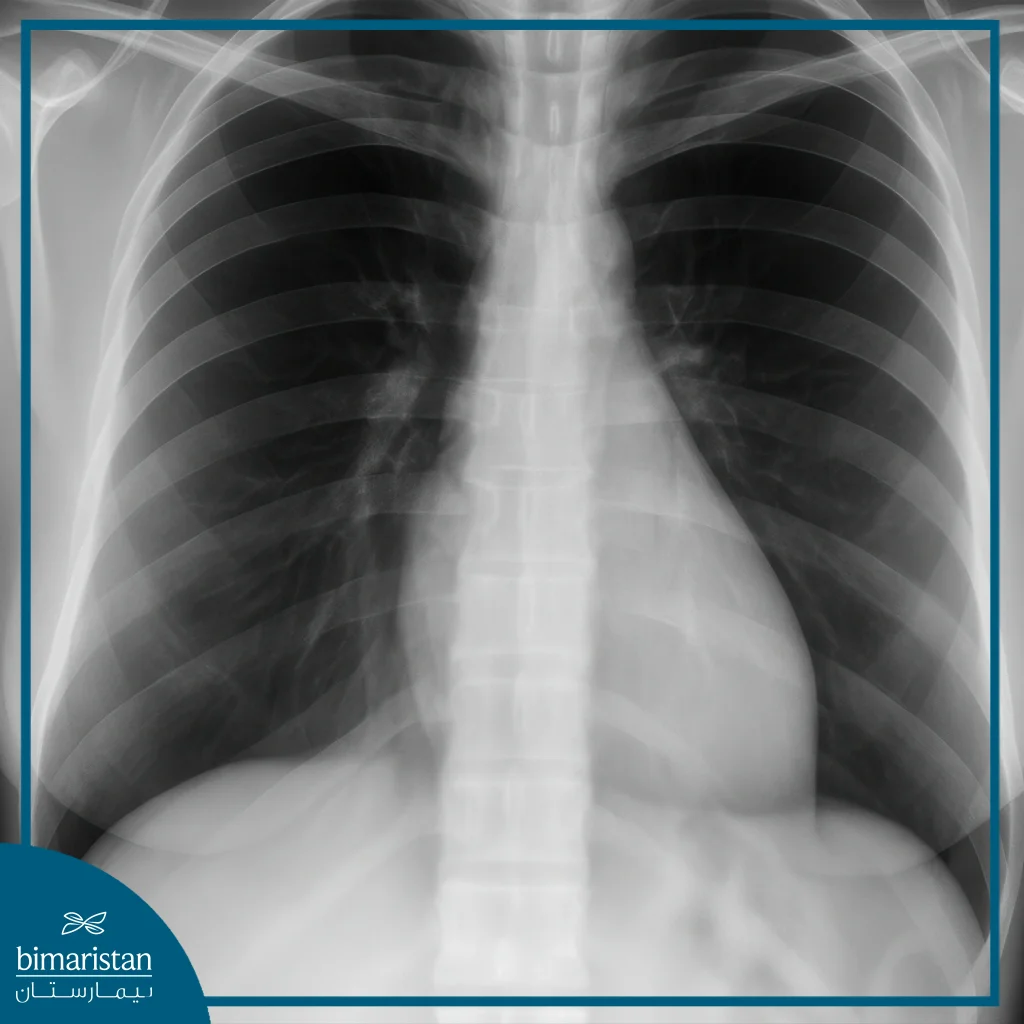
- Chest X-ray: Used as a first step and shows abnormal shadows that confirm the presence of a mass.
- CT Scan: This is the most accurate test to determine the exact shape, size, and location of the cyst as well as its relationship to surrounding structures.
- Magnetic resonance imaging (MRI): Helps to characterize the content of the cyst (fluid, tissue) and determine its exact nature.
- Functional examinations: Such as breathing or swallowing tests that ask if there is pressure from the cyst on the bronchi or esophagus.

Mediastinal cyst treatment methods
Methods of treating mediastinal cysts vary according to their size, nature, and the extent to which they cause symptoms, and treatment is done according to the accurate assessment of the specialized doctor and The most important methods of treating mediastinal cysts:
- Periodic observation: If the cyst is small and non-invasive, periodic follow-up with imaging.
- Surgical removal is done in the following cases:
- The presence of respiratory symptoms or pressure on the esophagus or bronchi
- The physician suspects the nature of the cyst (fearing it may be a tumor)
- The patient desires to have the cyst removed
The most important surgical methods used in the treatment of mediastinal cysts are conventional and laparoscopic, which are less invasive and faster recovery, as well as needle biopsy to determine the nature of the cyst before performing a surgical procedure.
It is very important for the patient to make an early diagnosis when suspecting any symptoms related to mediastinal cysts and to consult a specialized doctor immediately because early detection allows determining the exact nature of the cyst and helps in choosing the most appropriate treatment before the development of complications. Ignoring the symptoms or delaying the diagnosis may lead to increased pressure on neighboring organs or worsen the condition, making it difficult to treat, so it is important to adhere to periodic examinations and continuous medical follow-up to ensure the best results and maintain overall health.
Sources:
- Barrios, P., & Avella Patino, D. (2022). Surgical indications for mediastinal cysts – A narrative review. Mediastinum, 6(31).
- Esme, H., Eren, S., Sezer, M., & Solak, O. (2011). Primary mediastinal cysts: clinical evaluation and surgical results of 32 cases. The Texas Heart Institute Journal, 38(4), 371-374.

