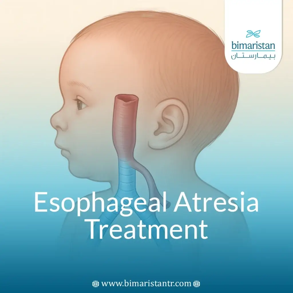
Congenital gastrointestinal and respiratory malformations are some of the most difficult medical challenges faced by newborns, especially when they affect the esophagus and trachea.
Over the past decades, esophageal atresia treatment and tracheal fistula management have undergone a paradigm shift supported by significant surgical and technical advances that have enabled doctors to significantly improve the life chances of affected children.
Minimally invasive surgery and modern laparoscopic techniques have become the mainstays in the treatment of complex conditions that previously posed a high risk to infants’ lives. In addition, neonatal intensive care has evolved to provide precise and specialized pre- and post-operative follow-up, reducing complications and improving long-term outcomes.
Turkey is emerging as a leading destination in the field of pediatric surgery and neonatal medicine, such as esophageal atresia treatment and tracheal fistula treatment, as it combines advanced medical expertise, modern infrastructure and internationally specialized doctors. Turkish centers are distinguished by adopting the latest treatment protocols and advanced endoscopic surgical techniques that reduce pain and speed up recovery, in addition to providing care and psychological support services for parents, which contributes to an integrated treatment journey.
What is esophageal atresia, and what is a tracheoesophageal fistula?
Esophageal atresia and tracheoesophageal fistula are two congenital neonatal conditions, often occurring together, that affect swallowing and breathing from the first moments after birth. To understand the issue, we start by explaining each condition individually.
In the case of esophageal atresia, the baby is born with an underdeveloped esophagus. Instead of being a continuous tube from top to bottom, the upper part of the esophagus ends in a closed sac and is not connected to the stomach. This means that any food or liquids swallowed do not reach the stomach, but rather collect in the upper part of the esophagus, which leads to choking, vomiting and increased salivation.
In the case of tracheoesophageal fistula, the issue lies in an abnormal opening or passage connecting the esophagus and the trachea. This abnormal connection allows food, saliva, or even air to pass from the esophagus to the trachea or vice versa, which leads to the child choking during feeding or fluid entering the lungs, causing frequent pulmonary infections.
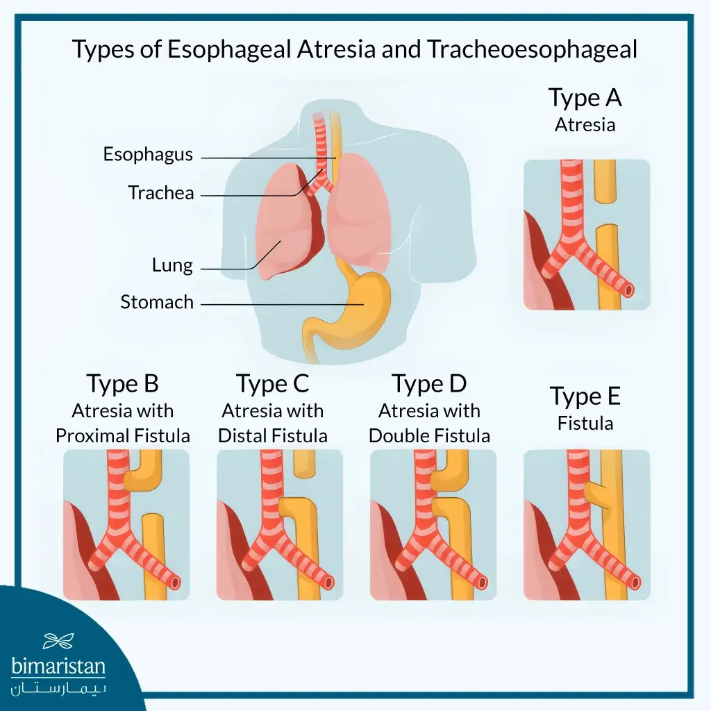
This is the most common form of the malformation, where the baby is born with the esophagus not connected to the stomach, while the lower part of the esophagus is connected to the trachea through a fistula, this leads to two issues at once: food does not reach the stomach and air may enter the gastrointestinal tract leading to flatulence.
There are several types of these malformations, which doctors categorize according to the form of separation and abnormal connection between the esophagus and trachea, the most common of which is the type in which the esophagus is incomplete and its upper end ends with a closed sac while the lower end connects to the trachea through a fistula, but there are other rare types, such as having atresia without a fistula at all or having a fistula only without atresia or having both upper and lower end fistulas.
How is esophageal atresia and tracheoesophageal fistula diagnosed?
The diagnosis of esophageal atresia and tracheoesophageal fistula is usually made in the first hours or days after birth, as there are characteristic symptoms that lead the doctor to suspect the presence of this condition.
Common symptoms in infants
Children with esophageal atresia or tracheoesophageal fistula often exhibit a range of characteristic symptoms:
- Hypersalivation or foaming at the mouth: This is caused by the inability to swallow salivary secretions, often due to a blockage of the upper end of the esophagus.
- Difficulty or failure to feed: The baby may refuse or start feeding and then suddenly stop with signs of shortness of breath.
- Frequent coughing and coughing when feeding: Especially in the presence of a fistula, milk may pass from the esophagus into the trachea and lungs.
- Choking or cyanosis: A blue discoloration of the skin due to lack of oxygen, it appears during or after breastfeeding, especially if milk enters the airway.
- Milk reflux: Milk coming out of the nose or mouth indicates a blockage in the normal swallowing pathway in the upper part of the esophagus.
- Flatulence: Occurs as a result of air passing through the fistula into the stomach, and is more pronounced in cases with tracheoesophageal fistula.
These symptoms draw the attention of the medical team, especially in cases of delivery in a well-equipped hospital, where they intervene quickly to accurately diagnose the cause.
Methods used in diagnosis
When esophageal atresia or tracheoesophageal fistula is suspected, a number of simple and accurate diagnostic procedures are used to confirm the condition and determine the type of abnormality.
- Nasogastric catheterization test: If the esophagus is not connected to the stomach, as in the case of esophageal atresia, the tube will stop in the upper chest and the catheter will bounce back into the mouth.
- X-rays: Chest and abdominal X-rays are taken after the tube is inserted to determine where it stops.
- Visualization of the esophagus using a contrast agent: In some cases, an oral contrast agent is used to precisely map the esophagus or fistula when needed, but this procedure is used with caution to avoid leakage into the lungs.
- Endoscopy or esophagogastroduodenoscopy: In unclear cases or if the exact location of the fistula needs to be determined prior to surgery, esophagogastroduodenoscopy may be used; this procedure shows the location of the fistula between the esophagus and trachea directly.
These conditions are diagnosed quickly and effectively after birth thanks to characteristic symptoms and simple clinical tests, enabling the medical team to make the right decision about the necessary esophageal atresia treatment and tracheoesophageal fistula treatment in a timely manner.
Tracheoesophageal fistula and esophageal atresia treatment methods
Surgical esophageal atresia treatment and tracheoesophageal fistula treatment are the only and primary solutions for the correction of esophageal atresia and tracheoesophageal fistula deformities, as they aim to restore the normal connection between the esophagus and the stomach and close the abnormal connection with the airway. The choice of surgical technique depends on several factors including the type of congenital deformity, the infant’s general health condition and the available medical equipment.
Open surgery for tracheoesophageal fistula and esophageal atresia treatment
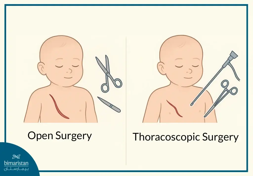
Open surgery is the traditional surgical option in which an incision is made on the right side of the chest to gain direct access to the esophagus and airways. During this procedure, the surgeon closes the abnormal connection between the esophagus and trachea, makes an anastomosis between the two parts of the esophagus when the distance between them is acceptable, and also treats any accompanying abnormalities, if any.
It is resorted to when advanced laparoscopic surgery techniques are not available and there are long distances between the separate parts of the esophagus or the presence of associated complex congenital malformations, as well as in critical cases that require rapid intervention or in cases where previous laparoscopic attempts have failed.
Laparoscopic surgery for tracheoesophageal fistula and esophageal atresia treatment
Laparoscopic surgery represents a breakthrough in esophageal atresia treatment for these cases, where the procedure is performed through small surgical incisions using a microscope equipped with a camera to see internal organs, miniature specialized surgical instruments and advanced magnification techniques, allowing the surgeon to perform the repair of the atresia and fistula without opening the chest. It is characterized by less pain after surgery, less visible surgical scars, a shorter recovery period, and a faster recovery. This method is used if there are specialized surgical centers equipped and an experienced surgical team, and it is also used in cases of medically stable infants and when the distance between the two ends of the esophagus is short.
The success of either surgical technique depends on early diagnosis, prompt intervention, and careful post-operative follow-up to ensure full recovery and avoid potential complications.
Timing of tracheoesophageal fistula and esophageal atresia treatment
The timing of surgical treatment for esophageal atresia and tracheoesophageal fistula depends on several factors:
- Stabilization of the baby’s general condition: If the baby is stable in terms of breathing and circulation, surgery is performed within the first 24-72 hours of birth.
- Presence of other abnormalities: If the baby has other congenital issues such as heart disease or kidney issues, the procedure may be delayed for a more complete evaluation.
- Presence of infections or complications: Such as pneumonia caused by fluid inhalation, this may require treatment first before surgery.
- In cases of long-distance atresia, Surgery may be postponed to give time for the esophagus to grow.
- After esophageal atresia treatment and tracheoesophageal fistula treatment, most cases are successful and the child regains the ability to swallow and feed normally. Some early or late complications may occur that require careful medical follow-up.
Possible complications after tracheoesophageal fistula and esophageal atresia treatment
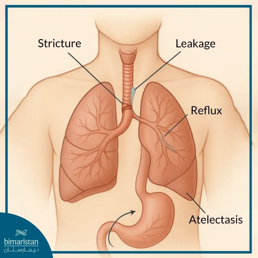
After esophageal atresia treatment and tracheoesophageal fistula treatment, most cases show positive results, as the patient regains full swallowing function and the ability to feed orally, however, early or late complications may arise, highlighting the importance of regular follow-up and long-term monitoring to ensure optimal improvement and manage any potential side effects.
- Leakage from the esophageal suture site: After the treatment of esophageal atresia and tracheoesophageal fistula, leakage may occur at the site of the surgical connection between the two ends of the esophagus, leading to leakage of saliva or food contents into the chest cavity, ranging from mild to severe, the treatment depends on the severity of the case and may require additional intervention.
- Pneumonia or respiratory disorders: May result from previous inhalation of milk or saliva, or due to fluid buildup in the lungs. Treatment includes antibiotics and respiratory therapy.
- Atelectasis: May occur as a result of lung compression during surgery or due to poor ventilation after surgery, usually temporary and improved with respiratory physiotherapy.
- Wound or chest cavity infection: Wound or chest cavity infection is an uncommon complication of esophageal atresia and tracheal fistula treatment, treated with antibiotics and may require surgical drainage in severe cases.
- Suture site stenosis: Suture site stenosis is one of the most common complications of esophageal atresia and tracheoesophageal fistula treatment in the medium and long term, leading to difficulty swallowing, especially with solid foods, treated through balloon dilation or under anesthesia, and may require repeated sessions.
- Gastroesophageal reflux disease (GERD): Common in children after treatment of esophageal atresia and tracheoesophageal fistula, may cause heartburn, vomiting, or developmental delays, treated with antacid medications and, in rare surgical cases, additional intervention.
- Fistula recurrence: The connection between the esophagus and the airway may re-form after surgery, often requiring additional surgical repair.
- Problems with the neurological movement of the esophagus: Some children may have a slow or abnormal movement of the esophagus, resulting in difficulty swallowing food or a feeling of blockage. This issue sometimes appears with age and is managed with progressive feeding and long-term monitoring.
- Developmental delays or feeding difficulties: Some babies may need more time to adjust to breastfeeding or solid foods; these babies are followed up with a nutritionist and developmental support programs.
Thanks to advances in surgical and intensive care techniques, the majority of complications after esophageal atresia treatment and tracheoesophageal fistula treatment can be effectively managed, and most children are able to grow and live a completely normal life after treatment.
Esophageal atresia and tracheoesophageal fistula care and tips for parents
After esophageal atresia treatment and tracheoesophageal fistula treatment, the child needs careful and continuous medical care, starting in the hospital and continuing after discharge home; the primary goal of care is to ensure esophageal healing, prevent complications, and support normal nutrition and growth of the child. In the hospital, the care phase after esophageal atresia treatment and tracheoesophageal fistula treatment begins in the neonatal intensive care unit (NICU). In the NICU, the baby is placed under continuous monitoring, including monitoring breathing, vital signs such as pulse, blood pressure, and temperature, assessing nutritional status and fluid intake, and placing a nasogastric or orogastric tube to feed the baby safely during the first phase of recovery.
Before starting oral feeding, silhouette imaging of the esophagus is usually performed 5-7 days after surgery, the imaging aims to confirm the integrity of the surgical joint, exclude the presence of leakage and assess the movement of the esophagus, where no oral feeding is allowed before confirming the results, when confirming the integrity of the esophagus, small amounts of milk are gradually introduced, while monitoring the child’s response to any signs of difficulty swallowing or reflux, in case of pulmonary complications, oxygen must be given as needed and respiratory physical therapy may be required, and antibiotics are used when there is pneumonia.
After the child is discharged from the hospital, a careful home follow-up phase begins, which includes:
- Regular medical follow-up: The pediatrician and surgeon should be visited regularly to assess growth and swallowing function, and periodic endoscopy or imaging should be performed as necessary to monitor the esophagus for narrowing or reflux.
- Nutrition management: Start with breast milk or appropriate formula in small, frequent amounts and gradually introduce solids under the supervision of a nutritionist. Maintain a semi-sitting feeding position to minimize reflux; in some cases, the baby may need to be fed through a temporary tube to support growth.
- Watch for possible complications: Watch for danger signs that warrant immediate medical attention, such as difficulty swallowing, refusal of food, coughing or choking while feeding, high fever, signs of infection, poor growth or weight loss.
- Prevention of respiratory infections: Adhere to the schedule of basic and additional immunizations such as the flu vaccine and RSV vaccine, avoid contact with people with respiratory diseases, and completely prevent secondhand smoke in the child’s environment.
- Psychological care and rehabilitation: Provide psychological support to parents, especially in the first months after surgery, join support groups for families with children with the same condition, and contact nutritionists and physiotherapists when needed to improve quality of life.
Important tips for parents after tracheoesophageal fistula and esophageal atresia treatment
Caring for a child after esophageal atresia and tracheal fistula treatment requires a lot of attention and support from parents, especially during the first recovery period. Here are some important tips to help parents deal with this stage with confidence and provide the best possible environment for the child’s recovery and proper development.
- Nutrition management: The size and concentration of meals should be graduated according to tolerance, maintaining an upright posture during and after feeding, and careful monitoring of pulmonary aspiration indices.
- Communicate with your medical team: Do not hesitate to consult your doctor if you notice any unusual respiratory symptoms, difficulty eating, or signs of infection.
- Commit to regular visits: Routine visits and checkups should be adhered to
- Comprehensive care: The child’s nutritional, respiratory, and psychological aspects must be taken care of to ensure the best results.
Thanks to modern medical advances, the results of this surgery are excellent in most cases, and adherence to aftercare ensures that your child has a healthy life and normal development.
Tracheoesophageal fistula and esophageal atresia treatment success rate and prognosis
Esophageal atresia and tracheoesophageal fistula treatment is a notable success story in neonatal surgery, with a survival rate of more than 90-95% in children born at normal weight and without other serious congenital malformations, especially in specialized centers and with intensive post-operative care.
Most children who undergo surgical treatment for esophageal atresia and tracheoesophageal fistula show a good recovery and are later able to feed and grow normally, as both swallowing and digestive functions gradually improve over time. Some children may require a short period of nutritional support or special care to ensure full adjustment.
In cases where surgery is performed in the early days of life, and the distance between the two ends of the esophagus is short, the results are often functionally excellent.
Is surgery enough, and when do we need a new medical intervention?
Despite the great success of the first surgery, some children may experience problems later that require follow-up or additional interventions. Here are some reasons:
- Esophageal suture site narrowing: A relatively common complication that causes difficulty swallowing or delayed feeding, it is often treated by dilating the esophagus using a balloon or special instruments, and may be repeated several times.
- Chronic gastroesophageal reflux: Occurs in a large percentage of children after surgery, initially treated with medication, but in some severe cases, additional surgical intervention, such as fundoplication, may be performed.
- Fistula recurrence: It is rare and causes similar symptoms as before surgery such as coughing after feeding or lung infections and requires a second surgery to close the fistula.
- Long distance between the ends of the esophagus: In some rare types of esophageal atresia, the distance between the two ends is long and cannot be connected directly.
These cases may require multiple surgeries or special techniques such as esophageal stretching or even creating a replacement esophagus using part of the intestine or stomach.
Long-term follow-up
Even after successful surgery and recovery, the child needs regular follow-up until childhood and adolescence, including assessing the child’s growth, motor and nutritional development, esophageal function, swallowing and respiratory status, in many cases the child does not notice any difference from his peers over time, especially if there is early follow-up and treatment of any issue.
In conclusion, the treatment of esophageal atresia and tracheoesophageal fistula is a complex surgical intervention that requires high expertise and careful follow-up to ensure the best results, and early diagnosis and specialized treatment contribute to improving the child’s quality of life and reducing potential complications, if you have any questions or need reliable medical advice, you can contact Bimarestan Medical Center where our specialized team is keen to provide support and care of the highest standards.
Sources
- Seattle Children’s Hospital. (n.d.). Tracheoesophageal fistula and esophageal atresia
- MedlinePlus. (n.d.). Esophageal atresia/tracheoesophageal fistula. MedlinePlus Genetics. U.S. National Library of Medicine
