Interventional radiology cancer treatments
Interventional radiology cancer treatments not only help diagnose tumors and determine their stage but also offer advanced, minimally invasive options to treat various types of cancer. With ongoing technological progress, these treatments are playing an increasingly vital role in modern cancer care.
Interventional radiology is a branch of classical radiology in which a radiologist’s team treats a diagnosed disease by inserting a needle through the skin or intravenously without surgery.
Radiological procedures have contributed significantly to advances in tumor diagnosis and treatment in recent years.
The most important advantages that interventional radiation therapy offers are for cancer patients.
The procedure can be performed painlessly through a small puncture and under local anesthesia, has high effects in eliminating the tumor, can be applied with other treatments, and patients can return to their normal lives in a short time after treatment.
In this article, we will explain the types of interventional radiology cancer treatments simply and comprehensively.
What is interventional radiology for cancer?
Interventional radiology cancer treatments are a minimally invasive option for patients seeking alternatives to surgery. Whether you’re not a candidate for surgery or need complementary care alongside chemotherapy, radiation, or immunotherapy, these treatments can help target tumors directly and manage cancer-related pain effectively.
Types of cancers that can be treated with interventional radiology
- Bile duct cancer
- Bladder cancer
- Bone cancer
- Breast cancer
- Colorectal cancer
- Kidney cancer
- Liver tumors
- Lung cancer
- Skin cancer
- Pancreatic cancer
- Most cancers
Most interventional radiology procedures for cancer require an incision as small as the tip of a needle, which minimizes pain and the risk of complications.
Interventional radiology cancer treatments offer a faster recovery compared to traditional methods like chemotherapy, surgery, or radiation. Most procedures are minimally invasive and completed in a single day, with many patients discharged within 24 hours, allowing a quicker return to daily life and activities.
Non-invasive cancer diagnosis with a needle
The most important contribution of interventional radiology cancer treatments to cancer diagnosis is its introduction of the concept of “image-guided biopsy”.
In the past, biopsies were only done through surgery.
For example:
Lung biopsy used to be necessary to surgically open the patient’s bones and take a piece of their lung.
Nowadays, the biopsy itself can be performed painlessly within 5–10 minutes under local anesthesia with a fine needle inserted into the tumor under the guidance of a CT scan or transcutaneous ultrasound, and the patient can go home within a few hours.
Thus, biopsy, which is very important in cancer diagnosis, can be performed quickly and safely, and treatment can be applied to the patient without wasting time through interventional radiology cancer treatments.
Radiotherapy procedures that gave relief to the patient
Diagnosis and treatment methods, which play an instrumental role in the fight against cancer, have given greater importance to patient comfort with technological advancements in recent years.
Interventional radiology cancer treatments can be divided into two groups: percutaneous ablations and transarterial procedures.
Treating tumors with transcutaneous interventional radiology
Direct destruction of the tumor is done with special needles placed in the tumor.
The most common ablation methods are microwave, freezing, nanoknife, and radiofrequency.
Brachytherapy cryoablation
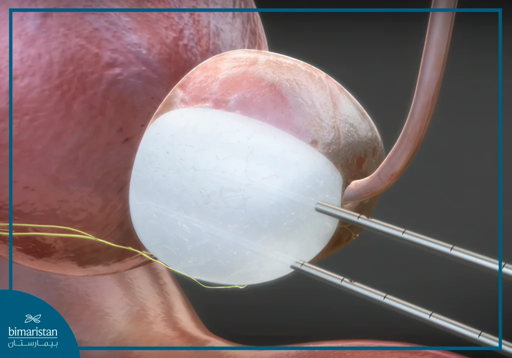
Cryoablation, commonly known as “snowball therapy,” is an innovation in interventional cancer treatment that provides great relief to the patient.
Harmful tissues or tumors in the body are destroyed by freezing, and the treatment works by targeting the cancer cells.
It can also be used in large tumors.
Cryoablation, which is performed by entering the body with special needles, destroys any tissue or tumor by freezing it.
Initially, an appropriate number of needles are placed in the mass under the guidance of information obtained from imaging devices such as ultrasound, computed tomography (CT) or magnetic resonance imaging (MRI).
While one needle is sufficient for a mass with a diameter of 1 cm, the number of needles increases as the diameter of the tumor grows.
In today’s cryoablation devices, 20-25 needles can be placed in the body.
They can be operated at the same time so that very large masses can be destroyed by the use of extreme cold.
The tumor is cooled to -80 degrees.
In cryoablation, argon gas is circulated through the needles and this creates a coldness of -20 to -80 degrees at the tip of the needle.
This cold destroys and kills the tumor or pathological tissue.
Cryoablation is a technique that targets only the tumor and has very distinctive features.
It can be applied multiple times because it only destroys the cancerous area and does not damage healthy tissue.
Take control of your health—discover advanced Interventional Radiology Cancer Treatments at Bimaristan Medical Center in Turkey. Minimally invasive, highly effective—schedule your consultation today!

Great success in risky cancers
Cryoablation can be used successfully in common and risky cancers such as prostate, kidney, lung, liver, and soft tissue tumors.
It is a method that can be applied alone or in conjunction with chemotherapy and radiation therapy.
Freezing in the brain, spinal cord, and areas where there are very important nerves is performed while preserving healthy nerve tissue with special techniques.
But it has no place in the treatment of tumors of the stomach, intestines, and gallbladder or in tumors close to these organs.
It can be performed with a needle under the guidance of ultrasound or computed tomography.
Radiofrequency in interventional radiology cancer treatments
It is the most commonly used method of “tumor destruction” procedures in the treatment of liver tumors, lung cancer, soft tissue cancer, and bone tissue cancer.
Cancer cells are burned by inserting a needle into the tumor.
Radiofrequency is one of the most commonly used methods.
Radiofrequency ablation is the most common; it is used in liver tumors, lung cancer, kidney cancer, and thyroid cancer treatment.
It is also used for bone metastases.
In many types of cancer, the tumor can spread to the bones in the final stage of the disease and cause severe pain.
These metastases can weaken bone tissue and lead to fractures, which can cause very severe pain.
In such cases, the pain can be relieved by the radiofrequency burning of the tumor tissue in the bones.
In the same session, bone strength can be increased by injecting a cement-like liquid into the bone.
These methods, especially in patients with a malignant spinal tumor who suffer from severe pain, can reduce the pain caused by the tumor and may also prevent some of the neurological issues that may occur due to the destruction of the spinal vertebrae.
Microwave burn
Similar to the action of radiofrequency, it eliminates tumors by burning or cauterizing.
Recently, it has become more favored and used more often than radiofrequency.
Its most common application is for liver tumors.
Nanoknife Nanoknife (Non-reversible Electroporation)
In this method, special needles are placed inside the malignant tumor area, and very high electric currents are given to the patient under general anesthesia for a short time.
In this way, large holes are opened in the walls of the cancer cells, and the cancer cells die from the radiation without the healthy cells being affected.
The most important advantage of this procedure is that critical structures such as vessels, nerves, and the gastrointestinal tract are not damaged during the ablation procedure.
Therefore, the Nanoknife method is especially preferred in tumors adjacent to such organs.
Nanoknife offers promising results in liver tumors adjacent to the bile ducts and large vessels in liver cancer, pancreatic cancer, and prostate cancer. Electroporation can be combined with surgery or other ablation methods if needed.
Read more about Gamma Knife’s role in treating tumors.
Treatment of tumors with transarterial interventional radiology
One of the key interventional radiology cancer treatments is endovascular therapy. This includes intra-arterial chemotherapy, used for patients who do not respond to standard chemotherapy. By targeting the blood vessels feeding the tumor, high-dose chemotherapy is delivered directly through a minimally invasive procedure. This approach offers fewer side effects and enhances the drug’s effectiveness on the tumor.
Chemoembolization therapy through vascular catheterization
It is especially used in patients with hepatocellular carcinoma that cannot be treated surgically.
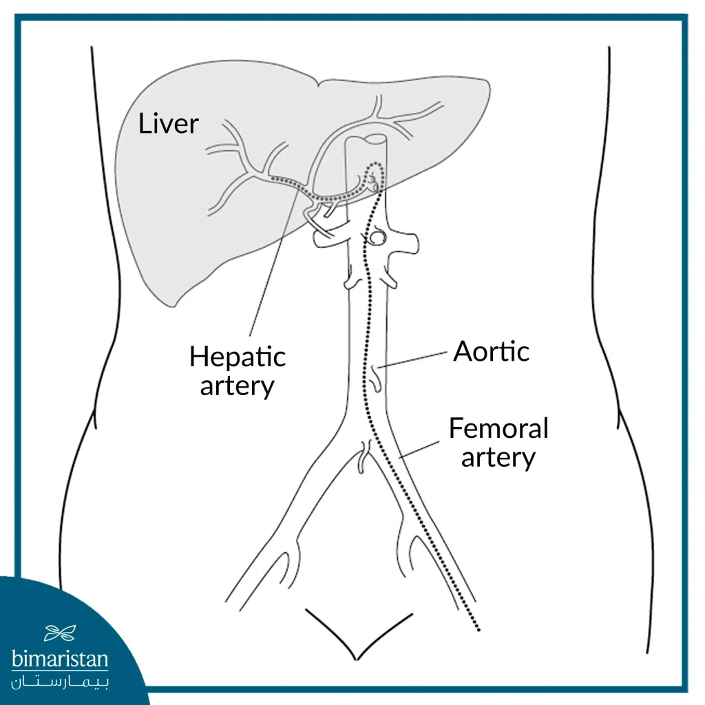
The radioactive material used in the imaging is mixed with chemotherapy and injected through a liver artery with a blood catheter.
This combination of drugs is a substance that is loved and absorbed by the cancer cells much more than healthy cells.
After this substance is injected into the tumor, the blood vessel is embolized with small particles, thus preventing the chemotherapy substance from moving away from the tumor, and due to the blockage of the vessels, hypoxia occurs in the tumor tissue.
This increases the effectiveness of the injected substance.
Thanks to this procedure, both the vessels that feed the tumor are blocked, and the chemotherapy drug is trapped in the tumor in a single session, with the targeted tissue receiving a much higher dose of chemotherapy than the dose of chemotherapy administered through conventional methods.
Catheter-based coagulation is one of the most recent major treatment modalities to emerge and is used for uterine fibroids and prostate enlargement without the addition of chemotherapy or radiation.

Radioembolization (Y-90) (Yttrium-90 Treatment)
In radioembolization, radioactive material is loaded into vascular embolization particles and injected directly into the liver artery or vein feeding the tumor.
In this method, very high doses of radiation therapy are given to the tumor, the vessel is coagulated as in the previous method, and healthy tissue is only slightly affected.
This procedure is applied in both liver and kidney tumors.
Chemosaturation used in interventional radiology cancer treatments
The method is only used in liver tumors where very intense amounts of the chemotherapy drug are delivered directly to the cancer cells.
During the procedure, the arteries and veins of the liver are isolated, and large amounts of chemotherapy drugs are prevented from entering the bloodstream and causing toxic effects on the patient.
The procedure is applied to the patient every month.
At Bimaristan Center, we help you choose the best interventional radiology cancer treatments.
Sources:
