The number of spinal fusion surgeries performed worldwide has significantly increased in recent decades, attributed to substantial advancements in surgical techniques and technologies. These procedures have emerged as a suitable option for treating various spinal problems due to their ability to significantly improve symptoms associated with these issues.
What are Spinal Fusion Surgeries?
Spinal fusion is a surgical procedure that involves connecting and stabilizing two or more vertebrae in any part of the spine. This stabilization is done by using screws, rods, or fusion by placing a bone-like material between the vertebrae. This reduces movement between the vertebrae, making the spine more stable. This helps alleviate pain and achieve the desired goal of the procedure.
Indication for Spinal Fusion Surgeries
Conditions that may necessitate spinal fusion surgeries include:
- Spinal deformities such as scoliosis and herniated discs
- Spinal disc slippage
- Spinal tumors
- Spinal injuries and fractures resulting from falls or car accidents
how is a spinal fusion done?
spinal fusion surgeries surgery is typically performed under general anesthesia. The surgeon may use various approaches depending on the location of the vertebrae to be fused, the reason for the surgery, and the patient’s overall health. Generally, the procedure involves the following steps:
- The surgeon makes an incision in the back, neck, or even through the abdomen, depending on the injury’s location and what the doctor deems most appropriate for the patient’s optimal outcome.
- Bone grafts are prepared either from a bone bank or from the patient’s own body, typically from the pelvic area. The surgeon makes a small incision near the pelvic bone, removes a small piece of the bone, and then closes the wound. Alternatively, the surgeon may use synthetic materials that mimic bone.
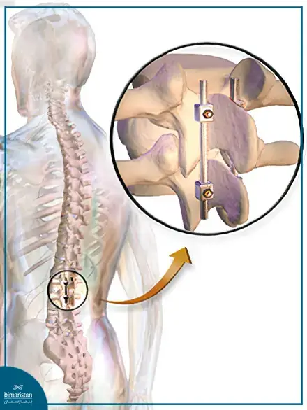
- Fusing and Stabilizing the Vertebrae, the surgeon places the bone grafts between the vertebrae and stabilizes them using metal plates, screws, or rods to hold the vertebrae in place until the bone graft heals and fuses the vertebrae together.
Types of Spinal Fusion Surgeries
Cervical Fusion Surgery for Vertebrae 1-2
Instability of the first and second cervical vertebrae can be caused by several conditions, including:
- Fracture of the odontoid process
- Rupture of the transverse ligament
- Ligament dysfunction caused by Rheumatoid Arthritis
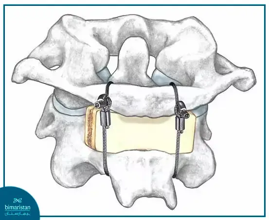
In such cases, cervical fusion surgery, which involves fusing the first and second cervical vertebrae (C1-2) and stabilizing them with a cable, may be necessary. The surgeon can perform this procedure using one of three techniques:
Gallie Technique
The surgeon passes a metal wire under the posterior arch of the first cervical vertebra (C1) and then loops the wire over C1 and under the spinous process of the second cervical vertebra (C2), The free ends of the wire are then tied together centrally to secure the bone graft. This technique is relatively safe and easy to perform.
Brooks Technique
This method involves placing one central or two lateral bone grafts between the posterior arch of C1 and the lamina of C2. The vertebrae are stabilized by passing two metal wires under the laminae of C1 and C2 and tying the free wire ends behind the bone graft. While this technique provides greater rotational stability, it presents challenges such as difficulty passing the wire under the laminae and the risk of spinal cord injury, particularly if spinal canal stenosis is present due to another condition.

Sonntag Technique
The Sonntag technique combines the simplicity and safety of the Gallie technique with the biomechanical advantages of the Brooks technique.
In this procedure, a metal wire is passed under the lamina of the first cervical vertebra (C1) and then tied below the base of the spinous process of the second cervical vertebra (C2), similar to the Gallie technique. However, the bone graft is placed between the posterior elements of the vertebrae as in the Brooks technique. The free ends of the wires are tied under the base of the C2 spinous process. Unlike the Brooks technique, the Sonntag technique does not involve passing the wire under the lamina of C2. Instead, an incision is made at the bottom of the graft, which is then secured to the superior surface of the C2 spinous process.
Cervical Fusion for Other Cervical Vertebrae
Anterior cervical fusion is the most common procedure in spinal surgeries due to its several advantages over posterior cervical fusion. These benefits include effective decompression and the use of advanced devices, allowing for spinal canal decompression without disturbing the spinal cord. The procedure involves placing the patient in a supine position and making an incision on the side of the neck, usually preferring the left side to avoid damage to the recurrent laryngeal nerve, though this puts the thoracic duct at risk. The surgeon then proceeds with the necessary steps to stabilize and fuse the vertebrae.
Thoracic Vertebrae Fusion
In thoracic vertebrae fusion, Harrington rods are commonly used. Since their introduction, these surgeries have significantly improved. The rods are fixed to the spine at multiple points, allowing the use of screws, wires, and hooks to facilitate the procedure.

Lumbar Spine Fusion Surgery
Posterior Lumbar Fusion Without Using Screws
This procedure is relatively easy to perform and is used to treat specific lumbar spine issues, especially when the patient is not ready for using metallic screws. However, it is not recommended if there is significant instability in the lumbar spine. The procedure begins with a surgical incision to expose the lumbar spine and the vertebrae that need to be fused and stabilized. The lamina of the vertebra is often removed to reduce pressure on the nerve, and the transverse processes are trimmed. A bone graft is then placed in the appropriate position to achieve the needed fusion.
Lumbar Fusion Using Screws
This is the most commonly used method to achieve the required stability in the lumbar spine. Screws are inserted into the vertebrae and connected with plates or rods. The surgeon places the screws into the pedicles of the vertebrae above and below the part of the vertebra to be fused, ensuring that the exiting nerve root is not damaged.

Minimally Invasive Spinal Fusion Surgeries
Minimally invasive spinal fusion surgeries are a safe and effective treatment option for various minor spinal issues. These procedures stabilize the vertebrae through a small incision without cutting the surrounding muscles. Instead, a muscle retractor is used to access the spine, the surgeon is guided by X-ray imaging throughout the operation to help him place the spinal fusion screws accurately.
Benefits of Spinal Fusion Surgeries
Spinal fusion surgeries help alleviate pain and symptoms associated with various spinal problems, such as tingling or numbness in the arms or legs. They can also correct spinal deformities that primarily affect body posture, such as scoliosis, thereby improving the quality of life for patients and helping them feel comfortable again. Most patients who undergo these surgeries are satisfied with the results.
Risks of Spinal Fusion Surgeries
All surgical procedures carry some risks, but the level of risk varies from one surgery to another. The primary risks of spinal fusion surgeries are due to the proximity of the surgical site to nerves and other vital structures, which may lead to:
- Reduced mobility
- Swelling and redness
- Pain and general discomfort
- Injuries to nerves and blood vessels
- Bleeding that may require a blood transfusion – studies have shown that tranexamic acid and aprotinin can reduce blood loss in spinal surgeries
- In rare cases, paralysis or hematoma
Spinal Fusion Success Rate
The success rate of spinal fusion surgeries can be as high as 90%. However, the definition of success varies, including pain relief, improved neurological function, the ability to perform certain activities, achieving complete vertebral fusion, correcting spinal deformities, proper wound healing, and more.
Preparations Before Spinal Fixation Surgeries
Patients must undergo several laboratory tests and imaging studies as recommended by their physician to gather essential information about their general health and their suitability for surgery.
Postoperative Care
Patients typically stay in the hospital for two to three days after spinal fusion surgeries, although some may be able to return home the same day. Patients with a history of severe health conditions, such as blood clots or sleep apnea, or those who experience postoperative complications, may need to stay in the hospital for four or more days. After returning home, patients should follow specific guidelines provided by their doctor and physical therapist to promote healing.
A follow-up visit is recommended 7-10 days after spinal fusion surgeries to monitor the wound, address any patient concerns, and evaluate the progress of spinal fusion, pain improvement, and neurological function recovery. Further follow-ups should occur at six weeks and three months post-surgery.
Cost of Spinal Fusion Surgeries
The cost of spinal fusion surgeries varies depending on the patient’s condition, the type and technique of surgery, and the required tools. In Turkey, the cost ranges from $5,000 to $7,000, which is considerably lower compared to prices in the United States and Europe.
Spinal fusion surgeries have achieved significant success in the medical field, helping many patients regain the ability to perform daily activities and improving their overall quality of life. These surgeries have become an important treatment option for various spinal issues
Resources:
- AJR: Spine Fixation Hardware
- HSS


