
Lung and chest diseases are conditions that can directly affect the body’s ability to breathe and supply itself with oxygen, so the sudden onset of chest pain or difficulty breathing may be a sign of a medical problem that requires prompt evaluation and treatment. Chest disorders vary in severity and cause, with some being mild and temporary, while others may progress to become life-threatening if not properly diagnosed and treated.
Among these conditions, pneumothorax stands out as one of the common chest disorders that can occur suddenly in some people, whether as a result of a direct injury, certain lung diseases, or sometimes even for no apparent reason. Symptoms can range from mild discomfort to severe medical emergencies requiring immediate medical attention.
What is Pneumothorax?
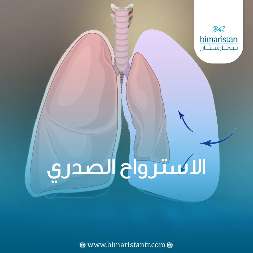
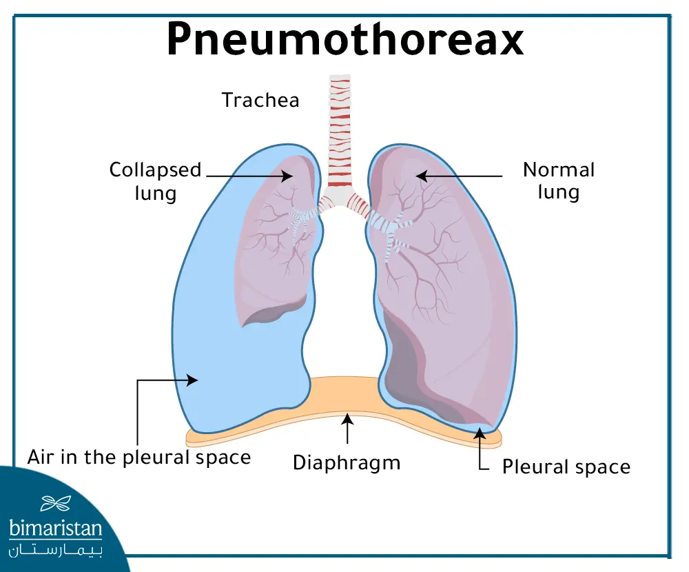
Pneumothorax, or a collapsed lung, is a buildup of air in the pleural space between the lung and the chest wall, which increases pressure on the lung and causes it to collapse partially or completely, making it difficult for the lung to expand normally during breathing. This can cause symptoms ranging from mild chest pain and slight shortness of breath to severe respiratory distress in some cases.
The severity of this condition varies with the amount and rate of air accumulation in the pleural cavity; it may be mild and resolve with observation, while in some cases it may progress to a life-threatening emergency requiring immediate medical intervention.
Types of Pneumothorax
There are two main types of pneumothorax: spontaneous and traumatic, each of which includes several subtypes that differ by cause and mechanism.
Spontaneous Pneumothorax
This type occurs without direct trauma to the chest and includes:
- Primary spontaneous pneumothorax: This occurs in people without any apparent lung disease and often appears suddenly.
- Secondary spontaneous pneumothorax: This occurs as a result of a pre-existing lung disease or disorder and is usually more severe than the primary type.
Traumatic Pneumothorax
It results from a direct injury to the chest or lung, and includes:
- Traumatic pneumothorax: This condition is caused by various chest injuries that result in air leaking into the pleural cavity.
- Post-procedural Pneumothorax: This may occur as a complication of certain medical procedures or thoracic surgeries.
Other Types
- Tension pneumothorax: This is considered one of the most serious forms and occurs when air accumulates in the pleural cavity, increasing pressure on the lung, heart, and blood vessels, making it a life-threatening medical emergency. Any type can progress to a tension pneumothorax if the air leak persists and the air cannot escape from the pleural cavity.
- Menstrual Pneumothorax: A rare condition that occurs in some women with endometriosis, often coinciding with the menstrual cycle. It results from the migration of endometrial-like tissue into the pleural cavity, which can lead to air leakage and lung collapse.
Causes of Pneumothorax
A pneumothorax occurs when air leaks into the pleural space surrounding the lung, increasing pressure on the lung and causing collapse. It may occur spontaneously, as a result of a direct injury, or as a complication of certain diseases or medical procedures.
Lung Diseases
Some lung diseases increase the risk of this condition due to weakened lung tissue or the formation of air bubbles within it. The most common of these are:
- Chronic Obstructive Pulmonary Disease (COPD)
- Asthma
- Cystic Fibrosis
- Pulmonary Fibrosis
- Pulmonary Tuberculosis
- Lung Cancer
- Severe Pneumonia
The rupture of small air bubbles on the lung surface is also one of the most common causes of primary spontaneous pneumothorax.
Injuries and Bruises
A pneumothorax may occur as a result of various chest injuries that cause air to leak from the lung, such as:
- Stab Wounds and Penetrating Wounds
- Gunshots
- Rib Fractures
- Traffic Accidents
- A fall or severe blow to the chest
Medical Causes and Treatment Procedures
Pneumothorax may sometimes occur as a complication of certain medical or surgical procedures, the most notable of which include:
- Lung Biopsy
- Bronchoscopy
- Insertion of Central Intravenous Catheters
- Mechanical Ventilation
- Some Thoracic Surgical Procedures
Risk Factors
There are several factors that may increase the risk of developing a pneumothorax, the most important of which are:
- Smoking
- Excessive Height and lower body weight
- A family history of a previous diagnosis
- Marfan Syndrome
- Changes in atmospheric pressure, such as deep-sea diving or air travel, in some cases
In addition, people who have previously had a pneumothorax are more likely to have another one than others.
Symptoms of Pneumothorax
The symptoms of pneumothorax vary with the amount and rate of air accumulation in the pleural cavity; symptoms may range from mild to severe, depending on the type of pneumothorax and the degree of lung collapse.
Sudden chest pain and shortness of breath are among the most common symptoms of pneumothorax; the pain is often sharp or stabbing and worsens with inhalation or coughing. The patient may also feel discomfort or pressure in the chest, along with varying degrees of difficulty breathing. Other symptoms may include:
- Rapid breathing
- Rapid heartbeat
- Dry cough
- Feeling tired or dizzy
- Cyanosis of the lips or face in severe cases
Symptoms usually begin suddenly and may be mild during simple breathing, while they appear more severe and dangerous during labored breathing or in patients with chronic lung diseases.
When is pneumothorax a medical emergency?
Pneumothorax requires emergency medical intervention when severe shortness of breath or a rapid deterioration in the patient’s condition occurs, especially if accompanied by cyanosis, altered consciousness, or signs of compressive chest pain, as a delay in treatment may lead to serious, life-threatening complications.
Diagnosis of Pneumothorax
The diagnosis of pneumothorax is based on the patient’s symptoms and a clinical examination, during which the doctor asks about chest pain, shortness of breath, medical history, and any previous lung diseases or chest injuries. The doctor will then examine the chest with a stethoscope and may notice weakened or absent breath sounds on the affected side. To confirm the diagnosis and assess the severity of the condition, the following tests may be used:
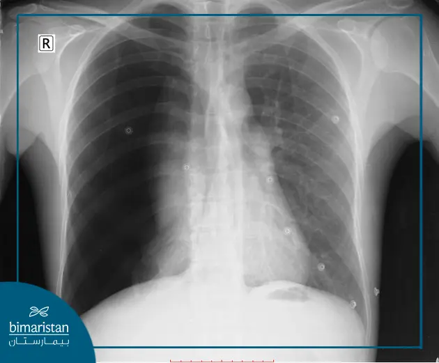
- Plain chest X-ray: This is the most commonly used test for confirming a pneumothorax.
- Computed Tomography (CT): Used to detect small pneumothoraces or to more accurately assess associated pulmonary diseases.
- Ultrasound imaging: Especially in emergency cases and for patients with chest trauma.
- Measuring the oxygen level in the blood, or arterial blood gas analysis: to assess respiratory function.
- Electrocardiogram (ECG): In some cases, to rule out cardiac causes of chest pain.
Treatment for Pneumothorax
Treatment for pneumothorax depends on the severity of the condition, the amount of air accumulated in the pleural cavity, and the patient’s overall symptoms and lung condition. Treatment options range from simple observation to interventional procedures and, in advanced cases, surgery. The primary goal of treatment is to remove the accumulated air, help the lung re-expand, and prevent the recurrence of pneumothorax as much as possible.
1. Monitoring and Oxygen Therapy
A simple pneumothorax may not require surgical intervention, as it can heal gradually with monitoring and oxygen administration to accelerate reabsorption of air from the pleural cavity. During this period, periodic X-rays are taken to confirm lung expansion and improvement in the patient’s condition.
2. Air aspiration with a needle
In some cases, air is aspirated using a needle or a thin catheter to remove air that has accumulated around the lung, particularly in patients with obvious symptoms or when the pneumothorax is moderate in size.
3. Chest drainage with a tube
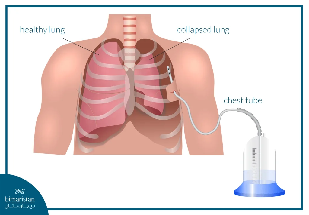
If the pneumothorax is large or causes severe shortness of breath, the patient may need to have a chest tube inserted into the pleural cavity to continuously drain air until the lung returns to its normal expanded state; the tube may remain in place for several days, depending on how the condition responds to treatment.
4. Pleural fixation
Pleural fixation may be performed as part of surgical treatment or as an option for patients who cannot undergo surgery, with the aim of reducing the risk of recurrence. It involves causing the two pleural layers to adhere to each other using substances such as talc powder or other pleural irritants.
5. Advanced Interventional Therapies
In some complex cases where air leakage persists despite conventional treatment, advanced treatment techniques may be used, such as injecting autologous blood into the pleural cavity to help seal the air leak or placing one-way valves via bronchoscopy to reduce air leakage and help the lung re-expand.
6. Surgery
Some patients may require surgical intervention, particularly in cases of recurrent pneumothorax, persistent air leakage, or failure of the lung to re-expand despite treatment. Surgery is often performed using video-assisted thoracoscopy (VATS), during which the leak site is repaired, and the air bubbles causing the condition are removed, with the option of performing a pleural seal to reduce the likelihood of recurrence.
Some of the most important conditions that may require surgery include:
- Recurrent pneumothorax
- The air leak has persisted for more than several days
- Involvement of both lungs
- Lung Failure to Expand Despite Chest Tube Placement
- Patients exposed to pressure changes, such as pilots and divers
- Certain severe chest injuries and contusions
Follow-up After Treatment
After recovery, your doctor may recommend that you avoid certain activities for a short time, such as air travel, scuba diving, or strenuous exercise, and attend regular follow-up appointments to ensure the pneumothorax does not recur.
Complications
A pneumothorax can lead to serious complications if it is large or not treated properly, especially in patients with chronic lung diseases or in cases of tension pneumothorax. The most notable potential complications include:
- Respiratory failure resulting from lung collapse and impaired normal oxygen exchange
- Low blood oxygen levels and circulatory problems in severe cases
- Hemothorax when expiration is accompanied by intrapleural hemorrhage
- A prolonged air leak from the lung
- Recurrence of chest pain after treatment
- Pressure on the heart and large blood vessels during chest compression, which may endanger the patient’s life
- Death in severe, untreated cases or when emergency care is delayed
Prevention
Not all cases of pneumothorax can be prevented, especially spontaneous cases that may occur for no apparent reason, but the risk of developing or experiencing a recurrence can be reduced by following some important preventive measures.
Some of the most important tips for preventing this condition are:
- Quitting smoking, as it is one of the most significant risk factors for developing and recurring chest pain
- Commitment to the regular monitoring and treatment of chronic lung diseases
- Follow safety instructions to reduce the risk of chest injuries and contusions
- Adherence to regular follow-up visits after treatment, especially for patients who have previously undergone thoracostomy
- Avoid deep diving or activities that involve significant changes in atmospheric pressure unless you have consulted a doctor
Some people may be more susceptible to the condition due to genetic factors or certain lung diseases, so regular medical follow-up remains important for early detection and to reduce the risk of complications.
In conclusion, pneumothorax is a condition in which severity can range from mild to life-threatening, so early diagnosis and appropriate treatment play a crucial role in preventing complications and improving the chances of recovery. With advances in modern diagnostic and treatment methods, most cases of pneumothorax can now be successfully treated, especially when patients adhere to medical follow-up and avoid risk factors that may increase the likelihood of recurrence.
Sources: