Pleural effusion (fluid accumulation around the lungs) is a common medical condition and may be an indicator of a serious disease such as cancer, so it requires prompt medical intervention to find out the cause and treat it.
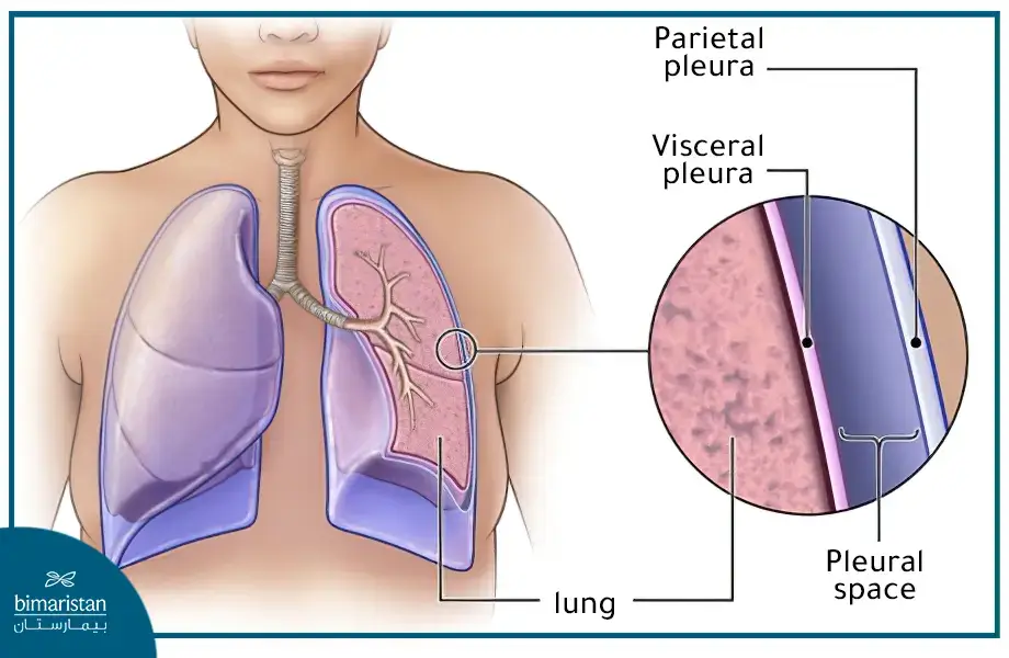
The lungs are surrounded by a membranous sac made up of two leaflets called the pleura. One of the leaflets adheres closely to the lung and is called the visceral pleura, while the other leaflet adheres to the inside of the rib cage and is called the parietal pleura. Between the two leaflets is a thin, fluid-filled cavity called the pleural cavity.

In this article, we will explore the causes and treatment of pleural effusion in Turkey.
What is pleural effusion disease?
Pleural effusion is caused by increased fluid accumulation in the pleural cavity, where the normal amount of fluid around the lungs is between 10–20 ml. This increase arises from an imbalance between hydrostatic pressure, which moves fluid out of the vessels, and oncotic pressure, which pulls fluid into the vessels.
Pleural effusion severity depends on the underlying cause of the effusion and its effect on respiratory capacity. Pleural effusion is never required to be the same amount on both sides, usually bilateral when caused by a systemic disease.
Types of pleural effusions
Special criteria have been established to categorize the types of pleural effusions according to the amount of protein present in order to facilitate the study and orientation towards the possible cause of the effusion, where two different types have been distinguished:
Transudative pleural effusion
This type of effusion is caused by an imbalance in fluid pressure or the transfer of fluid from the peritoneal cavity in the abdomen to the pleural cavity in the chest, and in rare cases it is caused by a medical error (such as during a bronchoscopy or when a catheter in the jugular vein in the neck moves and punctures the pleura).
A systemic disease usually causes an infiltrative effusion. The most common diseases that cause it include:
- Congestive heart failure
- Liver fibrosis
- Pulmonary embolus
- Nephrosis
- Protein deficiencies due to malnutrition, for example
Exudative pleural effusion
A high amount of protein characterizes this type of effusion, which is often caused locally within the chest cavity. The main diseases that cause it include:
- Pneumonia
- Pulmonary embolus
- Tuberculosis
- Tumors such as lung cancer
Causes of pleural effusion
There is a wide range of diseases that can cause pleural effusions, and knowing the cause of the effusion may be the key to treatment. Causes include the following:
- Circulatory heart disease, as in congestive heart failure and pericarditis.
- Pulmonary diseases, such as pulmonary embolism and pulmonary infarction.
- Hypoalbuminemia is caused by pancytopenia or malabsorption diseases.
- Tumors (malignant pleural effusion); bronchial cancer, metastases, lymphoma, chest wall tumors.
- Septic diseases, such as pneumonia, tuberculosis, and pyogenic bacteria.
- Connective tissue diseases, such as systemic lupus erythematosus and rheumatoid arthritis.
- Trauma may cause a hemorrhagic or chylous effusion.
- Other causes, such as Mediterranean fever and chronic hemolysis(kidney dialysis).
Pleural effusion symptoms
If you have a mild pleural effusion (small amount of pleural fluid), you may not experience any symptoms, but if the effusion is moderate or severe, or if it is accompanied by inflammation, you may experience one of the following symptoms:
- Dry cough
- A sense of heaviness and pain in my chest
- Respiratory distress (shortness of breath) during exertion or even at rest
- Fever; if the effusion is accompanied by inflammation
Signs of pleural effusion
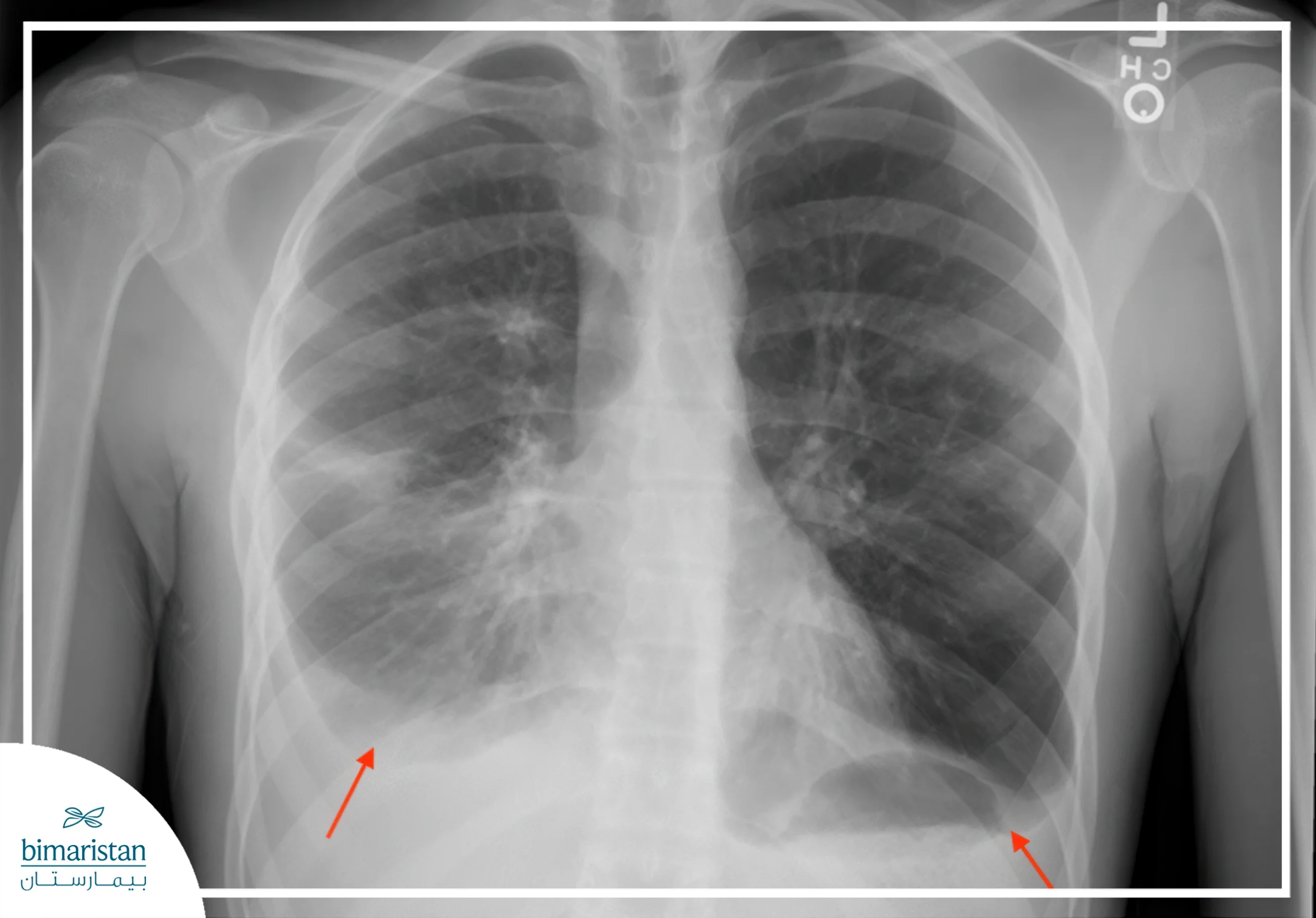
There are a number of important signs of pleural effusion, some of which are seen during the physical examination and some of which are detected on a chest X-ray (CXR). The following table shows the most important signs:
| Clinical assets | Radiograph assets |
| Limitations in chest movement during breathing | Absence of a free phrenic angle |
| Absence of sonic vibrations during chest palpation | Appearance of a hemorrhagic line (observed when the effusion is in large volumes exceeding 300 ml) |
| Percussion at the fifth intercostal space | Massive effusion with cardiac displacement |
| Reduced respiratory sounds |

Diagnosis of pleural effusion
Your doctor will start by asking about your symptoms and health history, then perform a physical examination, including listening to your lungs with a stethoscope. Tests that may be used to diagnose include the following:
- A simple chest X-ray: Often the first test performed to detect the presence of fluid in the pleural cavity.
- Ultrasound (Echo): Gives more detailed information about the amount and location of fluid in the pleural cavity.
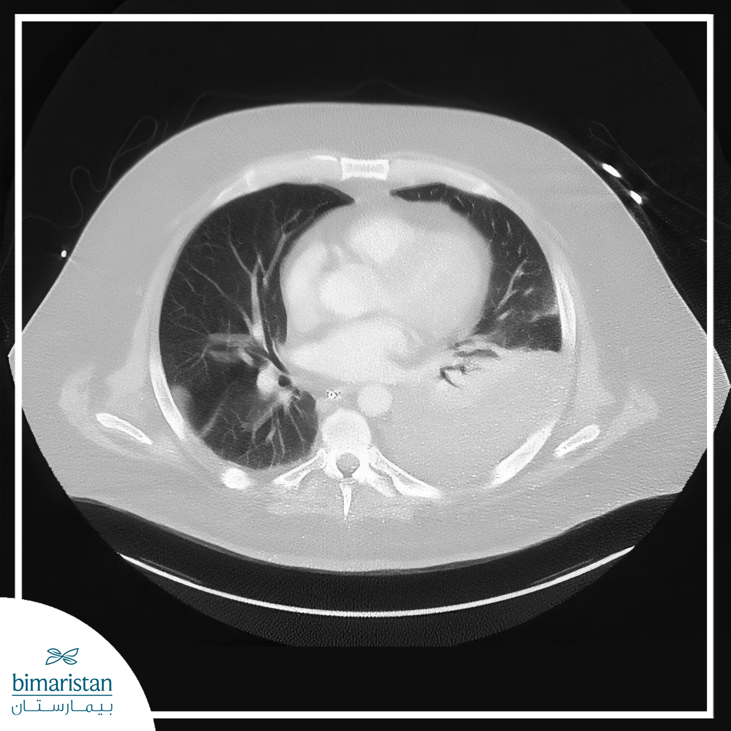
- CT scan: Provides more detailed information about the chest and helps determine the cause of the effusion.
- Pleurodesis: Inserting a needle into the pleural cavity to obtain and analyze a sample of fluid can help determine the cause of the effusion.
- Pleural biopsy: It is more useful in diagnosing tuberculous lesions than tumors because tuberculosis nests in the pleura, while a tumor may be elsewhere.
- Thoracoscopy: This can take a biopsy of the pleura, lungs, or even fluid.
- Blood tests: A sample of the patient’s blood may be drawn to check for signs of infection or inflammation.

Pleural effusion treatment in Turkey
In some cases, treating the cause is enough to get rid of the pleural effusion, such as antibiotic therapy in the presence of bacterial pneumonia, diuretic therapy in congestive heart failure, or cancer treatment in malignant pleural effusion…
In other cases, we have to remove the fluid from the pleural cavity to save the patient’s life or improve their breathing through:
Pleurodesis
In cases of mild to moderate effusions, the fluid may be removed from the pleural cavity using a special needle to relieve pressure on the lungs and improve breathing.
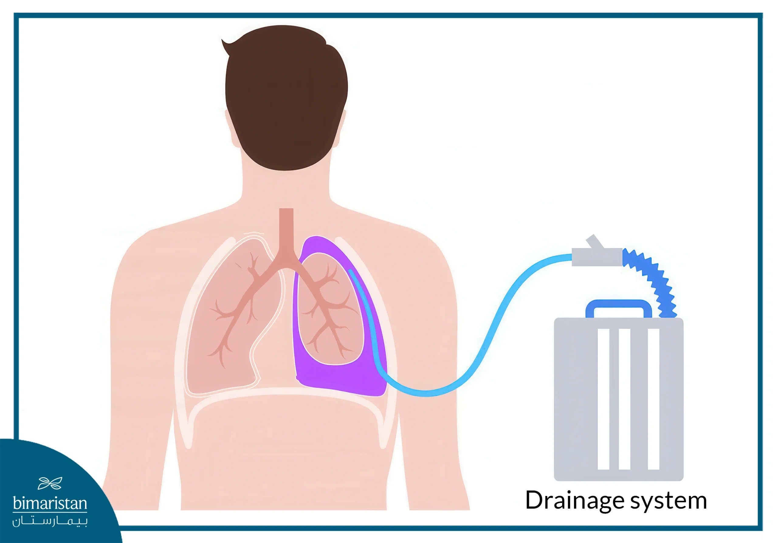
Chest drainage
A skin incision is made and a flexible tube is inserted into the pleural cavity to drain the fluid, after which a simple chest x-ray is performed to confirm that all the fluid has been drained.
We do not use chest drainage for effusions because the fluid may collect and cause infections as a result of prolonged placement of the chest drainage tube (about 5 days).
Therefore, we resort to chest blasting only in two cases:
- When the effusion involves the entire lung with a deviated heart
- If the patient’s general condition is poor

Pleurodesis
In special cases, after blowing the chest and making sure all the fluid is drained, we inject a chemical substance into the pleural cavity, this substance creates an inflammatory event that leads to the adhesion of the two pleura leaflets, which in turn leads to the absence of the pleural cavity and prevents the fluid from re-collecting permanently.
The chemical used in pleurodesis may be:
- Talcum powder (best)
- Bleomycin (does not cause pain)
- Tetracycline (may cause severe pain)
If the patient does not improve on pleurodesis, we insert a piece of gauze and scratch the pleura so that blood comes out and makes the surface rough, thereby inducing inflammation, and the two sheets stick together.
Read more about thoracic surgery in Turkey.
Complications of pleural effusion
It’s important to consult a doctor if you have any symptoms of an effusion, as untreated or severe cases can lead to serious complications such as:
- Lung tissue damage can lead to respiratory failure
- An infection in the pleural cavity that may turn into an abscess called empyema
- Pneumothorax after draining accumulated pleural fluid
- Pleural thickening (scarring of the lung lining)
In conclusion, people suffering from pleural effusion should seek medical help as soon as they experience symptoms. Medical treatment and appropriate medical procedures can help improve symptoms and minimize potential complications. It is also important to follow your doctor’s directions and maintain a healthy lifestyle to help prevent pleural effusions from recurring.
Sources:

