Urethral stricture is a common urinary condition that impacts quality of life and urinary function, and neglecting it may cause urinary retention or recurrent infections. Symptom severity and disease progression differ among patients, requiring precise evaluation and individualized care. Treating urethral stricture is crucial for better outcomes and preventing recurrence—especially through surgical urethral stricture treatment, which offers effective results. Selecting the right timing and surgical approach directly influences treatment success and long-term stability.
What is urethral stricture?
Surgical urethral stricture treatment addresses a condition in which the urinary tract narrows due to fibrosis or scarring from injury, infection, or previous medical intervention. This narrowing partially or completely obstructs urine flow, causing bothersome urinary symptoms and potential complications if untreated. The condition is more common in males, and its severity depends on the location, length, and underlying cause of the narrowing.
Causes of urethral stricture
The causes of urethral stricture are many and include:
- Direct injuries: Trauma to the perineum or pelvis.
- Chronic infections: Especially those caused by sexually transmitted infections such as gonorrhea.
- Medical procedures: Such as long-term urinary catheterization or cystoscopy.
- Previous urological surgeries: Including urethral or prostate surgeries.
- Congenital defects: In rare cases.
As transurethral laparoscopic surgeries become increasingly common, they can cause complications, such as transurethral resection of a bladder tumor, which can lead to urethral stricture or urethral progression if present. This is especially true when repeat laparoscopic procedures are performed, as there may be mechanical friction between the instruments and the urethra or injury to the urethral mucosa.
Symptoms of urethral stricture
Several clinical symptoms characterize urethral stricture:
- Weak urine flow with an initially slowed or intermittent stream of urine
- The sensation of not fully emptying the bladder, leading to frequent urges to urinate
- Chipping or splashing urine and straining to urinate
- Pain or burning during urination, as well as frequent urinary infections
- In severe cases, acute urinary retention and urine back into the kidneys may occur
Diagnosing urethral stricture: How to accurately assess the condition?
The clinical evaluation for surgical urethral stricture treatment begins with a thorough history taking, focusing on urinary symptoms, recurrent infections, or previous medical procedures such as catheterization or surgery. This is followed by a clinical examination of the perineum and lower abdomen to assess signs of urinary retention or a suprapubic mass. The most important tests used for diagnosis include:
Uroflowmetry test
It is a simple preliminary tool that measures the urine flow rate, showing slow or intermittent flow, which supports the presence of a stenosis. This test is complemented by the measurement of post-void residual urine (PVR), which indicates the efficiency of bladder emptying and is useful in assessing the severity of the obstruction.
Radiography
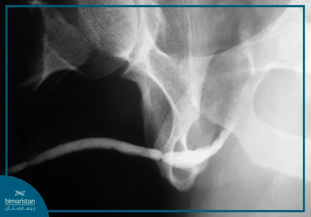
Retrograde urethrography (RUG) is used as the primary method to determine the exact location and extent of the stricture, and when needed, voiding cystourethrography (VCUG) may also be performed to evaluate for strictures in the posterior urethra, as in post-pelvic trauma cases.

Endoscopy
Cystourethroscopy and urethroscopy provide direct visualization of the stricture and assessment of its internal structure, which is particularly useful before making a treatment decision.
Additional imaging tools
In some cases, urethrography may be used to determine the thickness of the urethral wall and the extent of fibrosis, while in complex injuries, some centers use CT or MRI to obtain an accurate anatomical assessment.
When is surgery the best option?
Surgical urethral stricture treatment, such as urethroplasty, is considered the best option, especially after non-surgical methods have failed, with guidelines recommending starting with simpler options and progressing to more complex interventions based on the stricture’s nature.
Non-surgical options
For short (<2 cm) non-recurrent stenosis, surgical urethral stricture treatment guidelines first recommend dilation using a gradual dilator to open the urethra. If ineffective or if recurrence occurs, DVIU (Direct Visual Internal Urethrotomy) is performed under direct vision with a speculum. However, these treatments have limited effectiveness, especially for recurrent strictures, as studies show success rates drop to less than 20% after the second or third attempt.
When is it best to go for surgery?
- When expansion or DVIU fails after one or more attempts
- In the presence of a long stenosis (>2 cm) or recurrent stenosis despite treatment
- If the stenosis is complex, multiple, or associated with tissue fibrosis
Factors in choosing a surgical technique
A number of factors determine the method of surgery:
- Location of stenosis (anterior or posterior)
- Length and number of repetitions
- Type of fibrosis and surrounding tissue
- Previous surgeries
In these cases, open surgery offers success rates of more than 85-90% compared to previous temporary treatments.
Open urethroplasty to treat urethral stricture
Open surgery (urethroplasty) is the most effective line of treatment for advanced cases of urethral stricture, especially after the failure of dilatation or internal resection. It aims to remove the stricture and restore the normal passage of urine with high success rates.
Types of open surgery
One of the most common methods of urethroplasty:
End-to-End Anastomosis
This type is used in cases of short stenosis (usually less than 2 cm) in the membranous or bulbous urethra, where the entire stenotic segment is resected and the two healthy ends of the urethra are reconnected with fine sutures without tension. This procedure has a long-term success rate of more than 90-95% with limited complications.
Buccal Mucosal Graft Urethroplasty
This technique is used in cases of long or recurrent stenosis, especially in the bulbous or penile urethra, where a small portion of the oral mucosa is excised (usually from the inside of the cheek) and prepared as a patch, then the patch is implanted on the dorsal or ventral wall of the urethra according to the surgeon’s preference and the characteristics of the case, this method achieves success rates between 85% and 95% and is the best option when direct resection is not possible.
Skin Flap Repair
In cases of extensive damage to the lining of the urethra or complex scarring, skin flaps reinforced from the skin of the penis or perineum, where blood supply is maintained, are used to form an alternative urinary bypass. This technique is often used when grafting techniques have failed or in cases of complex urethral stricture resulting from severe infections or previous surgery. Despite its complexity, the results show a good success rate of 70%-90%.
Surgical treatment steps
- Preoperative evaluation: The location and length of the stricture are determined using retrograde urethrography (RUG) and cystoscopy to determine the appropriate surgical plan.
- Patient preparation and anesthesia: The procedure is performed under general or spinal anesthesia, and the patient is placed in a lithotomy position.
- Minimally invasive incision: The skin in the perineum is opened to access the narrowed area.
- Freeing the urethra from surrounding tissue: The urethra is exposed and freed from fibrotic tissue, and then the extent of the stricture is accurately assessed.
- Appropriate technology implementation
- In definitive urethroplasty: The stricture is completely removed, and the two healthy ends of the urethra are carefully reconnected.
- In block urethroplasty (BMG): A graft is taken from the inner cheek and attached to the ventral or dorsal wall of the urethra, depending on the severity of the stricture as assessed by the physician.
- In skin flap: A skin flap is prepared from the adjacent area and folded over the damaged part of the urethra to widen the canal.
- Urinary catheter placement: A 16-18 French silicone catheter is placed and remains in place for 10-14 days to ensure healing of the urinary tract.
- Post-operative care: Antibiotics include painkillers and recommendations for rest and abstinence from physical exertion. A radiologic examination is performed to make sure the canal has healed before the catheter is removed.
Follow-up after surgery and prevention of restenosis
After the surgical urethral stricture treatment with urethroplasty, the treatment does not end with the surgery, but the follow-up phase begins to ensure long-term success, aiming for full recovery, early detection of any relapse, and prevention of stricture recurrence.
Follow-up protocols after urethroplasty
After surgical urethral stricture treatment with open surgery, medical follow-up begins immediately after removing the urinary catheter, with the timing confirmed based on the urethral radiograph to ensure healing and absence of urinary leakage. Periodic visits are scheduled one month after surgery, then 3 to 6 months later, and annually once stabilized. These visits include clinical evaluation, uroflowmetry, and urinalysis to detect infection or hematuria, with additional radiographs or cystoscopy if symptoms appear.
Indicators of success and signs of recurrence
In surgical urethral stricture treatment, good urinary flow, absence of urinary symptoms, and stable test results are the most important indicators of a successful operation. Signs of recurrence include poor urine flow, a feeling of incomplete bladder emptying, or recurrent urinary infections.
Modern strategies to prevent recurrence
Recent research in surgical urethral stricture treatment is moving towards the application of advanced biological methods to promote urethral healing and reduce scarring, such as the use of platelet-rich plasma (PRP) and stem cells derived from adipose tissue. In some cases, urethral dilators coated with pharmaceutical compounds are used to help reduce postoperative fibrosis. These methods are still under study but show promising results in reducing recurrence rates in the long term.
Surgical urethral stricture treatment represents the basis for restoring normal urinary function, especially when non-surgical methods such as dilation and DVIU fail. Open surgery is considered the most effective option in most cases, with high success rates and long-term stability. The results of surgical urethral stricture treatment depend on the accuracy of the diagnosis, the choice of the appropriate technique, and regular follow-up after surgery. With the development of modern methods such as the use of PRP and stem cells, the chances of preventing recurrence and improving the patient’s quality of life in the long term increase.
Sources:
- Martins, F. E., McAninch, J. W., & Zinman, L. N. (2020). Urethral Strictures. In StatPearls. StatPearls Publishing.
- MedlinePlus. (2022). Urethral stricture. U.S. National Library of Medicine.
