Pectus carinatum causes many physical and psychological problems due to its shape and proximity to the heart and lungs, but currently, pigeon chest can be treated in Turkey by the most skilled doctors.
It is natural for children to find it difficult to adapt to school life at first, but having congenital chest wall deformities will pose an additional obstacle for them, and the solution may be simpler than you think!
In this article, we will learn about the cause of pectus carinatum and how to restore it to its correct position to regain the natural shape of the chest.
What is Pectus carinatum?
Pectus carinatum, also known as Pigeon chest or Chest bone protrusion, is a deformity in the front chest wall that leads to the protrusion of the sternum forward due to excessive growth of the cartilage that connects the ribs with the sternum. It is one of the non-life-threatening congenital deformities.
It is named “Pigeon chest” because the sternum protrudes making the chest looks similar to a pigeon’s chest. Pectus carinatum usually begins to appear suddenly during growth, specifically during puberty at the age of 11-15, and it continues to worsen until the bones stop growing, usually at the age of 18.
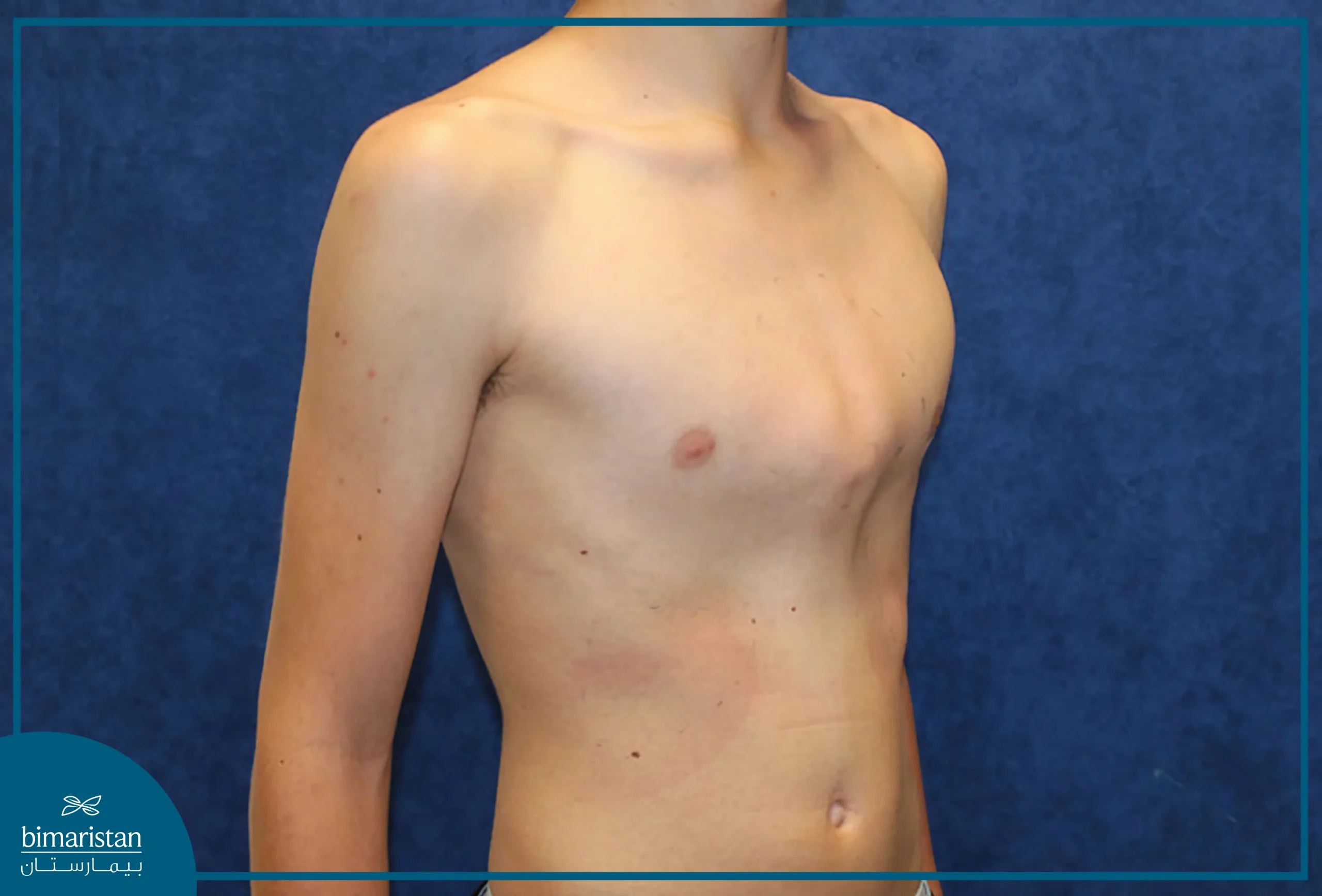
This deformity is less common compared to Pectus Excavatum (sunken chest) as it occurs in one child out of 1500 children and affects males more than females and is usually familial.
Types of Pectus carinatum
Pectus carinatum can be classified into several types; concerning the location of the deformity, it is classified into symmetric, located in the center, and asymmetric, such as right or left-sided chest bone protrusion.
Asymmetric pigeon chest is more common on the right side, while it is rare on the left side.
Carinatum Chest can be further divided into:
- Chondrogladiolar prominence (the most common type): Here, the middle and lower areas of the chest protrude forward, where the ribs are long and flexible, making correction easy.
- Chondromanubrial prominence: This condition affects the upper areas of the chest and is usually symmetric. It is difficult to treat because the affected ribs are shorter and less flexible.
Chest bone protrusion can also be classified based on the cause and time of appearance into:
- Postoperative Carinate Chest: Such as sternum protrusion after open-heart surgery when the sternum does not heal properly.
- Congenital Carinate Chest (the most common): It is present since birth and its cause is unknown, it appears at the age of 11-15 and is associated with growth mutations.
Pectus carinatum causes
The exact cause of chest pectus carinatum is still unknown. It may be caused by a disorder in the cartilage that connects the ribs with the sternum; when the cartilage grows faster than normal, it pushes the sternum outward.
Another cause of chest bone protrusion is excessive growth of the sternum after major surgeries that require cutting the sternum (such as open-heart surgery), which leads to swelling of the sternum and its protrusion outward.
The presence of several cases of pigeon chest within the same family raises suspicions of genetic causes.
Pigeon chest may be part of rare genetic disorders; some individuals with Marfan syndrome or Noonan syndrome also suffer from pectus carinatum as one of the symptoms of the syndrome.
Is Pectus Carinatum Dangerous?
Most patients with Pectus Carinatum are asymptomatic and do not have any problems except for their outward protruding chest, but 1 out of 10 children with a chest deformity usually suffer from Scoliosis.
Among the common symptoms of pigeon chest are chest pain and discomfort, usually observed in the supine position, in addition to shortness of breath during exertion in some patients and asthma-like symptoms in a quarter of teenagers with pigeon chest.
Some patients may also experience rapid heart rate or recurrent respiratory infections. Unlike sunken chest, the position of the heart in pigeon chest is not affected, but it is associated with an increased incidence of mitral valve prolapse.
It is worth noting that the symptoms worsen as the child gets older, making the treatment of pigeon chest more difficult.
Diagnosis of pectus carinatum
Pectus Carinatum is usually diagnosed clinically after examining the patient’s chest and seeing the protrusion of the chest bones with the naked eye, then the doctor asks the patient about their symptoms and examines them physically.
The primary test for diagnosis is a simple chest X-ray that shows the shape of the chest bones. Computed tomography (CT) or magnetic resonance imaging (MRI) may be performed to assist in diagnosis in some special cases.
We may need to perform some additional tests to ensure the safety of the heart and lungs, and some blood tests may be done to rule out genetic causes such as Marfan syndrome and Noonan syndrome.
Pectus carinatum treatment in Turkey
Patients with Pectus Carinatum in Turkey are treated using the latest surgical techniques with the aim of improving the shape of the chest and the quality of life. The optimal treatment method is selected depending on the severity of the condition; treatment of Pectus Carinatum may not be necessary, or it may be better not to treat it if the injury is mild (mild pectus carinatum) and does not cause symptoms.
In mild cases, exercise and weightlifting help build muscles in the chest wall, covering up the defective shape of the chest wall.
If you are worried about your protruding chest or it causes respiratory problems for you, you can treat Pectus Carinatum in Turkey conservatively or surgically, depending on the severity of the condition. Surgical treatment is only applied in severe or complicated cases or when the cartilage becomes hard and does not respond to conservative treatment, and this includes the Ravitch procedure and another procedure with better results called the Abramson procedure.
Non-surgical Treatment of Pectus Carinatum
Most cases do not require surgery; in moderate and mild injuries, a chest brace for pectus carinatum is used to correct the protrusion of the sternum in the same way that braces work, by continuous pressure on the protruding cartilage forward, and this continuous pressure helps reshape the cartilage correctly gradually.
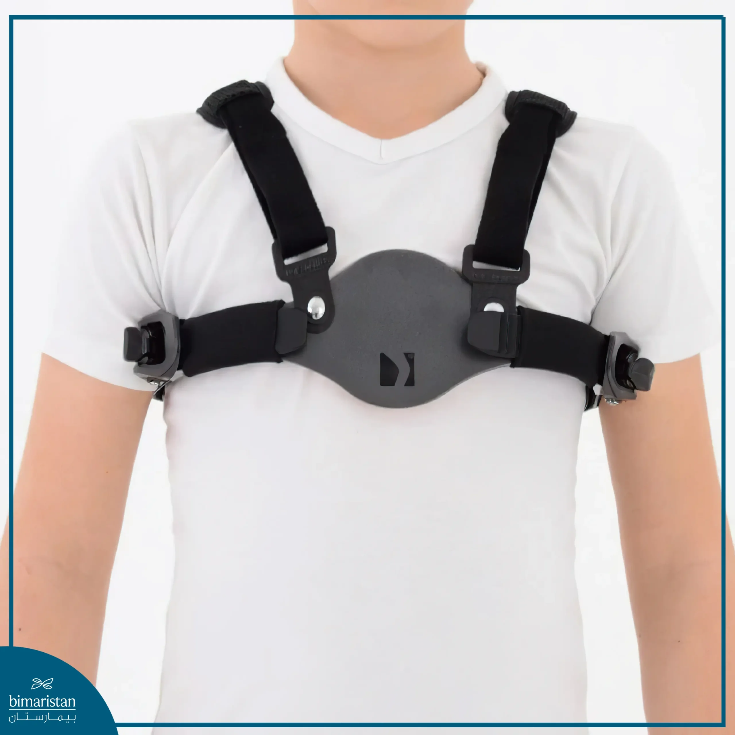
At the beginning, the brace is worn for 23 hours a day for a period of three to six months, which is called the corrective phase. Then the maintenance phase begins, where the patient wears the brace for 8-10 hours a day for three to six months.
The effectiveness of the brace in treating pigeon chest increases in early ages before the cartilages become hard, allowing monitoring of the patient and performing surgery if conservative treatment with the brace fails. The doctor determines the amount of pressure applied to the chest wall by the brace before using it, and results can be seen within a few months.
The price of a chest brace device generally ranges from $500 to 1500 US Dollars. It varies depending on the country of origin and the quality of the device.
Ravitch procedure
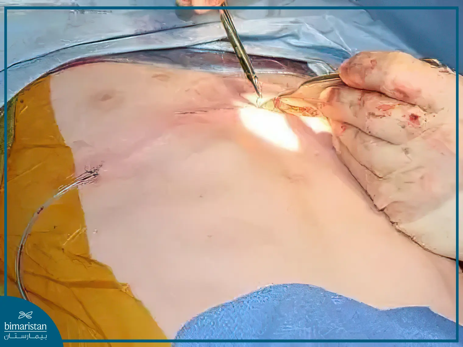
There are many surgical techniques that can treat pectus carinatum in Turkey, but the Ravitch procedure is considered the traditional method. This procedure involves making a horizontal incision in the middle of the chest to remove the abnormal cartilage from the front and push the sternum backward. Stainless steel struts are then placed across the front chest wall to support the sternum.
The struts are not visible from the outside and are later removed through surgery.
Although this surgical procedure offers acceptable results in treating pigeon chest, it requires large incisions, creating large skin and muscle flaps, excising costal cartilage arches, and cutting the sternum bone, making it a challenging procedure. Additionally, surgical scarring leaves a mark after the operation.
Abramson procedure
A new minimally invasive surgical technique for treating pectus carinatum through compression (instead of excision) called the Abramson procedure has recently emerged. Studies have shown its significant effectiveness when selecting suitable patients.
This procedure involves two intersecting incisions on both sides, each 3 cm long on a line in the middle of the armpit at the highest level of prominence. The surgeon continues by further dissecting to expose the rib at the level of the anterior axillary line, then performs a blunt dissection under the skin towards the sternum on both sides with long curved forceps to form a tunnel under the skin.
A convex metal rod designed to fit the shape of the chest prominence is then placed in this tunnel, and the incisions are closed. This metal implant corrects the chest prominence over time and treats pigeon chest, and here is a video that explains the whole procedure.
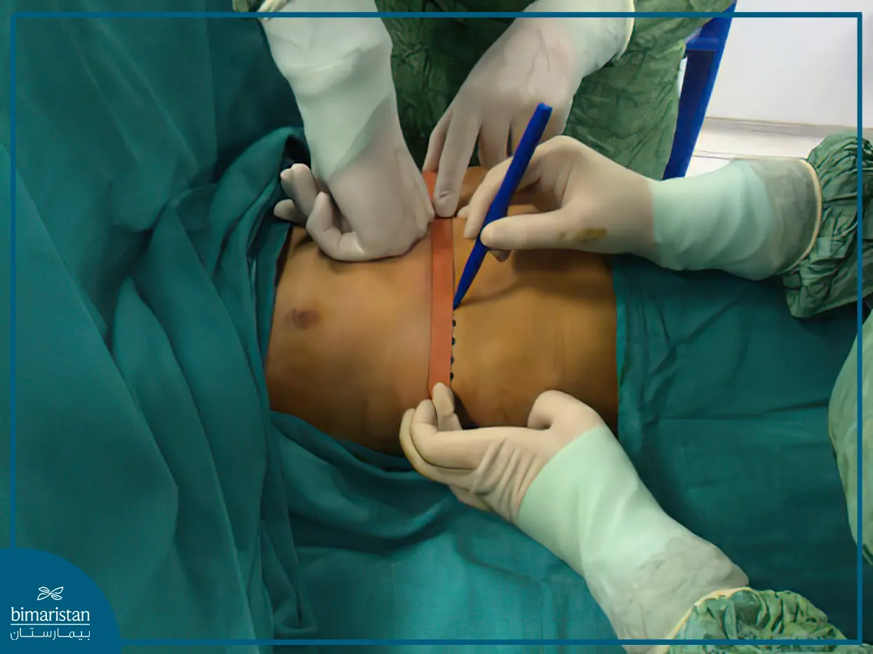
This technique allows the treatment of pectus carinatum without the need for wide skin incisions, excision of bones or cartilages, or even creating skin and muscle flaps! It also avoids the occurrence of chest wall stiffness that may occur after a period of surgical excision.
The body adapts well to the metal implant, but we recommend massaging the skin covering the implant to avoid any adhesions to the superficial fascia or excessive pigmentation in the skin or formation of seromas.
Anesthesia and care during surgery
During surgery, three types of catheters will be placed in the patient:
- A supraclavicular catheter on the child’s back for continuous local anesthesia.
- A urinary catheter to drain urine from the bladder.
- An intravenous catheter to administer fluids and medications.
The patient may need nasal oxygen during the operation, and sometimes the surgeon places a tube to drain fluids from the surgical area. All these treatments are temporary and will be stopped when no longer needed (usually before discharge from the hospital).
Post-Surgery Follow-Up
It is expected that the child will get out of bed and start walking on the day after surgery. They should practice deep breathing to maintain lung health and avoid pneumonia.
Children can be discharged from the hospital when they can take oral painkillers, eat and drink without difficulty, and do not have a fever or signs of infection.
If a surgical drain was placed during the operation, the child may need to keep the drain in place after returning home. The nursing staff will teach you how to care for the drain.
The drain is usually removed at the first clinic visit.
Risks of Pigeon Chest Treatment
The brace treatment for pigeon chest is very safe, but a few patients may experience irritation or skin breakdown where the brace touches. Patients are advised to stop using the brace and see a doctor at the first sign of irritation.
As for surgical treatment of pigeon chest, although the Ravitch and Abramson procedures are safe, like any other surgical procedures, they may carry some risks if the operating hands lack sufficient experience, including:
- Bleeding
- Infection
- Pneumothorax (air around the lung)
- Pleural effusion (fluid around the lung)
- Pericarditis (inflammation around the heart)
- Displacement of the metal implant
- Recurrence of pigeon chest
If you notice any protrusion in your or your family member’s chest bones, contact us for a free consultation and plan for treatment in Turkey. Bimaristan Medical Center will conduct a comprehensive evaluation of your case to detect any accompanying abnormalities or deformities and treat them simultaneously with pectus carinatum treatment in Turkey.
