Hydrocephalus treatment remains fundamental in reducing neurological complications and improving overall quality of life. Clinical data indicate that the congenital incidence of hydrocephalus ranges from approximately 0.3 to 2.5 per 1,000 live births, with a global prevalence of roughly 85 per 100,000 individuals. This significant burden highlights the necessity for effective and safe therapeutic approaches tailored to each patient’s age and anatomical characteristics. Within this framework, hydrocephalus treatment continues to advance—from programmable valves and anti-siphon systems to analog innovations, wireless intracranial pressure monitoring, and new minimally invasive techniques.
What is hydrocephalus?
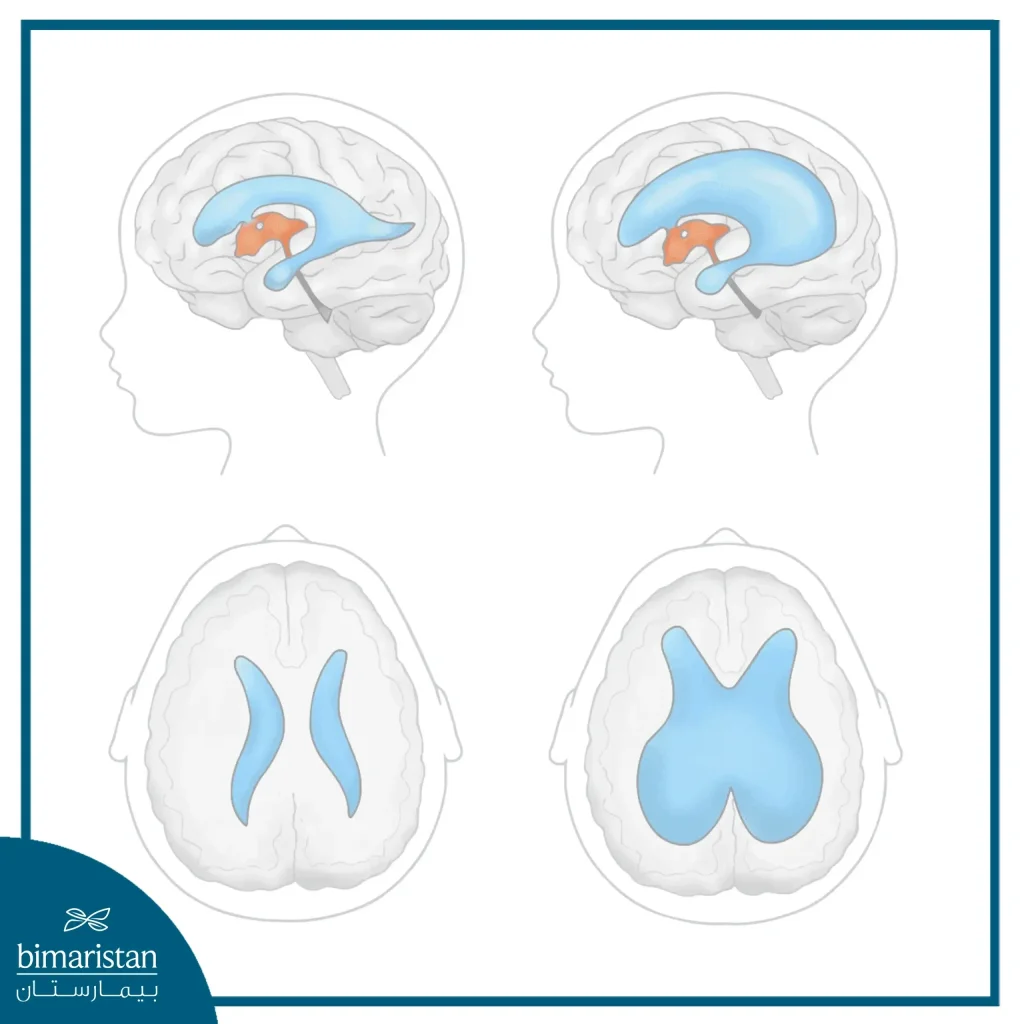
Hydrocephalus is a condition in which cerebrospinal fluid accumulates in large quantities, putting pressure on the brain and central nervous system, which in turn manifests as neurological effects that can be fatal if not treated in time. The causes and symptoms of hydrocephalus may be similar across different types of hydrocephalus (congenital, acquired, and normal). Still, each type has its own unique characteristics, and hydrocephalus treatment is often tailored to the specific cause of the condition.
What is the goal of hydrocephalus treatment?
Hydrocephalus treatment primarily aims to relieve pressure on the central nervous system and reduce the accumulation of cerebrospinal fluid within the ventricles or cavities of the brain, thereby alleviating neurological symptoms and minimizing the risk of long-term disability. In addition, hydrocephalus treatment aims to improve the aesthetic appearance of the patient, as hydrocephalus often causes shame for the individual due to the constant staring of people at the size of his head, making him lose his confidence and make him feel embarrassed and ashamed, so treatment works to improve the aesthetic appearance of the head, which enhances the quality of life of this person.
What are the best and newest treatment options available?
With the development of neurosurgical sciences, hydrocephalus treatment options have become vast and varied, including surgical solutions, endoscopic solutions, shunts, and others, making it easy to solve this issue when there is a specialized medical team sufficiently trained to perform one of these procedures.
Ventriculo-peritoneal shunt (VPS)
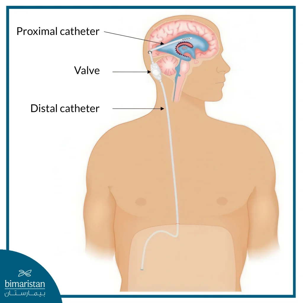
A ventriculoperitoneal shunt (VPS) is a standard neurosurgical procedure to treat hydrocephalus. The goal of this treatment is to divert cerebrospinal fluid (CSF) from the ventricles of the brain to the abdominal cavity (peritoneal cavity), where it is better absorbed.
This process requires a system with three main parts:
- Ventricular Catheter: This is a very thin, flexible tube that is inserted into one of the ventricles of the brain (often the lateral ventricle) to drain excess cerebrospinal fluid.
- Valve: This valve senses the amount of intracranial pressure, and when it rises to a certain level, the valve opens to allow the cerebrospinal fluid to flow towards the abdominal catheter and then towards the stomach, and closes to prevent the reverse flow of the fluid.
- Peritoneal Catheter: This is a long tube that is passed under the skin from behind the ear, through the neck
- This tube carries the fluid flowing from the valve into the abdominal cavity.
Success rates and long-term causes of failure
Ventriculo-peritoneal shunt is the most effective treatment for hydrocephalus, saving lives and improving quality of life in most patients. Unfortunately, the shunt is not a perfect fail-proof device, with relatively high long-term complication and failure rates.
The long-term success of the ventriculo-peritoneal shunt depends mainly on regular medical follow-up of the patient, through regular visits to the neurologist, even if no symptoms appear. The person must be monitored clinically and assessed for symptoms that may indicate shunt failure (headache, nausea, visual changes, personality changes, seizures). The person must undergo follow-up imaging tests, as these help determine the size of the ventricles using images, to ensure that the ventricles are of normal size and that the shunt is functioning properly. These procedures, in addition to family follow-up, greatly increase long-term success rates and make the shunt the first choice in the treatment of hydrocephalus.
Are there other shunts to treat hydrocephalus?
The ventriculoperitoneal shunt (VPS) is not the only type of shunt to treat hydrocephalus, as there are other types of shunts that relieve CSF pressure on the brain:
- Ventriculo-atrial shunt (VAS): Drainage of cerebrospinal fluid from the cerebral ventricles into the right atrium of the heart, where the cerebrospinal fluid mixes directly with blood to be filtered.
- Lumbo-abdominal shunt (LPS): The cerebrospinal fluid is drained from the lumbar region of the spinal canal into the abdomen, where it is absorbed.
Programmable valves and anti-siphon
Programmable valves are advanced valves that contain an internal mechanism that can adjust their opening pressure from outside the body without the need for another surgery, as the adjustment is done using a special magnetic device. These valves are more useful if the patient suffers from symptoms of overdrainage or underdrainage caused by normal valves, as the doctor can easily adjust the valve pressure in the clinic to solve the issue, thus avoiding the patient from undergoing another surgery later, in addition, programmable valves have the advantage that the doctor can check the valve setting using an X-ray or an imaging device.
Anti-siphon is an addition to the normal valve to prevent too much cerebrospinal fluid from draining (a common issue when standing due to the effect of gravity). Anti-drainage significantly reduces the risk of hyperdrainage, especially in patients who stand and sit a lot, prevents chronic hyperdrainage complications such as ventricular contracture and subdural hematoma, and finally, anti-drainage aims to improve the quality of life by eliminating the headaches associated with standing.
Comparison of VPS-(VAS)-(LPS) conversions
| Type | Destination | Main uses | Main advantages | Key disadvantages/risks |
|---|---|---|---|---|
| VPS | Abdomen (peritoneum) | The first choice for most cases of hydrocephalus | Minimal cardiac complications | Blockage, infection, or abdominal issues |
| VA Shunt | Heart (right atrium) | When the abdomen is unavailable (infection, previous surgeries), or repeated failure of VPS | Doesn’t rely on abdominal absorption | Serious cardiopulmonary complications |
| LP Shunt | abdomen | Normal pressure hydrocephalus (NPH), or idiopathic high intracranial .pressure | It does not require a brain puncture and is feasible for non-obstructive hydrocephalus | Futile for obstructive hydrocephalus, post-operative headache, and risk of overdrainage |
Endoscopic third ventriculostomy (ETV)
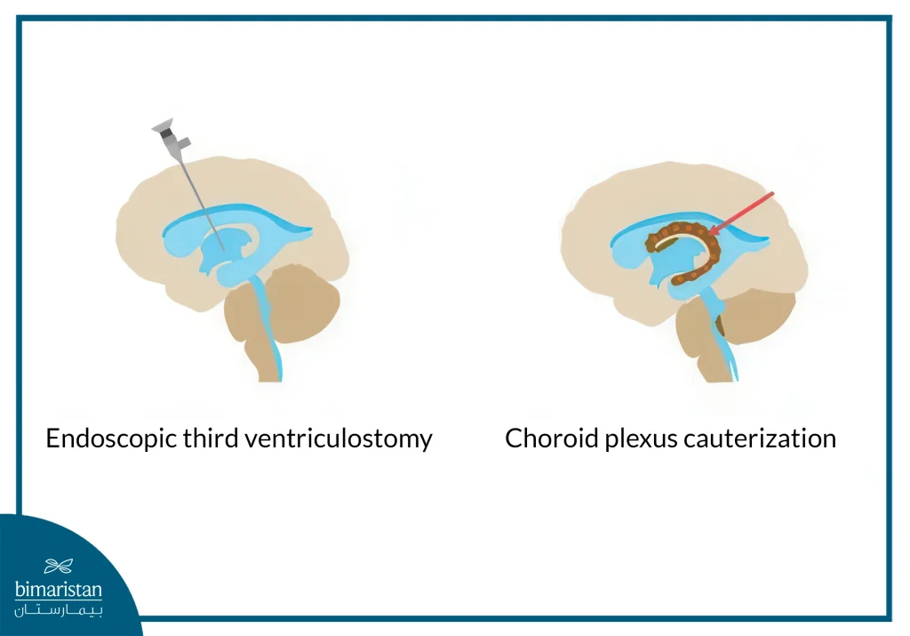
A minimally invasive surgical procedure in which a tiny scope with a camera is inserted into the ventricles of the brain, its goal is to make a small opening in the floor of the third ventricle to create a new passage for cerebrospinal fluid to flow and be absorbed naturally rather than relying on shunts.
Endoscopic third ventriculotomy + choroid plexus cautery (ETV+CPC)
This procedure aims to create an opening in the floor of the third ventricle by cauterizing part of the choroid plexus, which produces cerebrospinal fluid (CSF), using the same endoscope used to create the opening.
Comparison of shunt, ETV, and ETV+CPC for hydrocephalus
| Type | Shunt | ETV (opening the third ventricle) | ETV + CPC |
|---|---|---|---|
| Scope of use | All types of hydrocephalus (obstructive, ischemic, and normal pressure). | Very successful for obstructive hydrocephalus only (e.g., Sylvius stenosis). | For infants with obstructive, post-hemorrhagic, or post-digestive hydrocephalus. |
| Optimal age range | All ages (from preterm babies to the elderly). | Adults and older children (over a year). | Infants under one year of age (especially <6 months) where ETV alone failed. |
| Long-term success rate | Initially very high, but it decreases over time. Less than 50% of patients require repair within 2-5 years. | If successful for 6-12 months, the lifetime survival rate is high (less than 80%). | higher than ETV alone in infants (up to 60-70% compared to ~40% for ETV alone). |
| Lifestyle and follow-up | Lifelong monitoring, constant fear of device failure, and restrictions on certain sports. | Once success is confirmed, follow-up is less intensive (periodic imaging scans), and there are no restrictions on sports. | Similar to ETV, freedom of lifestyle follows confirmation of success. |
| Long-term cost | very high due to the cost of repeated operations, shunt replacement, and treatment of complications. | Low (only the cost of the initial operation and follow-up). | Low (similar to ETV, if successful). |
How do we choose the most appropriate treatment?
Many factors play a role in choosing the appropriate operation to treat hydrocephalus. It varies from one person to another based on age, type of hydrocephalus, and surgical history. Not only the patient, but also the doctor, as the surgeon’s experience plays a significant role in choosing the appropriate hydrocephalus treatment. To select the best and most suitable option for treating hydrocephalus, you can contact Bimaristan Medical Center for medical advice and guidance on determining the most suitable operation for treatment.
Hydrocephalus treatment today encompasses a broad spectrum of therapeutic options, ranging from programmable shunt systems equipped with anti-siphon mechanisms to neuroendoscopic procedures, advanced remote monitoring technologies, and promising minimally invasive approaches. The selection of the appropriate strategy depends on the patient’s age, the type of hydrocephalus, the surgical team’s expertise, and the availability of modern equipment. In Turkey, Bimarestan Medical Center provides comprehensive treatment coordination, offering referrals to highly specialized institutions to achieve optimal outcomes. The most critical stage involves an accurate evaluation that enables a personalized treatment plan aimed at restoring the patient’s autonomy and quality of life.
Sources:
- Warf, B. C., Weber, D. S., Day, E. L., Riordan, C. P., Staffa, S. J., Baird, L. C., & Fehnel, K. P. (2023). Endoscopic third ventriculostomy with choroid plexus cauterization.
- Chen, J., Xian, J., Wang, F., Zuo, C., Wei, L., Chen, Z., Hu, R., & Feng, H. (2025, April 12). Long-term outcomes of ventriculoperitoneal shunt therapy in idiopathic normal pressure hydrocephalus. BMC Surgery.
