Diabetic retinopathy is a frequent diabetes complication that may lead to vision loss if not promptly diagnosed and treated. Thanks to medical advancements in Turkey, diagnostic and therapeutic approaches have become more precise and safer, enabling effective vision preservation and slowing disease progression through laser therapy and advanced treatments under the care of expert ophthalmologists.
What is diabetic retinopathy?
Diabetes can cause damage to the small blood vessels located in the retina of the eye. The function of the retina is to receive light, convert it into electrical nerve signals, and send these signals to the brain for interpretation. When the vessels supplying the retina are damaged or obstructed due to complications from diabetes and high blood sugar, a condition known as diabetic retinopathy occurs. This disease frequently affects both eyes, impairing vision and potentially leading to blindness without noticeable symptoms or even minor vision changes.

What are the symptoms of diabetic retinopathy?
Diabetic retinopathy usually doesn’t show any warning symptoms, but when it begins to worsen, patients notice changes in their vision. By then, the condition has already become severe. Therefore, regular eye exams are essential to detect this condition and begin treatment to prevent its progression and blindness. Symptoms and complications of diabetic retinopathy may include:
- Partial or total loss of vision, or there may be a shadow or veil in the field of vision
- Blurry, double, or distorted vision or difficulty reading
- Permanent eye pain, pressure, or redness
- Blurry or spotty vision
What are the types of diabetic retinopathy?
There are two types of diabetic retinopathy, classified according to the progression of the disease: early diabetic retinopathy and advanced diabetic retinopathy.
Early diabetic retinopathy
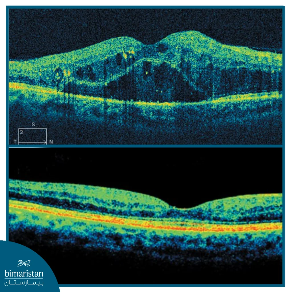
Also called non-proliferative diabetic retinopathy. It is a condition in which the walls of the blood vessels in the retina weaken, leading to small bulges (microaneurysms) that protrude from the walls of small vessels. In some cases, fluid and blood leak into the retina, causing the larger retinal vessels to dilate and become irregular in diameter. Additionally, we observe swelling of the central part of the retina, known as macular edema, which also requires treatment. The progression from mild to severe depends on the increasing number of blocked blood vessels.
Advanced diabetic retinopathy
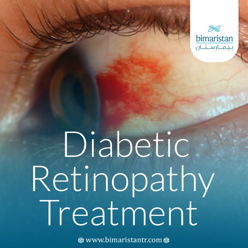
Also called proliferative diabetic retinopathy, it is more severe than non-proliferative diabetic retinopathy, characterized by the closure of damaged blood vessels and the growth of new abnormal blood vessels in the retina. This process results in the formation of scar tissue that pulls the retina, leading to its separation from the back of the eye. In some cases, it may rupture and leak into the vitreous body. If these vessels interfere with the flow of fluid outside the eye, pressure in the eyeball may increase, which can lead to optic nerve damage and ultimately cause glaucoma.
Risk factors for diabetic retinopathy in people with diabetes
People with diabetes have an increased risk of developing diabetic retinopathy if they experience:
- Poor control of blood sugar levels
- long-term exposure to diabetes
- High blood pressure
- High levels of fat
- Pregnancy
- Tobacco use
Diabetic retinopathy diagnosis
The most accurate way to diagnose diabetic retinopathy is a dilated eye exam, which is performed by an ophthalmologist after dilating the pupil with special drops that provide a clear view of the inner eye structure.
Dilated eye exam (fluorescein angiography)
It allows the doctor to detect subtle changes in the blood vessels and retina. During the exam, your doctor looks for:
- Retinal detachment
- Bleeding in the vitreous
- Changes in the optic nerve
- Abnormal or dilated blood vessels
- Blood or fatty deposits within the retina
- Growth of new blood vessels with the formation of scar tissue
Clinical screening tests
Several basic tests are performed to assess eye health and visual function, including:
Visual acuity test
It measures the clarity of vision and the eye’s ability to distinguish details both up close and at a distance. It also helps to detect any decline in visual ability.
Slit lamp examination and funduscopy
It allows the doctor to see the lens, retina, and internal tissues of the eye. It helps to detect issues such as cataracts or retinal changes.
Measuring Intraocular Pressure (IOP)
It is used to determine intraocular pressure (IOP) and detect glaucoma, a common complication in people with diabetes.
Advanced imaging scans
Several advanced imaging tests are used to diagnose diabetic retinopathy.
Optical Coherence Tomography (OCT)
A painless test that provides precise cross-sectional images of the layers of the retina, showing its thickness and the presence of fluid leaks. It’s also used to monitor the effectiveness of treatment over time.
Fluorescein angiography
Performed when needed to identify areas of blood leakage or microvascular blockage, especially in cases of blurred or distorted vision.
Digital fundus photography
It provides accurate images of the back of the eye (fundus) and is used to document changes and monitor disease progression. However, it does not replace a full eye examination.

Importance of regular checkups
People with diabetes are more likely to develop other eye diseases such as glaucoma and cataracts, so regular dilated eye exams are recommended to help:
- Early detection of retinal changes
- Preventing or delaying vision loss
- Carefully monitor the effectiveness of treatments and the progress of the condition
How is diabetic retinopathy treated?
Treatment is recommended when signs of macular damage or abnormal new blood vessels appear in the retina, a condition known as proliferative retinopathy. The disease cannot be completely cured, but early treatment can greatly help prevent or delay vision loss. The earlier the condition is detected, the more effective the treatment will be.
Control of blood sugar and risk factors
The key step at all stages is good control of blood sugar levels, along with blood pressure and cholesterol. Stabilizing these factors reduces the risk of retinal deterioration and prevents the condition from worsening even after treatment.
Non-surgical treatment options
Laser therapy (photocoagulation)
A laser is used to close leaks from tiny blood vessels within the retina.
- Helps stop or slow fluid leakage in macular edema
- In severe proliferative cases, Panretinal Photocoagulation (PRP) is used to limit the growth of new vessels
- Treatment aims to preserve current vision and prevent subsequent bleeding or retinal detachment
Laser treatment is most effective when performed before severe retinal damage has occurred, often requiring multiple sessions.
intravitreal injections
Anti-vascular endothelial growth factor (Anti-VEGF)
These drugs are one of the most effective treatments in the active stages of the disease, slowing the growth of abnormal vessels and reducing intraretinal leaks.
- Injected directly into the eye in a short session under local anesthesia
- Examples include: Ranibizumab (Lucentis), aflibercept (Eylea), bevacizumab (Avastin)
- May help improve vision and reduce swelling in the vision center
Corticosteroids (steroids)
- Used to treat diabetic macular edema when Anti-VEGFs are not enough
- They can be given as repeated injections or through long-acting implants such as Iluvien that release precise doses of medication into the eye for several months
- Some patients require regular injections to maintain treatment results
Surgical procedures
Preoperative preparation
There are several procedures that the doctor must perform before starting the surgery:
- Stop medications or herbal supplements under medical supervision
- Quit smoking at least one week before surgery
- Arrange for post-operative assistance
Vitrectomy
Vitrectomy is performed in advanced cases such as heavy vitreous hemorrhage or retinal detachment caused by scar tissue.
- It is done through tiny incisions in the eye to remove the cloudy vitreous and fibrous tissue that is pulling on the retina
- It can be combined with laser therapy during surgery to stabilize the retina and prevent recurrent bleeding
- The procedure is performed under local or general anesthesia, depending on the case
Types of laser therapy during surgery
- Laser focal therapy: Used to stop the leakage of blood and fluid from specific vessels in the retina, usually in one session.
- Panretinal Photocoagulation (PRP): Targets the peripheral areas of the retina to shrink abnormal vessels, usually performed in two or more sessions.
Diabetic retinopathy surgery in Turkey
Diabetic retinopathy surgeries in Turkey are performed in centers equipped with advanced imaging systems like OCT and PRP lasers, allowing high precision during intraocular procedures. The type of surgery depends on the severity of the disease, as monitoring and drug treatment are sufficient in the early stages, while surgery is necessary when there is a vitreous hemorrhage or retinal detachment.
Preparing for diabetic retinopathy surgery
- Stop medications or herbal supplements under medical supervision
- Quit smoking at least one week before surgery
- Arrange for post-operative assistance
After diabetic retinopathy surgery
- Wear a protective eye patch for several days
- Avoid smoking or strenuous activity
- Maintain a healthy diet and carefully regulate blood sugar
- Gradually resume daily activities within a few days
Potential risks of diabetic retinopathy surgery
As with any surgical procedure, rare complications may arise, including:
- Infection or bleeding inside the eye
- Irritation or allergy to anesthetics
- Recurrent bleeding or need for repeat surgery
However, success rates are very high in specialized centers in Turkey thanks to the use of precise equipment and surgeons with long experience in retinal surgery.
Thanks to major medical advancements, Turkey is now a top destination for diabetic retinopathy treatment using cutting-edge laser and microsurgical techniques. At Bimaristan Medical Center, we offer expert care under the supervision of elite ophthalmologists to deliver optimal outcomes and safeguard your vision.
Sources:
- National Eye Institute. (n.d.). Diabetic retinopathy. U.S. National Institutes of Health.
- NHS. (2025). Diabetic retinopathy. NHS.
