Aortic vs mitral valve stenosis represents one of the most critical comparisons in understanding conditions that impair the heart’s ability to function efficiently. Both aortic and mitral valve stenosis are prevalent disorders that hinder blood flow, yet they do so through distinct mechanisms and present with different clinical features. In this article, we will explore the key differences between aortic and mitral valve stenosis, focusing on their underlying causes, characteristic symptoms, diagnostic approaches, and current treatment options.
What is the function of the aortic and mitral valves?
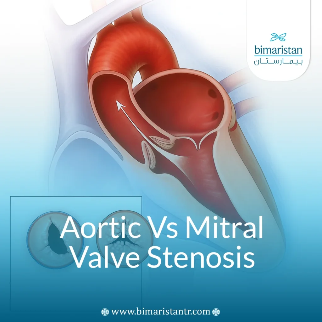
The heart is divided into four chambers: The right atrium and left atrium at the top, and the right and left ventricles at the bottom. Between these chambers are valves, composed of thin, strong folds of tissue known as leaves or cusps, which open and close to facilitate blood flow. Deoxygenated blood flows from the body into the right atrium, then passes through the tricuspid valve into the right ventricle. The right ventricle then pumps the blood through the pulmonary valve into the lungs, where blood receives oxygen. The oxygenated blood returns to the left atrium, passes through the mitral valve into the left ventricle, and then flows through the aortic valve into the aorta for distribution throughout the body.
The mitral valve usually has two cusps and separates the left atrium from the left ventricle, allowing blood to move from the atrium to the ventricle while preventing backflow. The aortic valve typically has three cusps and is situated between the left ventricle and the aorta, enabling blood to flow from the heart to the body while preventing backflow from the aorta into the ventricle. For valves to function properly, they must be properly shaped and flexible, open fully to allow blood to pass through, and close tightly to prevent any backward leakage of blood.
What is Valvular stenosis?
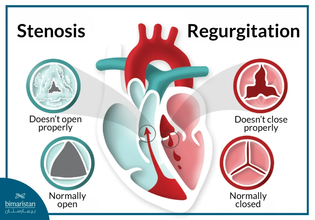
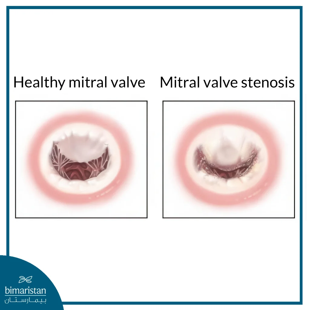
Valvular stenosis is a condition in which the opening of a heart valve is too narrow to open fully. Thickening, hardening, or fusion of the valve’s folds may prevent it from opening fully, forcing the heart to work harder to pump blood through the narrowed opening. This extra effort leads to increased pressure within the heart, especially in the chamber that pumps blood through the valve, and over time can cause the heart muscle to enlarge and weaken, eventually leading to heart failure if left untreated.
Heart valve disease is generally categorized into two main types: stenosis, where the valve fails to open completely, and regurgitation, where the valve does not close tightly, allowing blood to leak backward. In cases of aortic and mitral valve stenosis, the reduced valve opening limits blood flow and increases cardiac effort, which may compromise oxygen delivery to the body. Regurgitation, on the other hand, is commonly linked to valve prolapse, where the valve bulges under pressure and fails to seal properly, allowing blood to flow back into the preceding chamber.

Aortic vs mitral valve stenosis: Differences in the causes
The underlying causes of aortic valve stenosis and mitral valve stenosis differ based on the valve affected, although some contributing factors may overlap.
Causes of mitral valve stenosis
Mitral valve stenosis is the result of several pathological factors, most notably rheumatic fever, which leaves scars on the valve leaflets and leads to valve stiffness over time. It can also be caused by calcium deposition with age or by congenital abnormalities in the valve’s structure from birth. In some rare cases, endocarditis or connective tissue diseases may cause the valve to malfunction, leading to stenosis.
Causes of aortic valve stenosis
Aortic valve stenosis is often caused by calcification associated with aging, where calcium deposits gradually accumulate, impeding the movement of the valve leaflets and preventing them from opening fully. Another significant cause is a congenital bicuspid aortic valve, which replaces the normal tricuspid structure and is more prone to early deterioration. Like mitral stenosis, rheumatic fever, and endocarditis can also lead to scarring and deformities that result in aortic valve narrowing.
Mitral valve stenosis is often associated with rheumatic fever, while aortic valve stenosis is mostly associated with calcification and age-related changes. However, both conditions may share common etiologies.
Aortic vs mitral valve stenosis: Differences in symptoms
The clinical presentation of aortic valve stenosis and mitral valve stenosis varies depending on the valve involved and the severity of the narrowing. Symptoms may be subtle initially and progress gradually.
Symptoms of aortic valve stenosis
These symptoms usually appear gradually as valve stenosis worsens, and include:
- Shortness of breath: Increases with physical exertion due to difficulty pumping blood from the left ventricle.
- Chest pain (angina): Due to cardiac ischemia caused by increased pressure on the heart.
- Fainting or dizziness: Especially with exertion, due to reduced blood flow to the brain.
- Severe exhaustion: The result of not getting enough oxygenated blood to the organs and muscles.
Symptoms of mitral valve stenosis
These symptoms are often associated with increased pressure in the left atrium and lungs, and include:
- Shortness of breath, especially when lying down or during sleep (paroxysmal nocturnal dyspnea)
- Chronic coughing or hemoptysis due to congestion of blood vessels in the lungs
- Swelling of the legs and ankles due to blood reflux and fluid retention
- Heart palpitations due to arrhythmias, especially atrial fibrillation
Aortic vs mitral valve stenosis: Differences in diagnostic methods:
While the diagnostic approach to aortic valve stenosis and mitral valve stenosis shares common tools, such as patient history, physical examination, auscultation, echocardiography, and electrocardiography, each condition has distinct diagnostic priorities.
- Aortic valve stenosis: Diagnosis focuses on measuring the area of the aortic valve opening and estimating the pressure gradient across the valve using echocardiography (Echo Doppler). Cardiac catheterization may also be performed to accurately determine the severity of the stenosis, especially before deciding to perform surgery or therapeutic catheterization.
- Mitral valve stenosis: The focus is on measuring the area of the mitral valve opening and assessing the pressure in the left atrium and lungs, as transesophageal echocardiography or cardiac catheterization techniques help determine the extent of pulmonary hypertension associated with the stenosis.
In this way, the doctor can accurately determine the severity of the stenosis and plan the optimal treatment for each case of aortic and mitral valve stenosis.

Aortic vs mitral valve stenosis: Treatment options
Management of aortic and mitral valve stenosis depends on the specific valve affected, the degree of narrowing, and the severity of symptoms. While mild cases may only require regular monitoring, advanced stenosis often necessitates medications, surgical intervention, or minimally invasive procedures.
Aortic valve stenosis treatment
Treatment for aortic valve stenosis depends on the severity of the condition and symptoms. Treatment methods include:
- Lifestyle changes: Eat a healthy diet, exercise regularly, avoid smoking, and maintain a healthy weight.
- Medications: Used to relieve symptoms or minimize risks, such as antihypertensives, antiarrhythmics, and diuretics, to reduce the load on the heart.
- Surgical or catheterization procedures:
- Valve repair or replacement: Can be performed through open surgery or less invasive methods, including mechanical or biological valve replacement.
- TAVR (Transcatheter Aortic Valve Replacement): Catheter-based valve replacement without the need for open surgery, suitable for high-risk cases.
- Balloon valvuloplasty: Expansion of the valve with a balloon, often for children or patients who cannot currently undergo surgery. It may be used to temporarily relieve symptoms and is considered a temporary option for adults.
Mitral valve stenosis treatment
Mitral valve stenosis requires careful monitoring to determine when symptoms begin to affect daily life, and treatment methods may include the following:
- Medications: To relieve symptoms and prevent complications, such as diuretics, blood thinners to prevent clots in atrial fibrillation, medications to control blood pressure and heart rate, and anti-infectives to prevent the return of rheumatic fever.
- Catheterization procedures
- Balloon Valvuloplasty: Dilating the valve using a balloon through a catheter to relieve stenosis.
- TMVR (Transcatheter Mitral Valve Replacement): Transcatheter mitral valve replacement in some cases.
- Surgery: Valve repair or replacement, which may involve conventional methods or robotic surgery, with the choice of a mechanical or biological valve depending on the patient’s specific needs.
In conclusion, understanding the differences between aortic and mitral valve stenosis is essential, as both are serious conditions that demand precise diagnosis and timely intervention. Recognizing how each type affects the heart differently allows for more effective management, helps prevent severe cardiac complications, and ultimately enhances the patient’s quality of life.
Sources:
- American Heart Association. (2024, May 23). Problem: Mitral valve stenosis.
- Penn Medicine. (2025, July). Aortic valve stenosis

