
Facial fractures are among the most serious and dangerous injuries, as they affect the bones of the upper jaw, the structures surrounding the nose and eyes, and the cheekbones. These fractures are often caused by severe trauma, such as traffic accidents or sports injuries, and can lead to significant functional and cosmetic problems if not properly diagnosed and treated.
The Le Fort classification is used worldwide to determine the type of injury in maxillary fractures and to develop an appropriate treatment plan in maxillofacial surgery, where treatment focuses on repositioning the bones and restoring normal occlusion, as well as facial, respiratory, and aesthetic function.
What are facial fractures?
Facial fractures known as Le Fort fractures (characterized by specific fracture lines described by the French surgeon René Le Fort following anatomical and experimental studies of the facial bones, which is why the classification bears his name) refer to a group of fractures affecting the middle third of the face, leading to partial or complete separation of the upper jaw from the base of the skull to varying degrees. These injuries involve a number of important facial bones and structures, namely:
- Nasal bones: Small bones that form the bony part of the nose and give it its external shape.
- Cheekbones: The prominent bones in the cheek area that contribute to the shape of the face and support the cheekbones.
- Orbital bones: The bony cavities that contain and protect the eye, along with the surrounding muscles and nerves.
- Upper jaw: The bone that supports the upper teeth and forms an essential part of the roof of the mouth and the nasal cavity.
- The alar plates of the sphenoid bone are deep bony structures located behind the upper jaw that connect the midface to the base of the skull and are important landmarks in the diagnosis of Le Fort fractures.
Classification of Facial Fractures
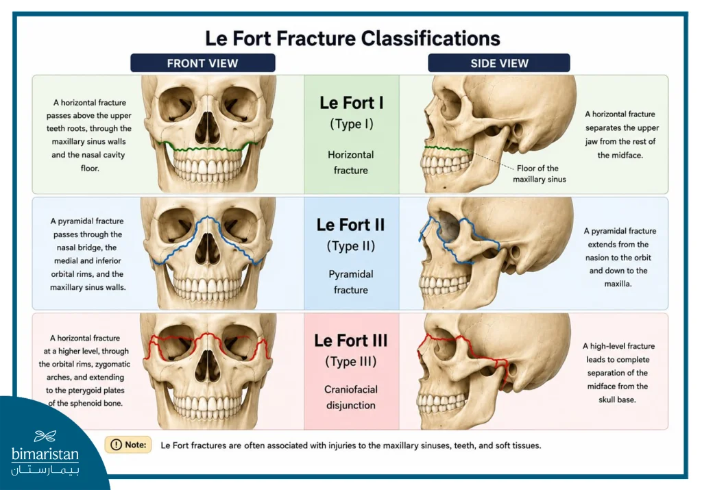
According to the Le Fort classification, facial fractures are divided into three main types, which differ based on the direction of the fracture and the severity of bone displacement.
Le Fort I
It is also known as a horizontal fracture of the upper jaw, in which the fracture line runs horizontally across the roots of the upper teeth and through the inferior alveolar sinus, resulting in the separation of the tooth-bearing portion from the rest of the upper jaw.
Common Symptoms
- Bruises inside the mouth
- Malocclusion
- Bleeding from the mouth or nose
- Pain and swelling in the upper lip
- Abnormal movement of the upper jaw
Le Fort II
It is known as a pyramidal fracture because of its triangular shape; the fracture extends from the nasal bones through the zygoma and the maxillary sinus to the upper jaw, resulting in a central separation in the middle of the face.
Common Symptoms
- Severe nosebleed
- Double Vision
- Bruises around the eyes
- Severe swelling in the middle of the face
- The nose and upper jaw move together
- Cerebrospinal fluid sometimes leaks
Le Fort III
This is considered the most severe type of Le Fort fracture and is known as craniofacial separation. In this type, the middle third of the face is almost completely separated from the base of the skull, and the fracture extends through the zygomatic, cheek, and nasal bones.
Common Symptoms
- Enophthalmos
- Difficulty breathing
- Obvious facial deformity
- Flattening or Elongation of the Face
- Severe bleeding and extensive swelling
- Severe malocclusion
- Visual and Neurological Disorders
Causes of Facial Fractures
Fractures of the upper jaw usually result from high-impact trauma, and the severity of the injury increases with the force and direction of the impact, especially when the midface is struck directly. The most common causes include:
- Traffic accidents.
- Falls from heights.
- Severe sports injuries.
- Direct blows to the face.
- Work-Related Injuries and Industrial Accidents.
Clinical Symptoms and Signs of Facial Fractures
Symptoms vary depending on the type and severity of the fracture, but the most common signs include:
- Nosebleed or bleeding from the mouth
- Malocclusion
- Difficulty Chewing and Speaking
- Severe pain in the center of the face
- Swelling and bruising around the eyes
- Double vision or blurred vision
- Abnormal Movement of the Upper Jaw
- Cerebrospinal fluid leakage through the nose in severe cases
- Numbness in the cheek or upper lip caused by injury to the infraorbital nerve
These injuries may be accompanied by airway obstruction; therefore, they are considered critical emergency situations in the treatment of facial fractures.
Diagnosis of Facial Fractures
The diagnosis of facial fractures depends on:
Clinical Examination
The doctor will assess:
- Bleeding and Swelling
- Stabilization of the Upper Jaw
- Alignment of the Maxillary Teeth
- The Presence of Facial Malformations
- Eye and Nerve Injuries
Radiography
Imaging is an essential step prior to any maxillofacial surgical procedure. Computed tomography (CT) is also considered the most accurate method for evaluating:
- Extension of the fracture lines
- Degree of Bone Displacement
- Sinus Infections in Pilgrims
- Associated injuries at the base of the skull
Treatment of Facial Fractures
Treatment depends on the severity of the injury and the patient’s condition, and aims to:
- Facial Skeletal Reconstruction
- Restoring the Natural Bite of the Teeth
- Maintaining the Airway
- Improved Function and Aesthetic Appearance
Surgical Fixation
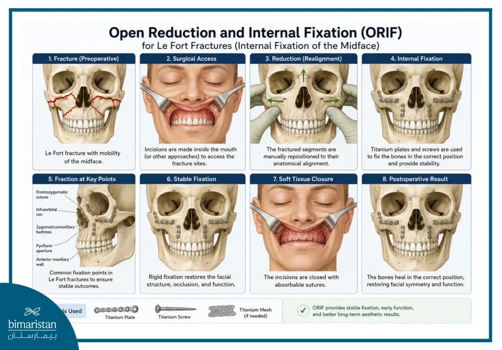
Open reduction and internal fixation (ORIF) is a fundamental surgical procedure for the treatment of facial fractures. It aims to restore the fractured bones to their natural position and fix them precisely to restore function and form. It is performed according to the following structured steps:
- The patient is given general anesthesia to ensure that they do not feel any pain during the procedure
- The surgeon makes an incision to gain direct access to the fracture site
- Broken bones are realigned to their correct anatomical position (open reduction)
- The bones are secured using precision metal plates and surgical screws to ensure stability
- Care is taken to ensure that the teeth’s natural bite and bone alignment are properly restored
- Surgical incisions are carefully closed to minimize scarring and speed up healing
- The patient is transferred to the postoperative follow-up phase to monitor recovery and detect any complications early on
Complications of Facial Fractures
Lo Fort fractures or their surgical treatment may lead to certain complications, including:
- Maxillary Malocclusion
- Vision Problems
- Sinusitis
- Sensory Nerve Damage
- Failure of the bones to heal properly
- Wound infections or abscesses
- Respiratory problems in severe cases
- Temporomandibular Joint Disorders
- Permanent Cosmetic Deformities Resulting from Delayed Treatment
Therefore, early diagnosis and careful follow-up are among the most important factors for successful treatment.
Postoperative Instructions for the Treatment of Facial Fractures
After completing treatment for facial fractures, the patient must follow these instructions:
- Avoid smoking and strenuous physical activity
- Maintaining Oral and Dental Hygiene
- Eat soft and liquid foods during your recovery
- Adherence to Prescribed Medications and Antibiotics
- See an ophthalmologist if you experience visual disturbances
- Regular follow-up appointments with a maxillofacial surgeon to monitor bone healing
The Cost of Facial Fracture Treatment in Turkey
The following is an approximate table comparing the cost of treating facial fractures and maxillofacial surgeries related to Le Fort fractures and maxillary fractures across several countries. Prices vary depending on the severity of the fracture, the number of bones involved, the length of the hospital stay, and the type of surgical fixation used.
| The State | Average Cost of Facial Fracture Treatment in U.S. Dollars |
|---|---|
| Turkey | $4,000 – $12,000 |
| The United States of America | $20,000 – $45,000 |
| Britain | $15,000–$30,000 |
| France | $13,000–$25,000 |
Le Fort fractures are complex injuries that require accurate diagnosis and prompt treatment to avoid long-term functional and cosmetic complications. The success of facial fracture treatment depends on the accurate assessment of the fracture pattern, the restoration of normal occlusion, and the precise surgical stabilization of the bones to ensure the return of normal facial function.
With significant advances in maxillofacial surgery and medical imaging, the outcomes of treatment for maxillary fractures and midface fractures have become more precise and safer, especially when treatment is performed at specialized centers with expertise in complex facial injuries. Today, Turkey is considered one of the leading countries in facial fracture treatment and maxillofacial surgery, thanks to the availability of specialized medical staff, modern surgical techniques, and comprehensive therapeutic care for both local and international patients.
Sources: