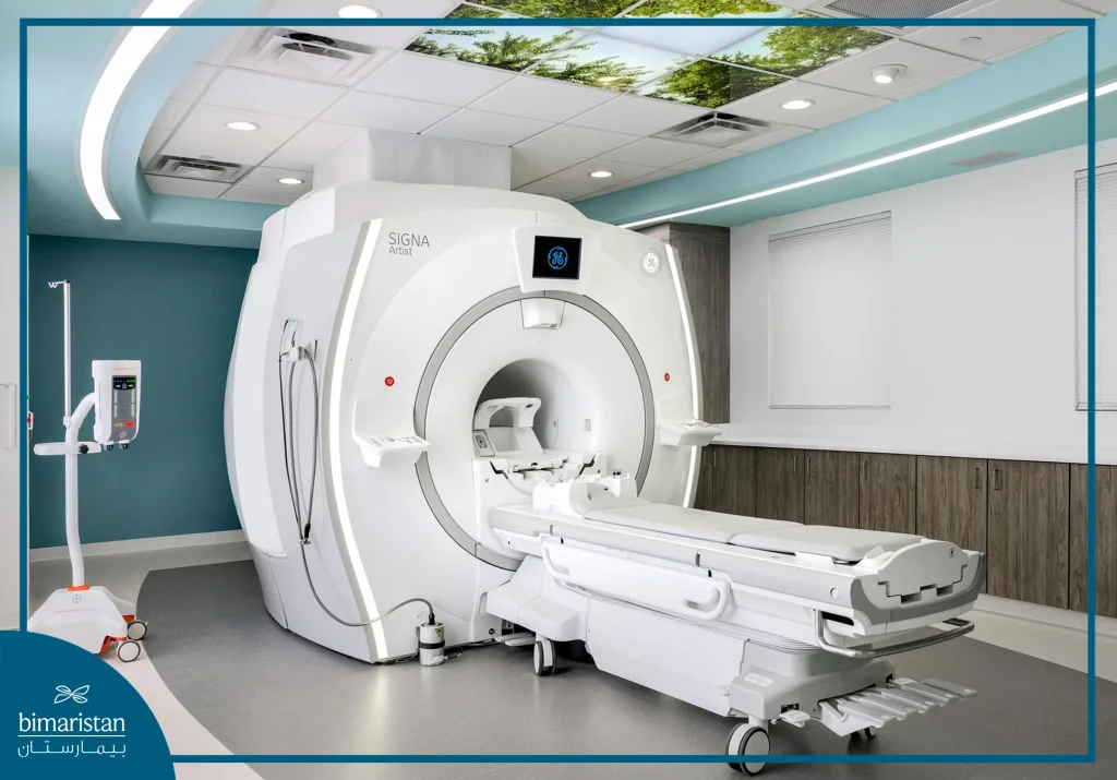
In this article, we will learn about some of the latest Cancer diagnosis and treatment methods in Turkey, of which the Positron Emission Tomography (PET) scanner is one of the most important.
Cancer diagnosis and treatment in Turkey
Since cancer has become so common in today’s world, medical studies have focused heavily on Cancer diagnosis and treatment methods. As a result of these studies, many new treatment methods and techniques have emerged and have begun to be used in Turkey immediately after their discovery.
Methods of diagnosing and treating cancer in Turkey
Cancer, one of the most widespread diseases in today’s world, has many types and poses a great risk to the health of patients. Different treatments are applied for each of these types of cancer. However, the most important prerequisite for successful cancer treatment is early diagnosis and treatment. Many new technologies have been developed to diagnose and treat brain tumors and Alzheimer’s disease. These technologies have increased the success rate of treatment with more accurate diagnoses. Considering all this, PET MR, which consists of a combination of MR and PET imaging, has gained a very important place in Cancer diagnosis and treatment methods.
You can read about the symptoms and treatments available for thyroid, bladder, uterine, and many other cancers on our website.
Magnetic resonance imaging with positron emission tomography
Magnetic resonance imaging (MRI) with positron emission tomography (PET-MRI) is one of the most common techniques used in the diagnosis and treatment of many types of cancer. A positron emission tomography scan, and a magnetic resonance imaging scan. PET MRI is an advanced imaging technique that has emerged using two different medical modalities together. This advanced imaging technique scans the entire body, and anatomical details are printed in high resolution. The two imaging modalities are performed in a single session, and all the anatomical and metabolic information of the body is obtained very accurately.
Many studies have shown its superiority over PET/CT, reducing radiation by 80% while obtaining a better quality tumor mass.
Magnetic Resonance Imaging (MRI)
MRI is a technique that has been used for many years to diagnose many diseases. It allows for a precise view of all the tissues, organs, and structures of the body.
Positron Emission Tomography (PET)
It is a medical technique used to measure the degree of activity of organs and tissues of the human body. Cancer cells’ degree of activity is higher by a certain percentage than normal cells. For this reason, PET MR is an advanced body scanning and imaging technique created by combining two separate medical technologies. This scanning and imaging technique is used to diagnose all types of cancer.
PET MR is a high-tech medical device that enables PET detectors placed in an MR machine to capture all PET images using MR. The most important purpose of this test is to determine if there are any cancer cells in the human body. As a result of the imaging, the points on the patient’s body where there are cancer cells are identified. In addition, very precise information is obtained about where the cancer cells are located in the body. Furthermore, information about the spread of the cancer and which tissues are most affected is revealed. Thus, a definitive diagnosis is made about the type of cancer, enhancing cancer diagnosis and treatment methods.
After an early and definitive diagnosis, depending on the type of cancer, treatment begins without wasting time. If cancer treatment is started early, the possibility of a successful outcome is greatly increased.

How is PET MR applied?
A radioactive substance is injected into the patient during the PET MR procedure. Many patients think that this radioactive substance given to the body is very harmful. However, the amount of radioactive material given is very small, and it is given to the body for a short time without any harmful effects. It is less harmful than PET combined with a regular CT scan. Before the PET scan begins, the patient is injected with a radioactive liquid into a vein. This process takes very little time. The fluid contains radio-labeled particles.
After this fluid is injected, the patient rests for half an hour in a lead-insulated waiting room. The purpose of this waiting is to spread the intravenously administered radioactive liquid throughout the body. After waiting, the patient is transferred and imaged by the machine. The PET scan on this machine lasts for about 20 minutes. The detailed, high-resolution images are examined by experts after the imaging is completed. After the scan, it is determined where the cancer cells have spread in the body and where they first or primarily started spreading from in the body.
What should I consider before applying PET MR?
There are a number of rules that patients should follow before undergoing an MRI scan in order for the imaging and scanning to be more successful. Patients should avoid strenuous activities and exercises 24 hours before the scan. However, the scan may take up to 6 hours.
The patient should not consume any food before the scan. This is because blood sugar must be kept at a normal level.
High or low blood glucose values affect the test results. The patient should also be psychologically ready for the test. All phases of the imaging take an average of 40-45 minutes. It is important that the patient has no complaints during this time. Some patients are afraid of enclosed spaces or injections.
All of these situations affect the test result and prevent healthy imaging. If patients have psychological issues related to claustrophobia, they should inform the specialist before the scan. Thus, by taking the necessary precautions, a healthy scan can be performed under general anesthesia, improving cancer diagnosis and treatment methods.
The difference between PET/CT and PET/MRI
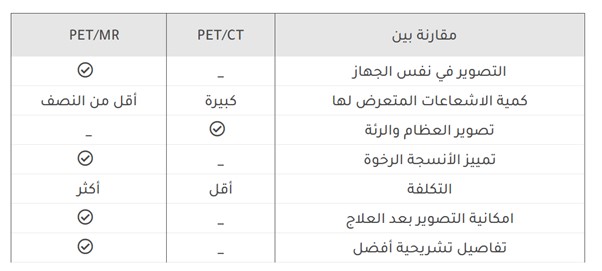
Let’s take a look at the differences between PET/CT and PET/MRI:
- CT/PET scanners cannot acquire data simultaneously as PET is imaged and then CT is imaged. In PET/MRI, they are imaged in the same machine.
- PET/CT has a large radiation dose. PET/MRI has lower radiation levels as the radiation ratio in MRI is less than half compared to CT, which is why it is used especially for cancer detection in children.
- PET/CT (positron emission tomography/computed tomography) can perform lung and bone imaging. However, PET/MRI cannot image the lungs because of their mobility and the bones because of their high density.
- PET/CT does not offer good contrast for tissue density difference. PET/MRI can discriminate tissue very well. Up to 50% of brain lesions are indistinguishable on CT.
- PET/MRI costs more than PET/CT and has a longer imaging time.
- PET/MRI distinguishes between living tissue and areas of necrosis after surgery or radiation therapy. PET/CT cannot distinguish between living and dead tissue after surgery or radiation therapy. It also improves the detection of low-grade or low-FDG lymphomas and increases the detection of tumor recurrence and newly developed distant metastases.
- PET/MRI shows a higher mapping ability for recurrent prostate cancer than PET/CT, according to a recent study presented at the SNMMI 60th Annual Meeting in Vancouver, British Columbia. Several studies confirm its superiority over other devices.

At Bimaristan, we offer the latest technologies for accurate cancer diagnosis and effective treatment.
Visit our website to explore modern solutions tailored to every type of cancer.
Sources:
