Congenital hip dysplasia, one of the most common bone conditions in newborns, presents a challenge for many families and medical teams alike. This article will review the various causes of this dislocation and explore the available treatment methods.
How can your little one be in the best possible health? And what steps should be taken to ensure full recovery? Let’s dive together into the world of this medical condition to discover the answers.
Congenital hip dysplasia
Congenital hip dysplasia, also known as Developmental Dysplasia of the Hip (DDH), refers to a health problem that affects the hip joint connecting the thigh and hip bones, disrupting joint function.
Most cases of congenital hip dislocation are present from birth and result from incomplete joint formation, causing the thigh bone to slip out of place. Therefore, doctors examine newborns shortly after birth to ensure there are no signs of congenital hip dysplasia.
Several genetic and environmental factors contribute to the development of congenital hip dysplasia in infants, estimated to occur in only 1-2 cases per 1,000 births.
In Turkey, a soft brace is usually sufficient to correct the problem. However, when congenital hip displacement does not cause symptoms and is not discovered until a later age, surgical treatment is primarily used to relocate the joint and restore its smooth and fluid motion.
Degree of congenital hip dysplasia
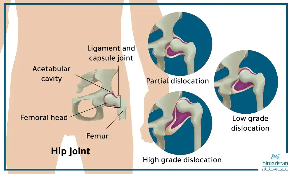
In all cases of developmental dysplastic hip in children, the joint socket is shallow, meaning that the spherical shape of the femoral head does not fit tightly into the acetabulum. This can lead to looseness of the ligaments responsible for stabilizing the joint. The severity of hip laxity in children with congenital hip dislocation is classified into several types:
- Subluxation: In mild cases of congenital hip dysplasia, the femoral head can move freely within the acetabular socket but does not fully detach from it.
- Low-grade dislocation: In this case, the femoral head is positioned within the acetabulum but may easily dislocate during a physical exam.
- High-grade dislocation: This is the most severe form of congenital dislocation, where the femoral head is completely out of the acetabular socket.
Causes of congenital hip dysplasia
The spherical femoral head fits perfectly into the acetabulum in a normal hip joint. However, in some children, the spherical head of the femur is not well stabilized, and the acetabulum is shallow, increasing the likelihood of the femoral head slipping and dislocating from the socket due to the pressure of the mother’s womb on the fetus or during childbirth. This congenital deformity is considered one of the main causes of congenital hip dysplasia in newborns.
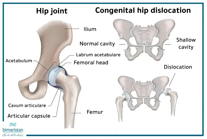
The occurrence of neonatal hip dysplasia increases when the uterus is small and hinders fetal growth, such as in first pregnancies, twin pregnancies, breech presentation, fetal macrosomia, or when there is a lack of amniotic fluid. Doctors have found that congenital hip dysplasia occurs more frequently in the following cases:
- Family history of hip dislocation or very loose ligaments.
- Female gender, as breech presentation occurs more often in females than males.
- Abnormal positions during childbirth, especially breech presentation.
Moreover, congenital hip dysplasia can occur after birth during the first year of life in some infants who are tightly swaddled with their lower limbs in a straight position. Therefore, parents should learn a safe way to swaddle their infants.
Symptoms of congenital hip dysplasia
The symptoms of congenital hip dysplasia may vary from one child to another and commonly include:
- The difference in the number of skin folds between the thighs.
- Loose or unstable hip joint on one side.
- Legs of unequal length.
- Limping or a waddling gait when the child starts walking.
- Pain in the hip.
Additionally, hip dysplasia can cause painful complications in teenagers and young adults, such as hip and back osteoarthritis or tears in the hip joint labrum, leading to accompanying pain during movement.
Diagnosis of congenital hip dysplasia in Turkey
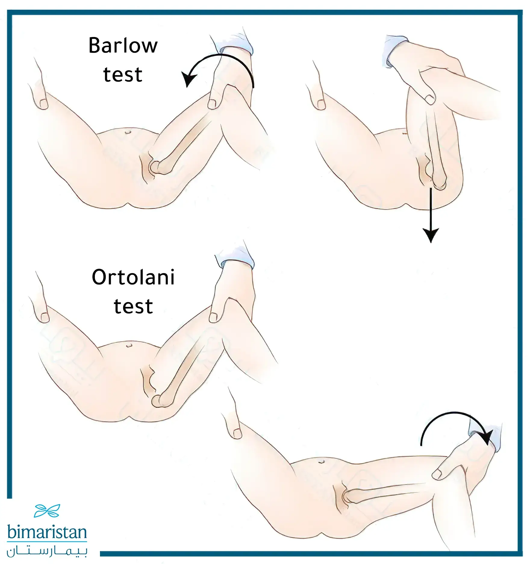
A pelvic exam is part of Turkey’s routine postnatal medical examinations to ensure hip joint health in newborns. The examination involves ruling out the presence of any abnormal “clicking” sound when moving the baby’s legs in various directions. Doctors currently perform the Barlow and Ortolani maneuvers without causing any discomfort to the baby.
The doctor may request a hip joint ultrasound between the fourth and sixth week if they suspect instability in the joint or if there is a family history of congenital hip dysplasia or breech birth. Although the hip may stabilize on its own before the imaging is done, it is essential to confirm by performing the ultrasound.
Mild cases of congenital hip dysplasia can be challenging to detect and may not cause problems until adolescence. Suppose the doctor suspects congenital hip dysplasia in children older than six months. In that case, they may request plain pelvic X-rays, a computed tomography (CT) scan, or magnetic resonance imaging (MRI) of the pelvis.
Early detection of congenital hip dysplasia in newborns is of the utmost importance, and studies have been conducted on its necessity for better treatment outcomes.
Congenital hip dysplasia treatment in Turkey
The treatment methods for congenital dysplasia in Turkey vary depending on the age of the affected child and the severity of the condition. The general goal of the congenital dysplasia of hip treatment is to relocate the femoral head back into the hip socket and stabilize it. At the same time, the joint grows naturally and is protected from further damage. Dislocation treatment may involve applying a soft brace, a rigid orthosis, or surgery in severe cases.
Pavlik harness
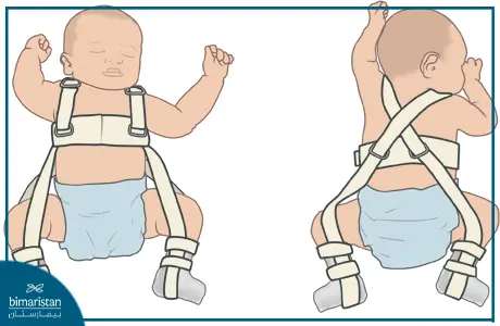
Children under six months diagnosed with congenital hip dysplasia are usually treated with a fabric brace called the Pavlik harness. This harness provides stability and keeps the femoral head securely in the socket, helping shape the socket and make it take the form of a ball. It also allows for free movement of the feet and easy diaper changes for the child.
Your child should wear this harness continuously for several weeks and only be removed by the doctor. The doctor may adjust the harness during follow-up visits and provide you with complete instructions on how to care for your child while wearing the harness.
Studies have shown that the Pavlik harness’s effectiveness decreases if its application starts after the baby is over four months old or if the congenital hip dislocation is severe.
Usually, the application of the harness is sufficient for treating congenital hip dysplasia. However, in some cases, the hip joint may remain partially or completely dislocated even after the child reaches six months of age.
Spica cast
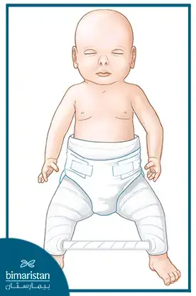
In some cases, the brace may not suffice, and the doctor may need to perform a closed reduction procedure under anesthesia to reposition the femur into its usual place. Afterward, a spica cast is applied to stabilize the child’s bones correctly and prevent joint movement for several weeks.
Caring for your child while they are wearing this cast requires special instructions. The doctor will teach you how to perform daily activities, maintain the cast, and recognize any problems that may occur.
Surgical treatment
If a closed reduction fails to reposition the femur after the child reaches six months of age, open surgery becomes necessary. The surgeon makes an incision in the child’s hip to gain a clear view of the joint, bones, and soft tissues.
Sometimes, the femur may be shortened to fit the acetabular socket. Simple X-rays are taken during surgery to ensure the femoral head is in its normal position. Following this, a spica cast stabilizes the pelvis until the bones settle and the wound heals.
The child may need to undergo multiple surgical procedures during their growth period, as the hip joint can be prone to dislocation again. It is worth noting that in sporadic cases, the entire hip joint may be replaced with an artificial joint through a surgical procedure or with the help of a robot, or it may be treated with stem cells.
How do I know if my child has congenital hip dysplasia?
Parents can recognize the possibility of congenital hip dislocation in their child through certain signs and symptoms. Here are some indicators that may suggest the presence of this condition:
- Different leg length: You may notice that one leg is shorter than the other when the child is lying on their back with extended legs.
- Restricted thigh movement: The child may have difficulty moving one leg normally or have limited movement compared to the other leg.
- Asymmetry in skin folds: The skin folds in the thigh or buttock area may appear uneven.
- Abnormal sounds: When moving the baby’s leg, a “clicking” or “popping” sound can sometimes be heard, which may indicate a hip dislocation.
- Tilting when walking: If the child has started walking, you may notice a tilt or deviation while walking due to hip instability.
If you notice any of these symptoms, you must consult a doctor for necessary tests such as X-rays or ultrasound to confirm the diagnosis and take appropriate action. Early detection and proper treatment can help prevent more severe complications later on.
The success rate of congenital hip defect surgery in children
Hip dislocation surgery in children is generally a successful procedure, with a success rate of over 90% in specialized and experienced centers.
However, it is essential to note that this rate can vary depending on:
- Surgeon’s experience: The surgeon’s expertise and skills play an essential role in the success of the surgery.
- Child’s health: The child should be in good overall health and free from any chronic conditions affecting wound healing.
- Following the doctor’s instructions: It is essential to follow them carefully before and after surgery to ensure the best results.
- Child’s age: Surgery outcomes are generally better when performed early.
- The severity of hip dislocation: In cases of severe hip dislocation, the surgery may be more complex, and the success rate may decrease.
References:
- Developmental Hip Dysplasia. Boston Children’s Hospital.
- Developmental Dysplasia of the Hip From Birth to Six Months. James T. Guille, MD, Peter D. Pizzutillo


