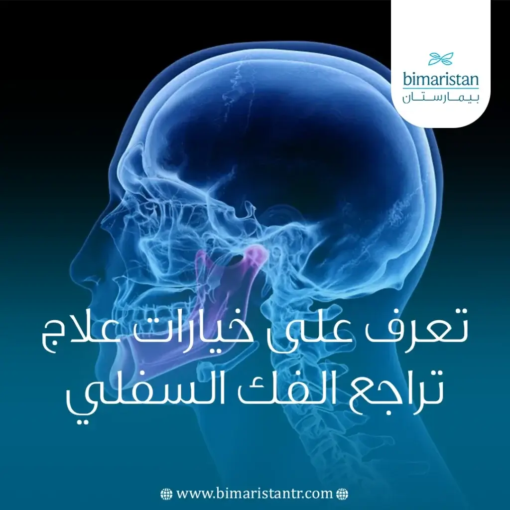
Retrognathia treatment is a common focus in orthodontics and maxillofacial surgery, as a receding lower jaw can affect facial balance and bite function. This condition is not only an aesthetic concern; it may also cause chewing difficulties, speech problems, and, in some cases, breathing disturbances during sleep.
The severity of retrognathia ranges from mild cases that can be treated with orthodontics to complex cases requiring surgical intervention. This article will review the possible causes of this condition, diagnostic methods, and the latest treatment options to improve the function and aesthetic appearance of the jaw.
Normal Anatomy of the Lower Jaw
The mandible is the largest and strongest bone in the face, and it is the only movable bone in the skull. It is connected to the skull via the temporomandibular joint and consists of a horizontal body and two ascending branches that terminate at the mandibular condyle. The mandible supports the lower teeth and plays a pivotal role in chewing, speaking, and swallowing, and it also significantly contributes to defining the shape of the lower third of the face. Its growth depends primarily on mandibular condyle development and on the balance between functional and muscular factors, making it sensitive to any disturbance during periods of growth.
What is Retrognathia?
Retrognathia is a skeletal condition in which the lower jaw is positioned behind its normal location relative to the base of the skull or the upper jaw, resulting in a Class II skeletal relationship. The primary cause is often underdevelopment of the lower jaw rather than protrusion of the upper jaw. Clinically, the patient presents with a convex facial profile, a clearly recessed chin, and relative protrusion of the upper teeth; this may be accompanied by malocclusion of varying degrees depending on the severity of the condition and the stage of development.
What causes the lower jaw to recede?
Mandibular retrusion is a multifactorial condition in which genetic, environmental, and functional factors interact in its development. The most important causes and risk factors can be summarized as follows:
- Genetic factors: A family history of Class II malocclusion or a small lower jaw increases the likelihood of developing the condition.
- Skeletal growth disorders: underdevelopment of the mandibular condyle or a disorder of mandibular cartilage growth during childhood and adolescence.
- Musculoskeletal imbalance: Weakness or imbalance in the masticatory muscles and the muscles responsible for jaw growth can affect normal jaw development.
- Chronic mouth breathing: This is associated with changes in the position of the tongue and lower jaw, which hinders the normal forward growth of the jaw.
- Non-functional oral habits, such as thumb-sucking or tongue-thrusting, affect the growth of the teeth and jaws.
- Anatomical abnormalities or syndromes, such as Pierre Robin syndrome or congenital torticollis, may affect the symmetry and growth of the lower jaw.
- Environmental factors during growth: malnutrition or general growth disorders that affect bone development.
These factors may act individually or in combination, leading to varying degrees of clinical mandibular retrusion, particularly during periods of active growth.
Other common causes of lower jaw retraction
A receding lower jaw may sometimes result from medical conditions, congenital abnormalities, or injuries that affect the growth or shape of the facial bones. The most common causes include:
- Pierre-Robin syndrome: A congenital condition characterized by a small lower jaw and a tongue that falls backward, which can cause breathing difficulties in some cases.
- Hemifacial microsomia: A developmental disorder in which one side of the face fails to develop normally compared to the other, resulting in a noticeable asymmetry of the jaw.
- Nagier syndrome: A rare condition that affects the growth of facial bones, particularly the lower jaw, and may also be associated with abnormalities in the limbs, such as the hands and arms.
- Treacher Collins syndrome: A congenital disorder that affects the facial bones, including the lower jaw and cheekbones, resulting in a small or receding jaw.
- Surgical procedures to remove tumors: Removing tumors from the jaw area may result in loss of bone or changes in its structure, causing the lower jaw to recede.
- Facial injuries or fractures: Fractures or injuries sustained during childhood can affect the growth plates in the jaw, leading to abnormal growth or underdevelopment later on.
These conditions vary in severity and impact, but all may lead to changes in the shape of the lower jaw and its position relative to the rest of the face, potentially requiring early medical and orthodontic evaluation.
Pathological Mechanisms and Functional Effects of Mandibular Retraction
Retraction of the lower jaw leads to a series of functional and structural changes that affect occlusal balance and the functions of the orofacial system:
- Its effect on occlusion: A retruded mandible results in a Class II skeletal relationship, with relative protrusion of the upper teeth and the development of a deep bite or excessive anterior overlap, due to an imbalance in the relationship between the upper and lower jaws. This imbalance impairs masticatory efficiency and leads to an uneven distribution of occlusal forces.
- Association with breathing disorders: A receding lower jaw is often associated with a narrowing of the posterior oral pharynx, which may contribute to partial airway obstruction, particularly during sleep. Studies have shown a link between mandibular retrusion and an increased risk of obstructive sleep apnea, due to the narrowing of the airway space behind the tongue.
- Effects on the Muscles and the Temporomandibular Joint: The posterior position of the lower jaw leads to a change in the function of the masticatory muscles, resulting in a muscular imbalance between the muscles that elevate and depress the jaw. The temporomandibular joint may also be subjected to abnormal functional stress, which can cause symptoms such as joint pain, clicking sounds, or limited movement.
These changes develop gradually and may affect not only oral function but also overall facial development and the neuromuscular balance of the mouth and face.
Clinical symptoms and signs of mandibular retraction
Cases of mandibular retraction present with a range of clinical signs and symptoms that affect both form and function:
- Facial features: A pronounced profile with a receding chin and a sagging lower third of the face.
- Chewing and speech problems: poor occlusion and difficulty pronouncing certain sounds.
- Sleep disturbances: In advanced cases, these may manifest as snoring or sleep apnea.
- Temporomandibular joint pain: This includes pain, clicking, or difficulty moving the jaw.
The severity of these symptoms varies from patient to patient, depending on the degree of deterioration, age, and the functional restorations present in the oral cavity.
Diagnosis of Mandibular Retraction
The diagnosis is based on a comprehensive assessment that includes:
- Clinical examination: Assessment of facial structure, the chin, and functional occlusion.
- Cephalometric analysis: It illustrates the structural relationship between the jaws and the base of the skull and confirms Class II malocclusion.
- Radiography: panoramic X-rays and CBCT scans to evaluate bones, joints, and airways.
- Differential diagnosis: To rule out similar conditions such as maxillary protrusion or purely functional issues.
This diagnostic integration is essential for developing a precise treatment plan tailored to the condition’s severity and stage of development.
Retrognathia treatment
Retrognathia treatment depends on the severity of the condition, the patient’s age, and whether the problem is structural or functional; treatment is often a multi-stage, multidisciplinary process:
- Conservative treatment: This is primarily used in growing patients and involves functional orthodontic appliances designed to stimulate or redirect lower-jaw growth, such as fixed or removable appliances. Functional guidance of growth is also used by improving tongue position and regulating muscle function to reduce the impact of factors that cause malocclusion.
- Orthodontic Treatment: In mild to moderate cases, removable orthodontic appliances can be used to improve the bite by moving the teeth without significantly altering the skeletal structure, thereby correcting the discrepancy between the jaws and improving both function and aesthetics.
- Surgical treatment: This is the treatment option for severe cases after growth has completed, and involves mandibular advancement surgery to reposition the lower jaw. Indications for surgery are determined when there is a clear structural abnormality that cannot be corrected orthodontically, or when the condition significantly affects function or appearance, such as chewing difficulties, breathing disorders, or obvious cosmetic deformity.
- Multidisciplinary Treatment: Managing complex cases requires collaboration between an orthodontist, an oral and maxillofacial surgeon, and sometimes a speech-language pathologist or sleep medicine specialist, with the goal of achieving stable functional and aesthetic outcomes. This integrated approach is considered the most effective in improving long-term outcomes and reducing complications.
The selection of the optimal treatment plan depends on a careful assessment of each individual case, taking into account future growth as well as functional and aesthetic outcomes.
The optimal timing for therapeutic intervention in mandibular retrusion
The timing of treatment initiation is a critical factor in determining the success of therapeutic interventions for mandibular retrusion, as the treatment approach differs significantly between children and adults depending on their stage of development:
- In children, Treatment during the period of active growth offers the most effective opportunity to modify the structural growth of the lower jaw using functional appliances or growth guidance. At this stage, chondral activity in the mandibular condyle can be leveraged to stimulate mandibular growth or to improve its direction, potentially reducing the need for future surgical interventions.
- In adults: Once skeletal growth is complete, the ability to conservatively modify the structural anatomy of the lower jaw is limited, so treatment is often limited to orthodontic correction or, in severe cases, requires surgical intervention to reposition the jaws and achieve proper occlusion.
Preventing the recession of the lower jaw and limiting its progression
Prevention of mandibular retrusion focuses on reducing factors that affect jaw growth from an early age, with the aim of improving the chances of natural growth and reducing the need for complex therapeutic interventions later on:
- Early Detection: Early diagnosis during childhood is a key component of prevention, as it allows identification of abnormalities in the lower jaw’s growth pattern at an early stage. Regular follow-up visits with an orthodontist help detect signs of Class II malocclusion before they worsen, allowing for timely intervention using effective orthodontic and functional treatments.
- Correcting harmful habits: Correcting unhealthy oral habits—such as thumb-sucking, tongue-thrusting, and chronic mouth-breathing—plays a significant role in preventing the progression of malocclusion. Addressing functional causes, such as nasal airway obstruction or breathing disorders, also helps restore the natural balance of jaw and tongue position, supporting proper lower-jaw growth and reducing the likelihood of structural abnormalities.
Mandibular retrusion is a skeletal disorder that clearly affects oral function, aesthetic appearance, and quality of life, and it requires a thorough understanding of its multiple causes, diagnostic methods, and management strategies. The scientific literature has shown that early intervention during the growth phase is the most influential factor in improving treatment outcomes, while the need for surgical intervention increases in severe cases after growth is complete. Furthermore, a multidisciplinary approach remains the optimal choice for achieving stable functional and aesthetic outcomes.
In recent years, countries such as Turkey have emerged as leading centers in orthodontics and maxillofacial surgery, combining clinical expertise with modern technologies and making them important destinations for both treatment and education. Ultimately, the success of managing these cases depends on accurate diagnosis, individualized treatment planning, and timely intervention, ensuring improved function and aesthetics while minimizing long-term complications.
Sources: