The best ways to treat tracheal stenosis in Turkey. It is most often formed due to pressure from the ventilator tube after a long stay in intensive care
Tracheal stenosis (Windpipe stenosis)
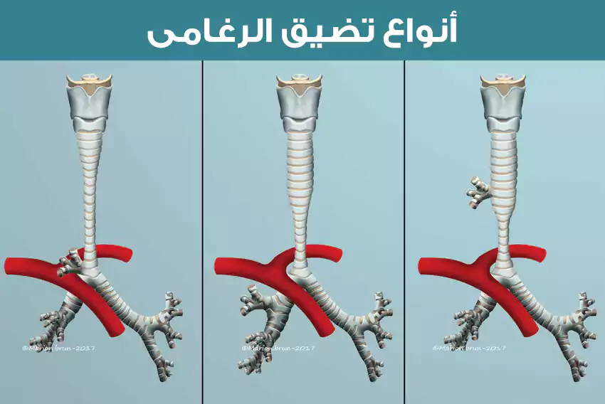
Tracheal stenosis refers to an abnormal narrowing of the trachea that limits your ability to breathe normally.
It can also be referred to as subglottic stenosis.
The subglottis is the narrowest part of the airway, and more often than not, this narrowing occurs at this level of the airway.
Causes of tracheal stenosis
Tracheal stenosis is commonly caused by an injury or disease such as:
- Damage to the throat or chest
- Infections (viral or bacterial), including tuberculosis
- Autoimmune disorders such as sarcoidosis, papilloma, granulomatosis, and amyloidosis
- Benign and malignant tumors
- Radiation therapy to the neck or chest
Symptoms of tracheal stenosis
In congenital tracheal stenosis, mild cases can often be interpreted as asthma or recurrent bronchitis. In mild cases, symptoms may not appear until late childhood or early adolescence, when they manifest as difficulty breathing when exercising. In more severe cases of this condition, you may notice the following symptoms:
- Stridor (high-pitched breathing sound)
- Cyanosis of the skin, with noticeably blue lips
- A whistling sound while inhaling
- Stressful shortness of breath (dyspnea)
In other cases of acquired tracheal stenosis, symptoms may not appear for several weeks after the injury occurs. The common first symptom is difficulty breathing. Like the congenital form, you may notice stridor, wheezing, or shortness of breath.

Tracheal stenosis diagnosis methods
Several testing methods can be used to help your doctor determine whether you have tracheal stenosis. These tests are useful in detecting and diagnosing other bronchial diseases. Bronchoscopy is considered the gold standard for diagnosing tracheal stenosis because it allows your doctor to directly visualize the shape of your trachea.
However, there are some risks associated with this method because using an endoscope will further obstruct your airway, making it more difficult to maintain oxygen levels. Therefore, you should discuss your risk factors with your doctor before undergoing endoscopy.
Other methods your doctor may use are X-rays, CT scans, ultrasound, MRI, and lung function testing.
X-rays
Standard X-rays are good at identifying structure, air plumes, injuries, and other preliminary data. Other, more sophisticated X-ray machines can be used to better define the diagnosis; however, X-ray diagnostics result in much more radiation exposure than other methods.
Computed Tomography (CT)
A CT scan can be a valuable tool for your doctor to determine whether you have tracheal stenosis. But it has some difficulties in localizing the soft tissues that cause it. Some techniques are used in such a way as to create a “virtual endoscopy” to minimize the need for a bronchoscopy. However, CT scans are not a great way to diagnose less severe degrees of stenosis.
Ultrasound
Ultrasound can be helpful in determining the amount of air space in the trachea. This allows your doctor to determine whether or not further testing is needed. However, due to the amount of cartilage around the trachea, the accuracy of the test can be questioned due to shadow effects caused by sound waves reflecting off the cartilage. This makes it so that only doctors with high skill levels are able to diagnose based on this method.
MRI scans
An MRI scan is also a great alternative method to help diagnose tracheal stenosis in both adults and children and is considered the ideal way to make this diagnosis. The main disadvantage of MRI is the length of time the procedure takes and the interference that can occur from normal breathing during the scan. Techniques are constantly being developed to improve the use of this method in its diagnosis.
Pulmonary function test (PFT)
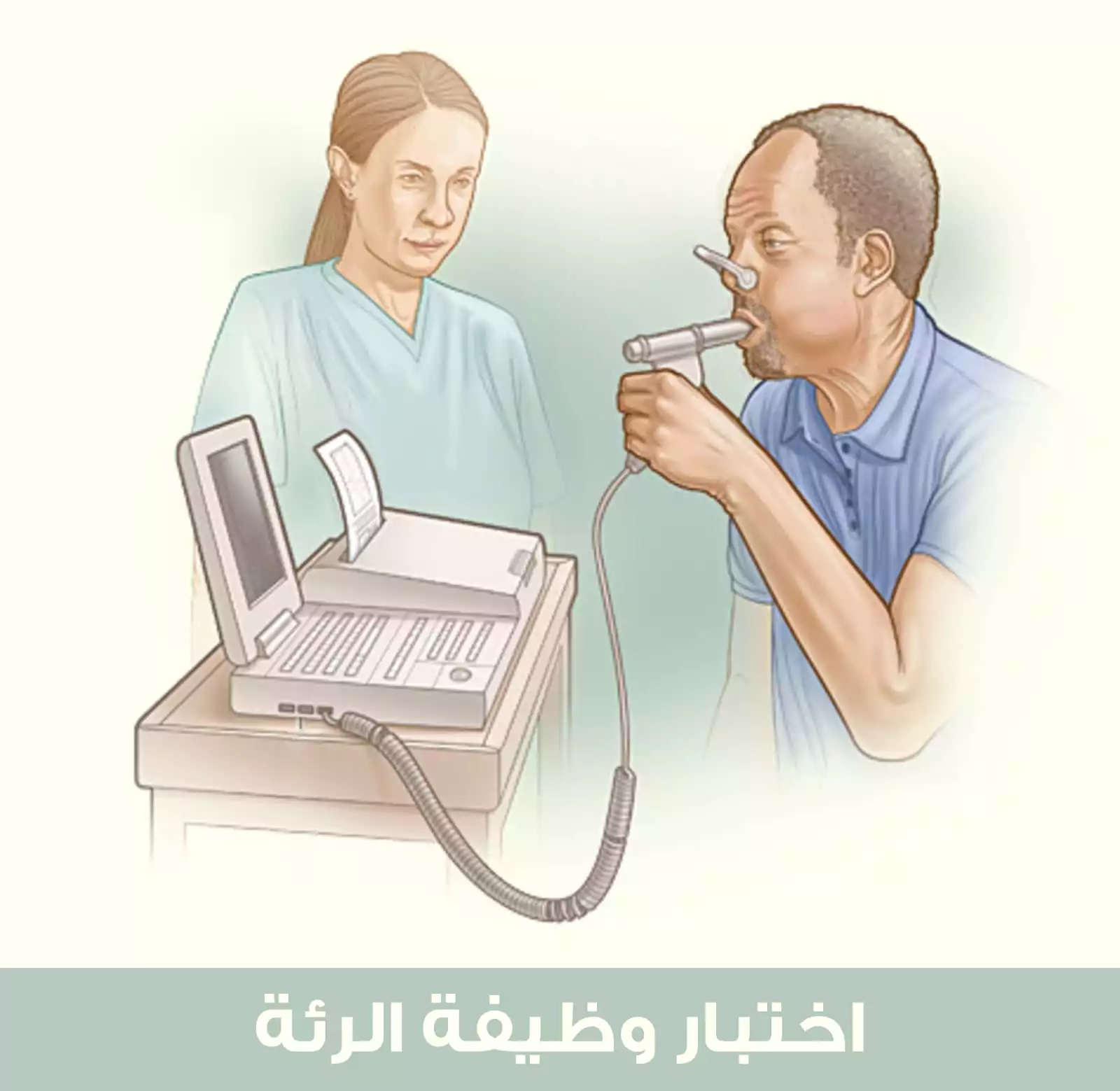
A lung function test can be performed in some doctors’ offices; if not available, you will be referred to a pulmonology lab. This test can be used to determine the extent to which your stenosis is impairing your breathing. This will be helpful in discussions with your doctor about treatment options.

Tracheal stenosis treatment in Turkey
Your otolaryngologist will develop a treatment plan based on the results of your evaluation. Treatment options, some of which are done using minimally invasive surgical techniques, include
Laser surgery, which can remove scar tissue, if this is the cause of the stenosis. This provides short-term relief but is usually not a long-term solution, as there are some studies that consider it a promising treatment plan.
Tracheal stenosis is treated with an airway stent, called a tracheobronchial stent, in which a mesh tube is placed in the airway to keep it open.
Tracheostomy, or tracheal dilation, involves the use of a small balloon or dilator to expand the airway. This may also not be a long-term solution.
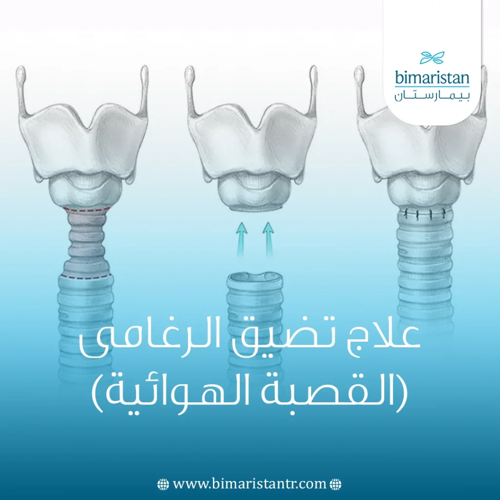
Complete removal and reconstruction of the trachea may provide long-term relief. The damaged part of the trachea is removed, and then the remaining limbs are joined.
Sources:

