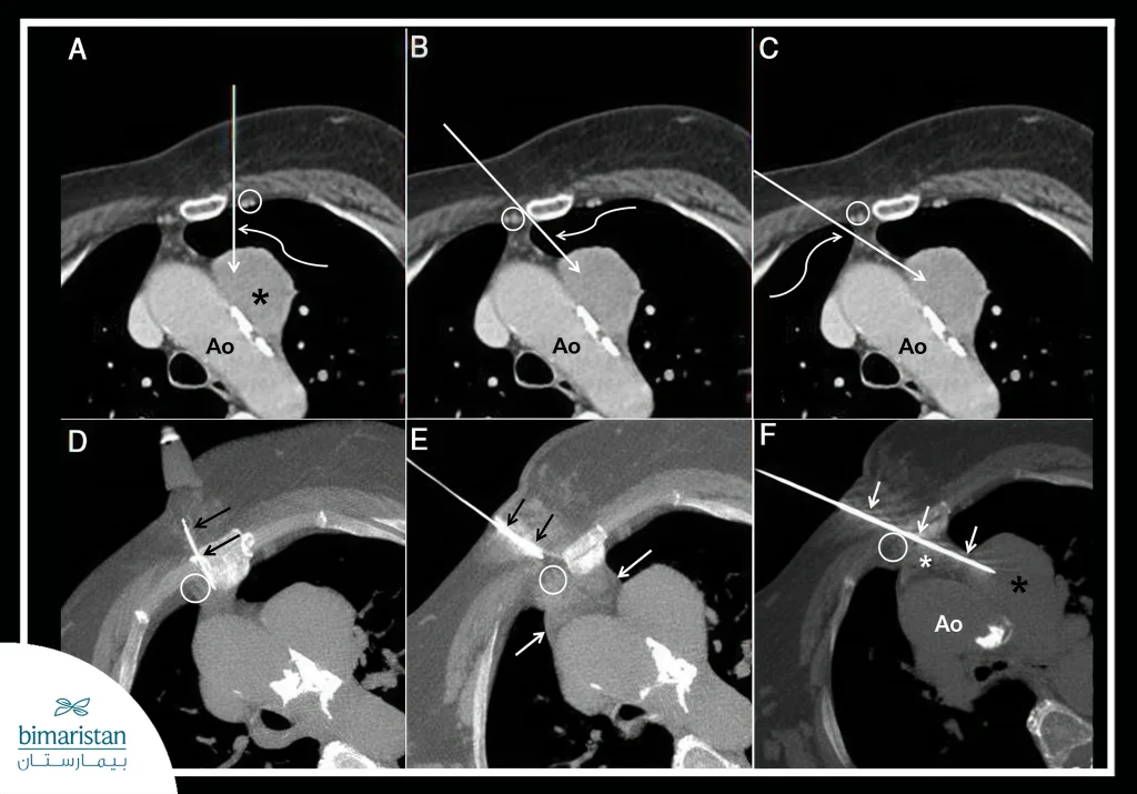
The mediastinum is the anatomical space between the lungs that contains the heart, major blood vessels, esophagus, and thymus. A mediastinal mass is defined as an abnormal growth or collection in this area, which may be tumors, cysts, abscesses, or other collections. Early mediastinal mass diagnosis is essential to determine their nature, whether benign or malignant, which helps in choosing the right treatment, minimizing the risk of complications, and improving the chances of recovery.
What are mediastinal masses?
Mediastinal masses are abnormal collections in the area between the lungs and include multiple types such as tumors, cysts, and abscesses. These masses can be solid or cystic—solid masses are cohesive and made up of cells or tissue, while cysts contain fluid or semi-fluid material within an envelope. Mediastinal masses are classified according to their location into three main sections: anterior, middle, and posterior. The distribution of mass types and causes varies based on this classification, which directly affects the patterns and methods of mediastinal mass diagnosis and treatment options.
Mediastinal masses, especially tumors or cysts near the heart, may lead to pressure on the pericardium or cause a pericardial effusion, which requires evaluation of the heart as part of the diagnostic tests.
Possible causes of mediastinal masses
Mediastinal masses appear for a variety of reasons, including benign and malignant tumors or immune-mediated diseases:
- Benign tumors: These include teratomas and cysts that grow noncancerously within the mediastinum.
- Malignant tumors: Mediastinal lymphoma and cancerous metastases that spread from other tumors.
- Immune or inflammatory diseases: Cause swelling or lumps due to inflammation of the tissues in the mediastinum.
- Enlarged glands: Like an enlarged thymus or lymph nodes, a scan shows an enlargement that looks like a lump on imaging.
When do we suspect a mediastinal mass?
We suspect you have a mediastinal mass in each of the following cases:
- Shortness of breath: Due to the mass pressing on the bronchi.
- Unexplained cough: Persistent or sometimes accompanied by blood.
- Chest pain: May be caused by the lump expanding or pressing on nerves or tissues.
- Difficulty swallowing or a change in voice: As a result of the mass affecting the esophagus or laryngeal nerve.
- In some cases, the mass is discovered accidentally during a chest X-ray for other reasons.
Steps to diagnose mediastinal masses
Mediastinal masses are diagnosed in several ways:
Medical history and clinical examination
During this stage, symptoms (such as coughing, shortness of breath, or chest pain) are investigated, taking into account age, gender, and risk factors such as family history of tumors or immune diseases.
Radiography
- Chest X-ray: This is the first step in determining the presence of a mass or changes in the mediastinum.
- Computerized tomography (CT): Provides detailed information about the size, location, and nature of the mass and its relationship to surrounding structures.
- Magnetic resonance imaging (MRI): Used to evaluate the extension of a mass, especially in soft tissues or nerves.
Laboratory tests
- A general blood test: To assess inflammatory status or the presence of anemia.
- Tumor markers: Alpha-fetoprotein (AFP) and beta-HCG for some tumors of the anterior mediastinum (e.g., fetal genital tumor).
Biopsy
This is the crucial step to determine the exact nature of the mass, which can be done by
- CT-guided needle biopsy.
- Surgical or open surgical thoracoscopy, depending on the case.
- Video-assisted thoracoscopy (VATS): A minimally invasive surgical procedure.

The importance of determining the nature of the mediastinal mass
Mediastinal mass diagnosis and determining the exact nature of the mediastinal mass is a pivotal step in developing the appropriate treatment plan, as treatment methods differ substantially between benign masses that may be monitored or surgically removed and malignant masses that often require multistage treatment, including surgery, chemotherapy, or radiation
Identifying the type of affected tissue (lymphoma, metastasis, or congenital tumor) helps assess the extent of the tumor and the presence of metastases or involvement of adjacent lymph nodes, which is important in staging the disease and estimating the overall prognosis. In addition, accurate diagnosis helps avoid unnecessary surgical interventions in some cases and directs doctors to use the least invasive and most effective treatment methods.
Difficulties in mediastinal mass diagnosis
Many difficulties and overlaps may confuse the doctor during the diagnosis of mediastinal masses:
- Difficulty accessing the block in some locations
- Mass interference with the heart or arteries
- Some masses have a similar radiographic appearance and require additional testing
The diagnosis of mediastinal masses requires the cooperation of a multidisciplinary medical team that includes chest physicians, radiology, oncology and surgery, radiography and biopsy are the cornerstone to reach an accurate diagnosis, and early detection plays an important role in determining the treatment plan and increasing the chances of recovery, especially when differentiating between benign and malignant masses, so it is advised not to ignore any unjustified respiratory symptoms such as cough or shortness of breath, especially if they persist for a while without improvement.
Sources:
- uanpere, S., Cañete, N., Ortuño, P., Martínez, S., Sánchez, G., & Bernado, L. (2013). A diagnostic approach to the mediastinal masses. Insights into Imaging, 4(1), 29-52.
- Almeida, P. T., & Heller, D. (2024, April 19). Anterior mediastinal mass. In StatPearls. StatPearls Publishing.

