Each year, more than half a million people visit emergency rooms due to kidney stone issues, and it is estimated that one in ten people will develop kidney stones at some point in their life. Kidney stones are common and are becoming more prevalent over time, with approximately 11 percent of men and 6 percent of women in the United States experiencing kidney stones at least once in their lifetime, potentially leading them to undergo various stone fragmentation techniques, including those using flexible ureteroscopy.
Doctors often resort to stone fragmentation via flexible ureteroscopy, which is an effective and minimally invasive method for treating and fragmenting kidney stones. Thanks to significant medical advancements in kidney stone treatment technologies, along with improvements in endoscopic procedures and laser technology, this method is effective and minimally invasive.
What are kidney stones?
Kidney stones are solid pieces that form in one or both kidneys and are associated with high levels of certain minerals in the urine. The majority of kidney stones contain calcium in their composition. They can be as small as a grain of sand or as large as a pea, and in rare cases, they can be as large as golf balls. Stones may be smooth or rough, usually yellow or brown.
Symptoms of kidney stones
Symptoms of kidney stones may appear intermittently, meaning they can occur for short or long periods and then disappear in waves, or they may be continuous at times. These symptoms include:
- Sharp pain in the back, side, lower abdomen, or groin area (inguinal region).
- Pink, red, or brown-colored blood in the urine (hematuria).
- Pain during urination.
- A persistent need to urinate.
- Foul-smelling or cloudy urine.
- Inability to urinate or only being able to urinate in small amounts.
- Nausea and vomiting.
- Fever and chills.
Causes of kidney stones
Possible causes include:
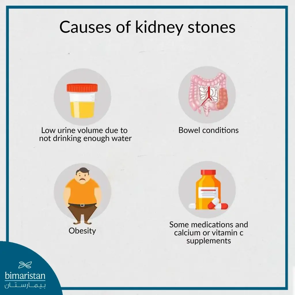
- Not drinking enough water daily, with the recommended amount being around 3 liters.
- Lack of exercise or excessive exercise.
- Obesity or undergoing weight loss surgery.
- Intestinal diseases
- Eating food that contains a lot of salt or sugar.
- Family history of stone formation may be significant in some cases.
- Consuming large amounts of fructose, which is abundant in table sugar and high-fructose corn syrup, is linked to an increased risk of developing kidney stones.

Diagnosis of kidney stones
The diagnosis of kidney stones is based on medical history, physical examination, and imaging. It is crucial to accurately determine the size and shape of the stones, which is often done using high-resolution computed tomography (CT) scanning from the kidneys to the bladder or through X-rays (KUB X-ray for kidneys, ureters, and bladder) that will precisely show the size and location of the stone. However, CT scanning is generally preferred for diagnosis. In some cases, doctors may also request an intravenous pyelogram (IVP), which is a particular type of X-ray of the urinary system performed after injecting a contrast dye.
What is the flexible ureteroscopy?
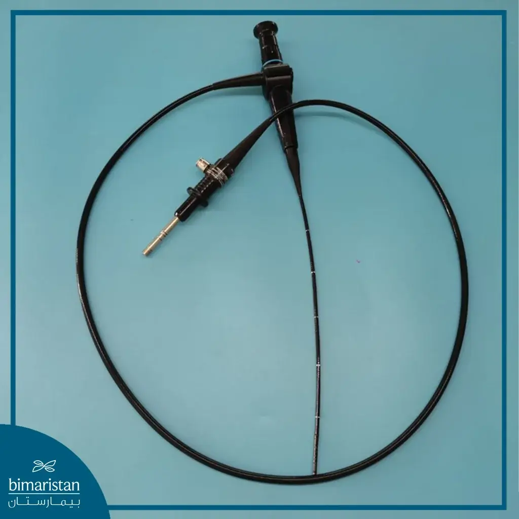
The flexible ureteroscope is a medical device used for diagnostic and therapeutic purposes in the upper urinary tract. It is designed with capabilities that allow it to reach difficult-to-access areas, enabling the fragmentation of stones and treatment of other conditions with minimal surgical intervention. Stone fragmentation via flexible ureteroscopy
Ureteroscopy with the flexible ureteroscope has proven to be an effective and viable method in many cases for fragmenting kidney stones, particularly in situations where shock wave lithotripsy cannot be used (such as in pregnant women and children).
During therapeutic ureteroscopy with the flexible ureteroscope, a guidewire is typically used to help guide the instrument and facilitate multiple passes through the ureter to reach distant urinary tracts. Then, laser energy is delivered through fine quartz fibers to fragment the stones. An attached tool is then used to extract the small stone fragments, all of which is done through the flexible ureteroscope, and these procedures are typically completed in a single day.

After the flexible ureteroscopy for the kidneys
On the same day of the flexible ureteroscopy procedure, internal ureteral stents are usually, but not always, placed to facilitate the healing process and ensure drainage, especially if therapeutic solid maneuvers were performed or if the ureter needed dilation to enable access to the stones. However, ureteral stents are generally not required post-surgery if ureteroscopy was performed for simple diagnostic purposes without ureteral dilation.
These internal ureteral stents may cause some lower urinary tract symptoms, such as frequent urination, urinary urgency, and mild to moderate blood in the urine. These symptoms are transient and resolve over time without needing any treatment.
The ureteral stents are then removed after a healing period that can range from a few days to 6-8 weeks, depending on the patient’s specific condition. This procedure is typically done in the clinic, either using an attached nylon thread or through cystoscopy.
The urinary catheter is removed on the same day or the morning after once the ability to urinate normally returns.
In most cases, patients are given preventive antibiotics and oral painkillers after being discharged from the hospital. Anticholinergic medications and alpha-blockers may be prescribed to reduce the severity and frequency of symptoms and discomfort often associated with the placement of ureteral stents, as tolerance varies significantly among patients.
Careful selection of the best stent length and optimal placement are key factors in minimizing or preventing these unwanted symptoms after stone fragmentation.
Follow-up after flexible ureteroscopy for the kidneys
Most patients return for follow-up after flexible ureteroscopy within one to two weeks to remove the stent if present and to undergo some necessary imaging to determine the effectiveness of the treatment and detect the presence of stones.
Advantages of flexible ureteroscopy in kidney stone fragmentation
- Precision: Provides high-definition, high-quality images and can magnify, store, and capture any part or condition in the urinary tract, aiding in the diagnosis, treatment, and follow-up of conditions.
- Effectiveness: Capable of fragmenting all types and sizes of stones thanks to the laser used with the flexible ureteroscope.
- Safety: The laser used with the flexible ureteroscope does not cause any damage or injury to the ureter, kidney, bladder, or urethra, and does not cause bleeding, infection, or stone escape.
- Flexibility: Can reach any location in the kidney or ureter with flexibility and precision and fragment stones regardless of their size, shape, or location, though it is preferably used in cases of large stones after the failure of traditional stone fragmentation methods.
- Speed: The flexible ureteroscopy reduces operation time and hospital stay duration, allowing the patient to return to normal activity more quickly.
Complications of kidney stone fragmentation via flexible ureteroscopy
Complications depend on the size and severity of the procedure, the doctor’s expertise, and the advancement of the medical center. Complications are rare when a highly experienced doctor uses the best techniques and tools in the hospital. These complications may include:
- Colic or pain
- Fever
- False discharges
- Mild or prolonged hematuria
- Urinary tract infection
Cost of kidney Stones Fragmentation Using Flexible Ureteroscopy
The cost varies depending on the patient’s specific medical condition. Still, it is often around 2,500 US dollars in the best hospitals in Turkey and under the hands of the most skilled specialist doctors. Turkey is one of the countries that many patients resort to for the treatment of such medical conditions due to the quality of the medical service, the development of hospitals and the technologies used, and the reasonable cost of treatment compared to the countries of Europe and the USA.
Endoscopy techniques, particularly flexible ureteroscopy, have achieved great success in the medical field and have been relied upon to treat many patients, helping them to get rid of kidney and ureteral stones as quickly as possible with minimal complications. In addition to its therapeutic benefits, it also has diagnostic value, aiding in the detection of many urinary tract diseases. It is worth mentioning that the size of the devices used in ureteroscopy will continue to decrease with the use of smaller optical fibers, enhanced digital imaging devices, improved accessories, and new power sources. As the size of the devices becomes smaller and their precision increases, they will also become more sensitive, thereby providing the best possible benefits to patients.
Resources:
