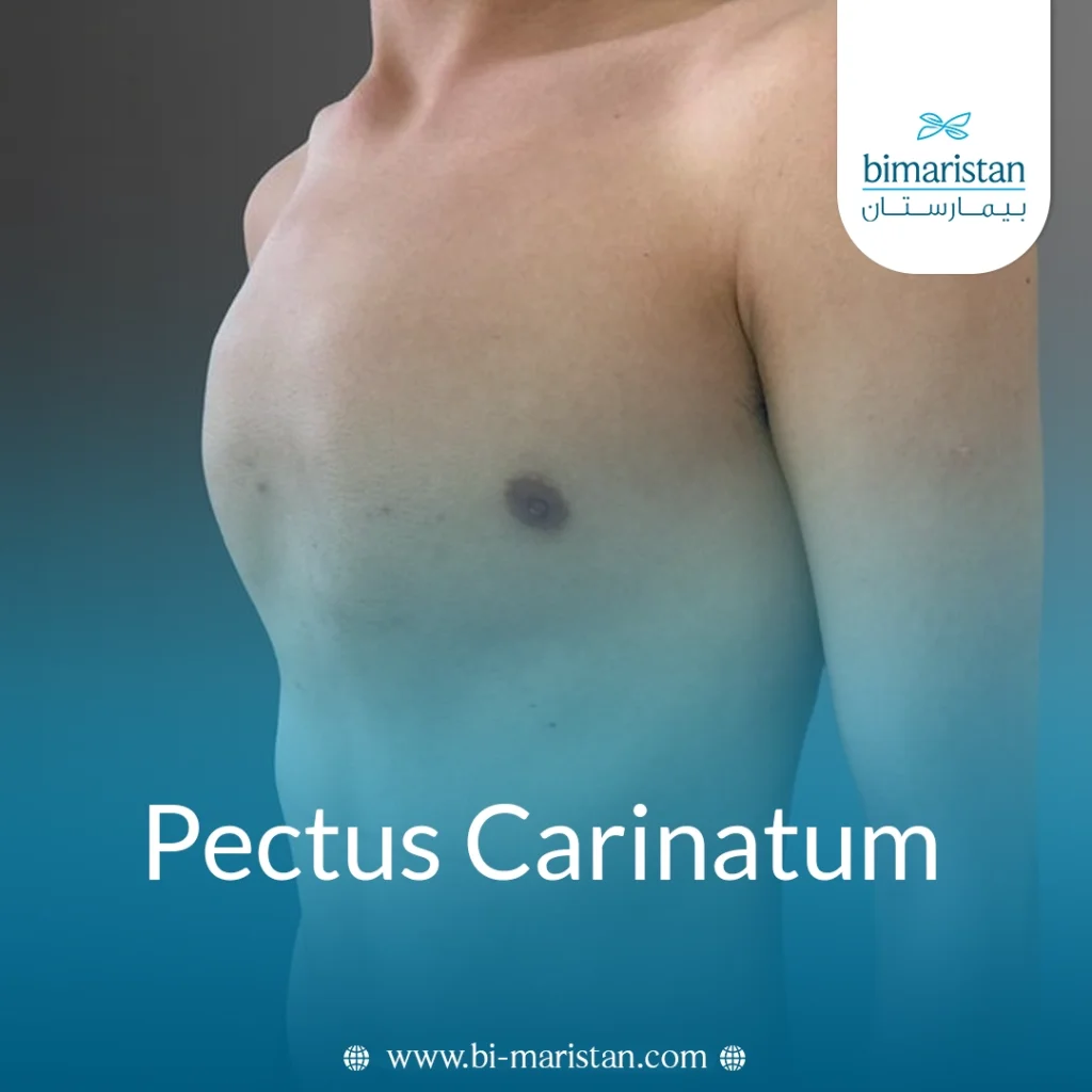

The shape of the chest wall is an anatomical feature that typically appears normal, but it may draw attention when growth patterns of bones and cartilage change, especially during childhood and adolescence. Among these changes is a Pectus Carinatum, characterized by a noticeable protrusion at the front of the chest that may become more pronounced with age.
Although this change may initially seem like a mere cosmetic issue, it can raise many questions about its nature and its impact on the body, both functionally and psychologically. Therefore, gaining a thorough understanding of pectus carinatum—including its causes, types, and treatment options—is an essential step toward appropriately understanding and managing this condition.
What is pectus carinatum, and what are the treatment options in Turkey?
Pectus carinatum is a deformity of the chest wall characterized by an abnormal outward protrusion of the sternum resulting from excessive growth of the cartilage connecting the ribs to the sternum. It is also known as pigeon chest. This abnormal growth causes the anterior chest to protrude, giving it a prominent or convex appearance.
This condition is the opposite of pectus excavatum, in which the sternum protrudes inward rather than outward; it is often present from birth but becomes more noticeable during adolescence as growth accelerates. It is also more common in males.
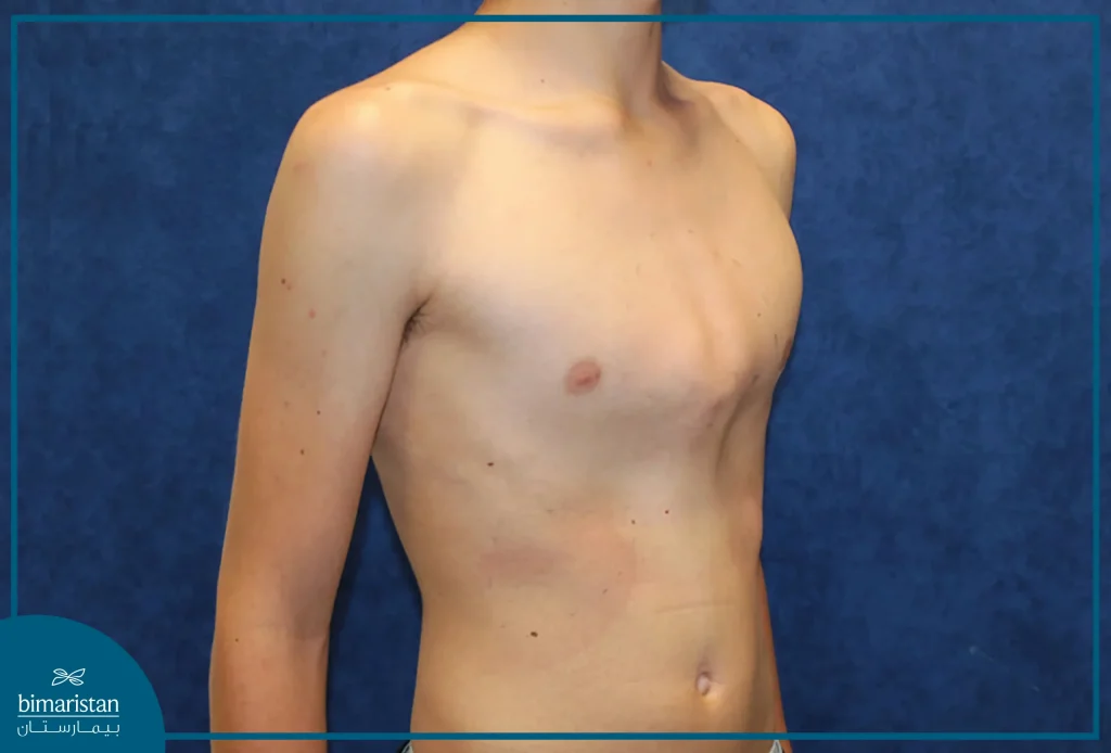
Types of pectus carinatum
Pigeon chest can be classified into two main types based on the location of the protrusion in the sternum:
- Sternal chondral type: This is the most common form, in which the lower part of the sternum protrudes outward.
- Upper sternum type: Characterized by a protrusion of the upper part of the sternum.
The deformity may be symmetrical on both sides of the chest or asymmetrical; in some cases, it may appear on only one side, with a reduction on the opposite side.
Causes of pectus carinatum
The exact cause of pigeon chest remains unknown, but most medical explanations suggest that it results from a disorder in the growth of the cartilage connecting the ribs to the sternum, leading to excessive or abnormal growth that pushes the sternum outward. It is believed that this disorder may be part of a malformation in the development of the chest wall or in the maturation of the costal cartilages.
Some studies suggest a possible genetic factor, as a proportion of those affected have a family history of chest wall deformities, supporting the hypothesis that genes play a role in the development of the condition, although no clear genetic cause has been identified to date.
pectus carinatum is also associated with certain other conditions or disorders, the most notable of which are:
- Marfan Syndrome
- Noonan Syndrome
- Spinal Curvature (Scoliosis)
- Down Syndrome
- Certain other metabolic or genetic disorders
- Asthma or bronchitis
- Mitral valve prolapse
Symptoms of pectus carinatum
The symptoms of pigeon chest vary from person to person depending on the severity of the condition; however, most people with this condition do not experience obvious symptoms, and the problem is often more cosmetic than functional. Possible symptoms and signs include the following:
- A distinct outward protrusion of the sternum and ribs
- Asymmetry of the chest, where one side may protrude more than the other
- Pain or discomfort in the chest area or tenderness at the site of the protrusion, especially during activity or in certain positions
- Shortness of breath during exercise
- Fatigue or reduced stamina
- Psychological effects such as feelings of embarrassment or low self-confidence
Diagnosis of Pigeon Chest
Pigeon chest is usually diagnosed through a clinical examination, during which the doctor relies primarily on observing the shape of the chest wall and assessing the degree of protrusion and asymmetry, as well as taking the patient’s medical history. The condition is suspected when there is a noticeable protrusion of the sternum or a change in chest shape, especially if it is asymmetrical or becomes more pronounced over time, warranting a more thorough medical evaluation.
To confirm the diagnosis and determine the severity of the condition, certain imaging techniques may be used, the most notable of which are:
- Frontal and Lateral Chest X-rays
- Computed Tomography for Assessing Chest Wall Details
- Magnetic Resonance Imaging (MRI) in Certain Cases
- 3D Imaging Techniques for Monitoring Breast Shape
Certain radiographic measurements can also be used to determine the severity of the deformity and monitor its progression, such as the thoracic cage dimension ratio, which is also used to assess the response to treatment later on.
Treatments for pectus carinatum
Pigeon chest does not go away on its own, so the decision to treat it depends on the severity of the condition, the presence of symptoms, and the extent to which they affect the patient physically or psychologically. In mild cases that do not cause discomfort, intervention may not be necessary, whereas moderate or severe cases require more effective treatment.
Non-surgical treatment (compression brace)
A compression brace is the primary treatment option in most cases of pigeon chest, especially in children and adolescents. This treatment involves applying gradual pressure to the sternum from the front and back, which helps realign it with its normal position over time, similar to orthodontic treatment.
This treatment is most effective when the chest wall is still flexible—that is, before growth is complete or during early stages of growth. The brace is custom-designed to fit the patient’s chest and requires periodic adjustments to accommodate changes that occur during treatment.
Compression brace therapy typically involves two main stages:
- Correction Phase: During this phase, the brace is worn daily for long periods—up to 23 hours—to gradually reshape the chest.
- Maintenance phase: During this phase, corset wear is reduced to about 8–10 hours per day to maintain the results achieved.
The course of treatment with the brace also requires regular follow-up to assess the patient’s adherence to its use, monitor changes in chest shape, and adjust the brace as the condition progresses, in order to ensure the best possible results.

Surgical Treatment of Pectus Carinatum
Surgical treatment is used in cases where the patient does not respond to compression brace therapy or when the chest deformity is severe. The goal of surgical intervention is to reshape the chest wall and correct the position of the sternum to achieve a more normal appearance. There are several surgical techniques used, which differ in the extent of intervention and the method of correction:
Ravitch Surgery
This is considered a traditional surgical method, in which the surgeon makes an incision down the center of the chest and then removes the abnormal cartilage connecting the ribs to the sternum. Afterward, the sternum is reshaped and returned to its normal position, then secured with surgical sutures, plates, or implants.
Minimally invasive surgery using a metal rod
This technique involves inserting a metal rod through small incisions on both sides of the chest; the rod is positioned in front of the sternum to apply pressure and push it back into place. The rod is secured to the ribs and left in place for up to a year, after which it is removed in a subsequent surgical procedure.
This method is the preferred option in many cases, especially for the most common type of pigeon chest (costal pectus carinatum), but is less suitable for the superior costal type due to the chest wall’s reduced elasticity.
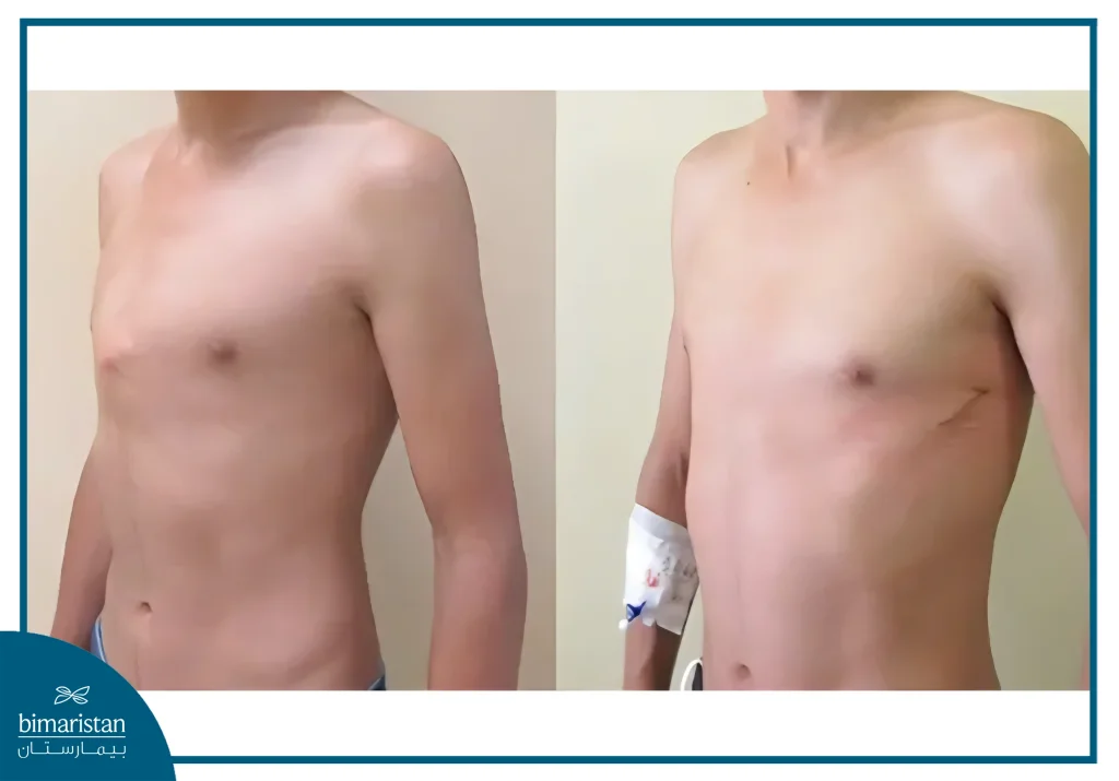
Complications of Pectus Carinatum Treatment
Treatments for pigeon chest are generally safe and effective, but may be associated with side effects that vary by treatment type. In non-surgical treatment with a compression belt, a small number of patients may experience skin irritation or superficial sores at the points of contact with the body. If these symptoms occur, you can temporarily stop using the belt and consult your doctor to have it adjusted properly.
As for surgical treatment, despite its effectiveness, it may be associated with complications, as with any surgical procedure. The most notable of these include:
- Pneumothorax (collapsed lung) or fluid accumulation around the lung
- Bleeding or infection
- Inflammation of the tissues surrounding the heart
- The sternum fixation devices have shifted out of place
- The Deformation Returns Once Again
Recovery Period After Treatment for Pectus Carinatum
The recovery period after treatment for pigeon chest varies depending on the type of treatment used—whether a compression brace or surgery—and each method requires ongoing follow-up to ensure the best possible results.
In non-surgical treatment with a compression brace, the patient needs to care for the skin daily in the areas where the garment contacts the chest to prevent irritation or sores. It is recommended to check the skin regularly and keep it clean and thoroughly dry. It is normal to feel mild pressure while wearing the compression garment; however, the onset of pain warrants a consultation with a specialist. Treatment is also monitored periodically: the condition is assessed shortly after starting to use the bandage, and then regularly thereafter to monitor the development of the chest shape and adjust the brace as needed. Even after treatment is completed, follow-up visits continue at intervals to ensure the results are stable.
As for surgical treatment, the patient may need to stay in the hospital for one to five days after the procedure, with follow-up appointments scheduled over the next few weeks to monitor recovery and ensure the procedure was successful.
In conclusion, pectus carinatum is a chest wall deformity that may appear simple on the surface, but it can have implications that extend beyond its external appearance, including functional and psychological aspects in some patients. Although this condition usually does not pose a direct threat to health, a thorough understanding of its nature helps in making the appropriate decision regarding its management or treatment. Treatment options vary, and the choice of the most appropriate method depends on the severity of the deformity, the stage of development, and the extent of the condition’s impact on the patient. Therefore, medical evaluation and regular follow-up are essential for achieving and maintaining long-term effectiveness.
Sources: