
Bile duct cancer is a rare malignant tumor that originates from the epithelial cells lining the bile ducts. It is the second most common primary liver tumor after hepatocellular carcinoma, and its incidence tends to increase with age. The prevalence of bile duct cancer varies considerably by geographic region, largely due to different risk factors in each area, such as exposure to liver parasites in Southeast Asia.
This disease is difficult to diagnose because it often lacks specific symptoms early on; clear signs such as jaundice do not appear until the bile duct becomes obstructed, which usually occurs in relatively advanced stages. Nevertheless, advances in computed tomography (CT) and magnetic resonance imaging (MRI) techniques, along with molecular testing for genetic mutations, have improved diagnostic accuracy and enabled more specialized treatment plans. This article discusses the causative factors, clinical manifestations, and modern diagnostic and therapeutic approaches for this tumor.
What is bile duct cancer?
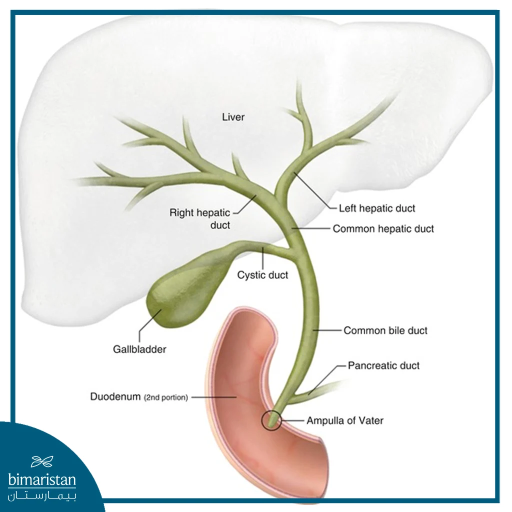
The bile ducts are small channels that connect three digestive organs: the liver, the gallbladder, and the small intestine. The primary role of the bile ducts is to transport bile produced by the liver to the gallbladder and the small intestine (duodenum), so that excess bile is stored in the gallbladder and secreted from it as needed. Bile is a fluid produced by the liver that consists mainly of water, bile salts, and cholesterol, along with other components. Bile helps the body break down fats and proteins during digestion.
Bile duct cancer, also known as cholangiocarcinoma, arises from the epithelial cells lining the ducts; it is a malignant cancer that metastasizes to nearby lymph nodes at an early stage. In later stages, it also spreads to the bloodstream, and the tumor may spread locally and invade adjacent structures and tissues, particularly large blood vessels such as the portal vein and the hepatic artery, making surgical resection difficult.
Types of bile duct cancer
Bile duct cancer can be divided into three types, depending on where the cancer occurs. These types are as follows:
- Intrahepatic bile duct cancer: It originates in the cells of the bile ducts located within the liver and can be classified as a type of liver cancer.
- Intraductal cholangiocarcinoma: It arises from cells in the bile ducts located just outside the liver.
- Distal bile duct cancer: It originates in the bile duct cells closest to the small intestine.
What are the causes and risk factors of bile duct cancer?
Bile duct cancer occurs when a defect in the genetic material (DNA) of the cells lining the ducts causes them to multiply uncontrollably, forming tumors that invade and destroy healthy body tissue. While the exact cause of this defect is unknown, scientists have identified a number of factors that increase the risk of developing this cancer, namely:
- Primary sclerosing cholangitis: An autoimmune disease that affects the bile ducts and causes inflammation; this inflammation progresses to fibrosis of the duct walls, narrowing and partially obstructing bile flow.
- Chronic liver diseases: Such as liver fibrosis; fibrosis associated with chronic liver disease increases the risk of developing bile duct cancer.
- Bile duct stones: These stones are similar to gallstones but smaller; they can cause inflammation, increasing cancer risk.
- Alcohol: Alcohol consumption causes various liver problems and increases the risk of bile duct cancer.
- Age: This type of cancer predominantly affects individuals over the age of 50.
- Congenital bile duct abnormalities: These increase the risk of developing cancer.
- Obesity
Symptoms of bile duct cancer
Symptoms of bile duct cancer usually appear late and do not manifest at the onset of the disease. They result from a blockage of the bile ducts, which prevents the drainage of bile, causing it to accumulate in the body, leading to a range of symptoms, including:
- Jaundice: Jaundice is one of the characteristic signs of liver and biliary tract diseases, and is caused by the buildup of bilirubin in the body. When the liver can’t eliminate bile, it builds up in the body, and bilirubin enters the bloodstream, turning the skin and mucous membranes yellow.
- Abdominal pain: In the early stages of the disease, cancer may be asymptomatic and cause no pain, but as the disease progresses, pain begins to appear, particularly in the upper right quadrant of the abdomen.
- Pale-colored stool: Bilirubin gives stool its dark brown color, and a bile duct blockage prevents bilirubin from reaching the intestines, causing the stool to appear pale.
- Itching: Bilirubin accumulation in the blood is associated with itching, particularly in cholangiocarcinoma.
- Dark-colored urine: When blood bilirubin levels rise, urine bilirubin levels also rise.
- Weight Loss
- General fatigue
Methods for diagnosing bile duct cancer
Diagnosis begins with a complete medical history, risk factor assessment, and symptom analysis to narrow the diagnosis. The doctor then performs a physical examination of the abdomen to check for any tumor masses or enlarged lymph nodes. Afterward, the doctor orders laboratory and imaging tests to confirm the diagnosis. Tests used to diagnose bile duct cancer include:
Blood tests
This type of test is performed to evaluate liver and bile duct function, as well as to screen for tumor markers, particularly the carbohydrate antigen (CA 19-9), a protein that is overproduced by bile duct cancer cells; elevated levels of this tumor marker in the blood indicate the presence of cancer. However, an elevated level of this marker does not necessarily mean cancer, as its concentration in the blood may also rise in the context of other diseases.
Endoscopic Retrograde Cholangiopancreatography (ERCP)
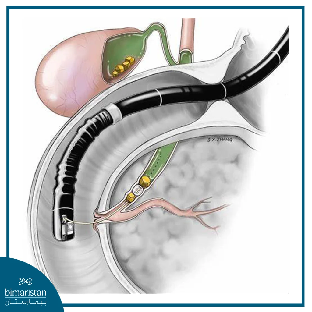
This procedure involves inserting a camera through the mouth into the esophagus and then into the area where the bile ducts connect to the small intestine. The camera then enters the bile ducts and provides the doctor with a clear image of their condition. This method is very important in diagnosing bile duct diseases, particularly bile duct cancer.
Ultrasound imaging
A simple, non-invasive test performed initially to examine the gallbladder and bile ducts and detect any gallstones or abnormalities in the gallbladder wall.
Computed Tomography (CT) and Magnetic Resonance Imaging (MRI)
These imaging modalities provide a clear view of the body’s internal organs and can reveal metastases to adjacent organs; they are also important for assessing local tumor invasion and its extent.
Magnetic Resonance Cholangiopancreatography (MRCP)
It is increasingly used as a non-surgical alternative to Endoscopic Retrograde Cholangiopancreatography (ERCP) for imaging the pancreas and bile ducts, providing clear three-dimensional images without contrast agents.
Percutaneous Transhepatic Cholangiography (PTC)
In this procedure, the doctor inserts a thin, hollow needle through the skin of the abdomen into one of the liver’s bile ducts. A contrast dye is then injected through the needle, and X-ray images are taken as the dye moves through the bile ducts. Like Endoscopic Retrograde Cholangiopancreatography (ERCP), this test can also be used to collect fluid or tissue samples or to place a stent (a small tube) inside the bile duct to help keep it open.
Biopsy
This is a procedure in which a portion of the tumor tissue is removed for examination under a microscope at the cellular level; the biopsy is taken using a needle, which is inserted into the bile ducts under ultrasound guidance or guided by endoscopic retrograde cholangiopancreatography (ERCP), and then sent to a pathology laboratory.
Treatment of bile duct cancer
Treatment for bile duct cancer varies from patient to patient, and the choice of treatment depends on the tumor’s location, stage, and extent of spread, as well as the patient’s overall health. Treatment aims to control the disease, remove as much of the tumor as possible, relieve symptoms, and improve the patient’s quality of life. With medical advances, numerous treatment options have emerged for bile duct cancer, including:
Surgery
Surgical treatment of this cancer is challenging, especially when the tumor invades nearby major blood vessels such as the portal vein and the hepatic artery. During surgery, the surgeon attempts to remove as much of the tumor as possible, and the surgical procedure may involve the removal of parts of the liver, the bile ducts, and the pancreas.
Liver transplant
Some patients with mucinous cholangiocarcinoma qualify for liver transplants, which can be curative. However, the disease may recur as soon as six months after treatment, making regular monitoring and follow-ups essential.
Chemotherapy
Chemotherapy relies on drugs that kill cancer cells. This type of treatment may be used in some patients with advanced-stage cancer to slow the growth and spread of the tumor, as well as to alleviate symptoms and improve the patient’s quality of life.
Radiation therapy
Radiation therapy directs a high-energy beam of X-rays or protons at the tumor to destroy cancer cells.
Targeted drug therapy
Targeted therapy focuses on specific abnormalities or changes within cancer cells; by disrupting these abnormalities, the therapy can cause the cancer cells to die.
Immunotherapy
Immunotherapy relies on the body’s immune system to fight cancer, but the immune system may not attack cancer cells because these cells produce proteins that help them hide from immune cells; immunotherapy works to disrupt this process. In bile duct cancer, immunotherapy may be an option in advanced cases when other treatments do not provide the desired benefit.
Radiofrequency ablation
Radiofrequency ablation uses an electric current to heat and destroy cancer cells; the doctor inserts one or more thin needles through small abdominal incisions, guided by an imaging test such as ultrasound. Once the needles reach the tumor, they are heated by an electric current to destroy the cancer cells.
Photodynamic therapy
During this procedure, a light-sensitive substance is injected into a vein and accumulates in rapidly growing cancer cells. A laser is then directed at the tumor, triggering a chemical reaction inside the cancer cells that causes them to die. Patients typically require several treatment sessions. Photodynamic therapy also helps relieve signs and symptoms and may help slow cancer growth. Exposure to sunlight should be avoided after treatment.
Percutaneous Biliary Drainage (PBD)
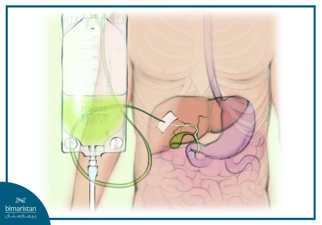
This procedure aims to restore bile flow and reduce its buildup in the body. It may involve inserting a thin tube into the bile duct to drain bile. Other options include bypass surgery, which reroutes bile flow around the tumor, and the placement of stents to keep the bile duct open when cancer causes it to narrow or collapse. Bile duct drainage helps alleviate the signs and symptoms of bile duct cancer.
In conclusion, bile duct cancer is one of the cancers that requires a personalized treatment plan for each patient, depending on the tumor’s location, stage, extent of spread, and the patient’s overall health. With the variety of treatment options available, ranging from surgery and liver transplantation to targeted and immunotherapies, it is now possible to control the disease and alleviate its symptoms in many cases. Early diagnosis and regular follow-up with a specialized medical team help select the most appropriate treatment and improve the patient’s chances of recovery and quality of life.
Sources: