Overview of bionic eye implant
Bionic eye implant in Turkey, Also known as retinal implantation, this procedure can be performed on patients with severe visual impairment or night blindness.
The procedure has been performed on 170 patients worldwide, including Turkey. Perhaps the most important results were achieved in patients with corneal cloud (a disease caused by inflammation in the cornea that then heals and prevents the passage of light). These patients obtained satisfactory results that radically improved their lives and returned them to normal life.

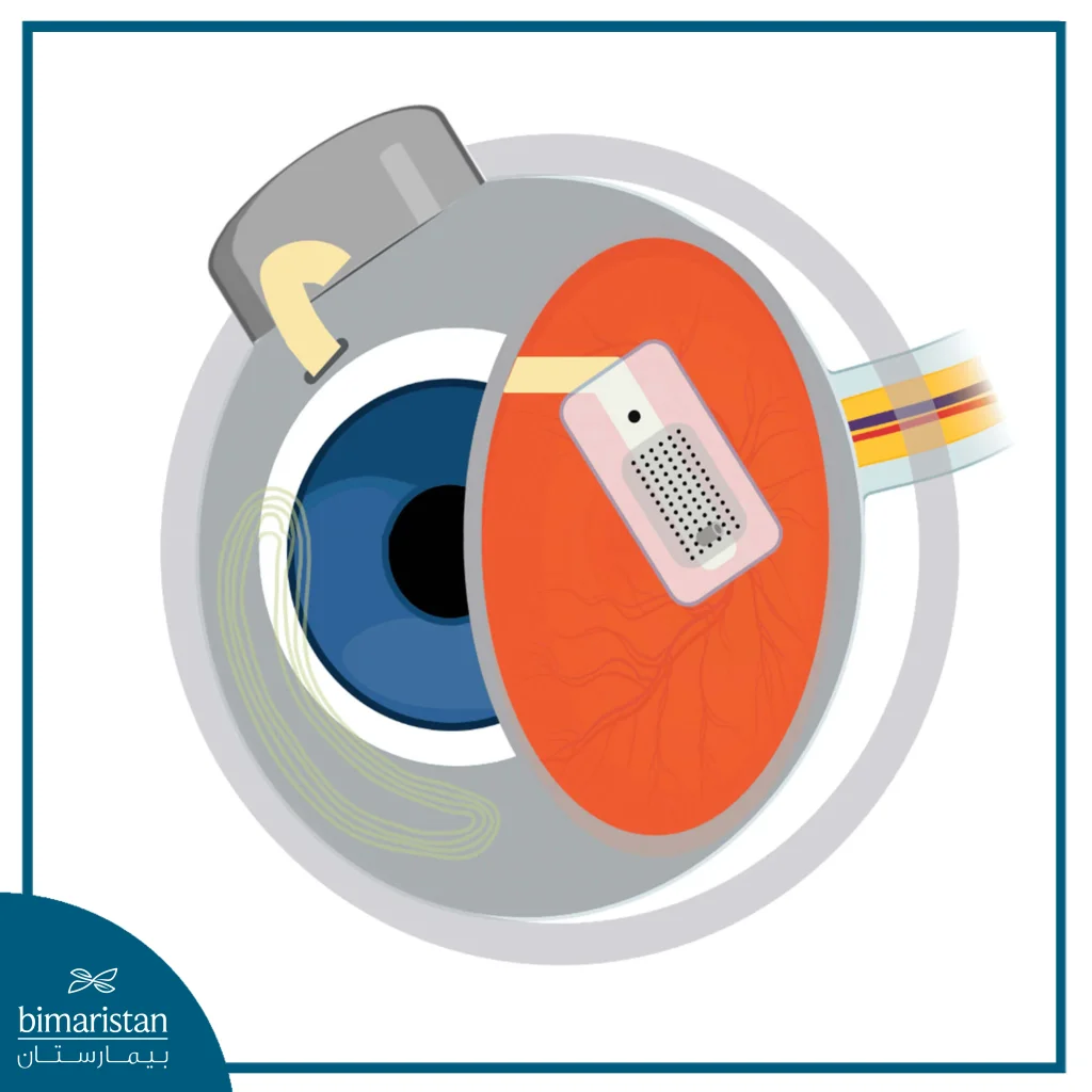
The bionic eye implant consists of two main parts. The first (the electronic part) is a collection of electronic tweezers in the eye’s periphery.
The other part is inside the eye on the surface of the retina.
The retina consists of an orange-colored layer that contains light-sensitive nerve cells. In the central area of the retina, there is a so-called central point on which the other part of the bionic eye implant is fixed.
The system’s goal is to convert light from electronic systems into neural signals through a camera that saves what it sees and sends it to the brain. The technology targets patients with new visual impairment due to retinal cell damage.
Bionic eye implants (retinal implants)
These implants are placed in the epiretinal tissue, often ensuring that the cells underneath the implants continue functioning.
There are two types of systems, especially those that are placed over the retinal macula.
Both have been introduced in the United States, Europe, and Turkey.
Thanks to this technology, many blind people can see again.
The procedure can also be performed on patients with night blindness, in which the patient loses the ability to see as darkness falls. A defect in the light receptors in the retina causes the disease.
This disease can lead to blindness, affect the eye’s sensitivity to light, reduce the speed with which the eye adapts to changes in the amount of light, and cause keratoconus. Read more about corneal transplantation for keratoconus.
After a bioelectronic retinal prosthesis, the patient needs at least 40 rehabilitation sessions to reach an acceptable level of vision. Unfortunately, he will not yet be able to distinguish colors and will not have a fully detailed vision of foreign objects.
For example, when the patient looks at himself in the mirror, he can see clearly, but he cannot distinguish fine details such as eye color or shape.
The function of the bionic eye implant is to alleviate the effects of vision loss and enable the patient to improve his life and self-reliance. For example, the patient can distinguish the door from the chair and other large objects while entering a room. The patient can also walk on the road and distinguish whether a car has passed, which facilitates many of the obstacles that the patient faces with total or significant vision loss.
Which patients are candidates for retinal implants?
Currently, bionic eye implants are only available for patients with night blindness or retinitis pigmentosa, especially patients with advanced stages of the disease.
These patients will undergo a light sensitivity test to see if the retina is sensitive to light or not.
In addition to other electrophysiological tests.
What conditions must be met for a patient to receive a bionic eye implant?
- Have advanced night blindness.
- The patient’s best level of vision is to see the movement of the hand near the eye or even less.
- Visual sensory cells and visual conduction cells must be intact.
- The patient must be over 25 years old.
- No contraindications to anesthesia.
- It must also be done with the patient’s consent.
- He also has to undergo rehabilitation sessions to follow up on his post-operation treatment.
It is impossible to perform the procedure on patients with other diseases that may cause vision disturbances.
How is bionic eye implant surgery performed?
The bionic eye implant surgery is performed under general anesthesia.
As mentioned above, the bionic eye consists of two parts: an incision is made in the white of the eye under the eye muscles, and an electronic receiver is installed using the same endoscopic surgery technique used for vitrectomy.
Three bionic receptors are then attached to three different areas of the white tissue of the eye through a tube no larger than 1 mm in diameter.

After these three receptors are stabilized, an electrode is attached to the yellow patch of the retina.
This is the point in the retina where vision best occurs.
After the receptors and electrodes are installed, each is tested individually to ensure no abnormalities.
The surgical instruments are then sutured, and the tissues are returned to their original state, making the operation indistinguishable from those who see the patient.
Recovery period after bionic eye implant surgery
Like any eye surgery, the eye needs about a week to recover.
During this time, the patient uses several treatments in the form of eye drops and avoids any work that stresses the patient or the eye in particular.
After the redness of the eye has completely disappeared, the patient begins his sessions with a specialized engineer who performs a calibration of both the receptors and electrodes. The patient’s vision will be calibrated to suit the patient’s vision.
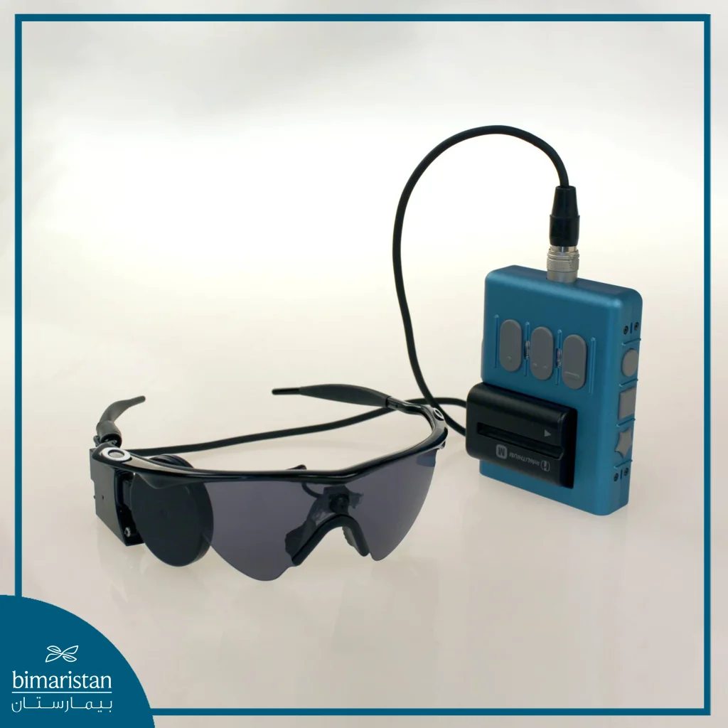
The patient must then wear glasses and carry a small bag containing an electronic device that records everything the patient sees.
Specialists then examine this, giving the patient the necessary rehabilitation to recognize objects again.
It should be noted that a study by the American Academy of Retinal Surgery, based on the results of three years of clinical trials, has proven the long-term safety and effectiveness of bionic eye implants. This study gives hope for the return of vision for many visually impaired people.
What is the percentage of vision after a bionic retina implant?
At first, the scanning is focused on large objects (coarse scanning) so that the patient can see large objects. Then, with time and more experience, the scanning becomes finer so that the patient can see smaller objects, and so on.
For example, as mentioned above, when entering a room, recognize large objects such as the presence and number of chairs, whether people are in the room, who they are, the presence of glass on the tabletop, and then other details such as shadows.
Details begin to increase gradually. However, fully recognizing details and having a normal human vision is impossible. However, the patient will notice significant progress from no vision to seeing things in detail, even if the vision is not 100% complete.
This will greatly improve his life and allow him to return to a normal life without dependence on anyone.
Sources:
