The spinal cord is an important neurological center, and disorders that affect it, including tethered cord, should receive special attention. Turkey has developed neurological treatment and surgery to become one of the leading countries in this field.
What is Tethered Cord Syndrome (TCS)?
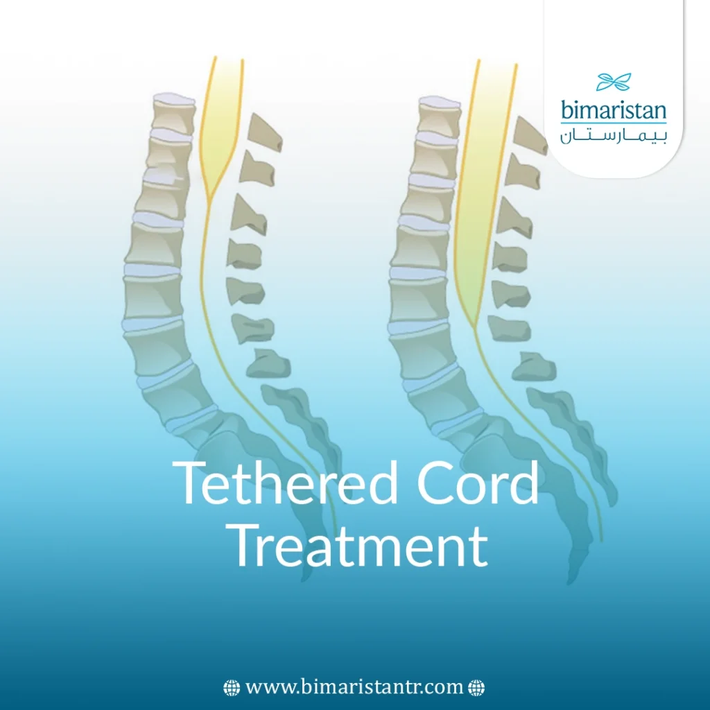
Tethered or pinned spine syndrome is a functional disorder caused by the stretching and elongation of the spinal cord due to its caudal fixation as a result of congenital and rarely acquired disorders (such as tumors) and is a term that can be applied to many diseases in which the spinal cord is pinned.
This abnormal fixation leads to an increase in spinal cord expansion over time and as the child grows, damage to delicate and sensitive tissue, and the onset of various neurological and systemic symptoms.
Due to people’s varying speeds of development, the onset of symptoms varies. Some show symptoms in early childhood, others do not show symptoms until adulthood, and some have mild movement limitations that do not cause symptoms.
What are the causes of Tethered Cord Syndrome?
Let’s understand that this syndrome is a functional physiological disorder resulting from the inability of spinal cord neurons to use oxygen, i.e., impaired oxidative metabolism. This is caused by a lack of oxygen supply (ischemic effect) caused by the stretching of the nerve cord, partly due to the malfunction of ion transport channels caused by the stretching of the cell membranes of axons and neurons.
Garceau first described “filum terminale syndrome” in 1953 in 3 patients, and two decades later, in 1976, Hoffman and colleagues coined the term “tethered cord syndrome” to describe the symptoms of their patients with elongated spinal cord and thickened filament terminale.
A doctor named Yamada et al. expanded the term to include patients with other malformations that cause spinal cord injury in 1981.
This syndrome used to be seen as a pediatric disease, but as mentioned earlier, the cause can be congenital or acquired and affects adults and children, although it is much more common in children.
Congenital (primary) causes
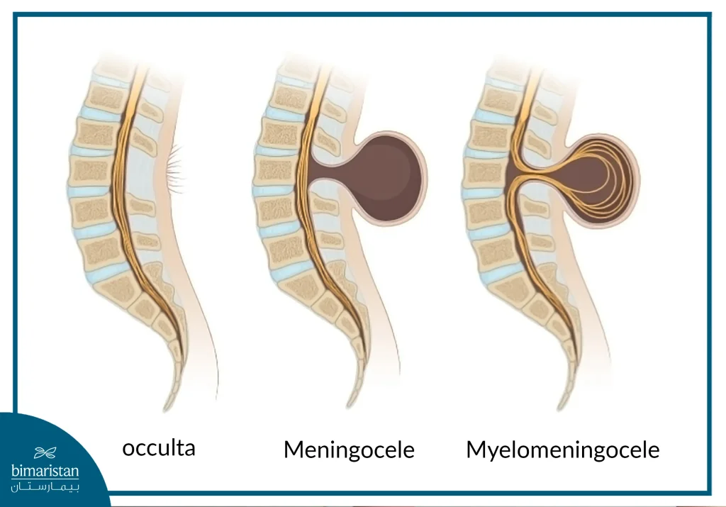
These tissues that connect and stabilize the spinal cord arise from several congenital anomalies, particularly spina bifida, a condition characterized by the defective closure of the neural tube (on the spinal cord side) during embryonic development.
Types of spina bifida associated with Tethered Cord Syndrome include:
Myelomeningocele: The incomplete formation of the bones of the neural canal in which the spinal cord resides, resulting in the nerve tissue protruding outward.
They also include a split spinal cord (diastematomyelia), a benign fatty mass or tumor (lipoma) that persists in the spinal cord.
Another fatty abnormality is a lipomyelomeningocele, where a lipoma emerges from the spinal canal under the meninges but is covered by normal skin.

The disorder can also be seen in people with the Arnold Chiari Malformation.
In many patients, the cause is mechanical due to the thickening and loss of elasticity of the terminal cord, a condition often found in children. (The terminal cord is a thread of tissue (glial cells) that connects the tip of the spinal cord to the sacrum.) This is due to abnormal fibrous tissue growth and its replacement by glial tissue, which leads to the attachment of the spinal cord at its caudal end.
Acquired (secondary) causes
There are many acquired causes of this syndrome, and these causes may be an exacerbation of an already existing condition in a child or adult, including Tumors, infections, trauma, or previous operations on the spinal cord.
There could be a genetic basis for this syndrome since most of the causes of this syndrome are congenital diseases. Still, we cannot directly link genetics with it because this syndrome is a functional disorder whose symptoms appear only when the spinal cord is stretched and stretched. Studies are still ongoing in this field as some researchers have found a genetic predisposition for this syndrome in some patients.
What are the symptoms of Tethered Cord?
The difference between a symptom and a sign is that a symptom is what the patient complains about (e.g., pain), while a sign is what the doctor sees or measures through clinical examination or various tests.
The symptoms of this disorder vary according to age, and of course, there are common symptoms.
Symptoms in children
They include several symptoms that vary in severity depending on the speed of growth or the severity of the disorder:
- Hair tufts, lumps, benign lipomas, skin discoloration (red spots or loss of skin color), or even hemangiomas in the lower back, usually on the midline.
- Pain in the lower back that radiates to the legs (may be associated with numbness in the lower extremities) is usually worse with movement and better at rest (we cannot define it because children cannot express the pain or its location).
- Delayed motor development, such as delayed walking.
- Asymmetry in leg strength (one leg is stronger than the other), an unsteady or abnormal gait, and sometimes leg twitches.
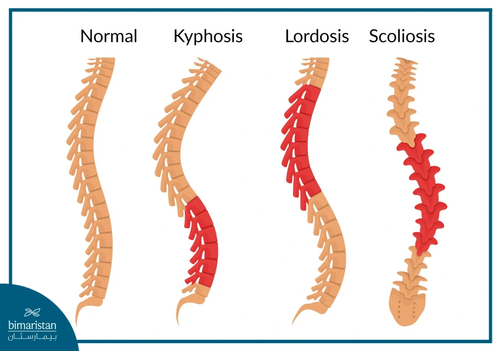
Severe cases can result in deformities of the legs or feet or spinal deformities such as scoliosis or lordosis.

This syndrome is often associated with uncontrolled urine and feces(urinary or fecal incontinence) and frequent urinary tract infections.
Symptoms in adults
Persistent pain in the back and legs that is often severe and can spread to the genital area or rectum.
Leaning forward slightly, sitting upright with legs crossed, or holding a moderate weight (such as a child or a stack of books) at waist level often aggravates back pain. This pain pattern is sometimes called the “3-B sign” for Binding, Buddha sitting, and Baby holding.
Sensory and motor weakness in the legs that may lead to numbness, atrophy, and fatigue after walking short distances.
Bowel and bladder dysfunction manifested by increased frequency or urgency of urination and constipation.
How is tethered cord syndrome diagnosed in Turkey?
The patient’s history should be taken in detail because it helps in the diagnosis greatly, and attention should be paid to the presence of any of the aforementioned symptoms or signs.
MRI is the best diagnostic method here, and CT or Echo can also be performed.
In some cases, electromyography ( EMG ) and nerve conduction studies can be used to assess muscle function.
An electromyogram is a test that records electrical activity in skeletal (voluntary) muscles at rest and during contraction.
Abnormalities on this test are only seen in patients with an advanced stage of tethered cord syndrome.
If you would like to learn more about the cost of tethered cord treatment and diagnosis, do not hesitate to contact us at your family bipartisan center in Turkey.
Tethered cord treatment in Turkey
Most pediatric neurosurgeons recommend Tethered Cord Treatment through detethering for infants or young children diagnosed with this syndrome, regardless of its origin. However, there is controversy over the surgical approach, with some advocating for preventive surgery while others suggest waiting until symptoms emerge or worsen.
The condition can be monitored and followed up if there are no symptoms despite the presence of evidence on the MRI.
Tethered cord treatment is typically surgical for neurological symptoms. Surgeons determine the best approach based on the cause, severity of the syndrome, and the patient’s surgical tolerance.
A laminectomy is performed in the vertebrae facing the site of fixation; the dura is opened, the attached spinal cord is released, and, if possible, the cause or mass that led to the fixation is removed.
However, this operation often relapses, and the patient needs several more operations as he grows older (especially in the case of children, as their spinal cord grows and they develop new adhesions as a result of this growth).
Frequent detethering surgeries and the formation of numerous surgical scars can cause a new cause of adhesions or functional issues in the spinal cord.
New ways for tethered cord treatment
Doctors at Johns Hopkins Hospital and several other studies have favored a new solution for recurrent spinal cord adhesions (known as Tethered Cord), as they found that, after several decortications, the procedure becomes almost useless in relieving symptoms.
Therefore, treatment is directed towards shortening the spinal cord (spine) through partial or total resection of certain vertebrae (most commonly T12).
This procedure has helped many patients in their daily lives and can even be considered a cure for this intractable issue.
Bimaristan Medical Center directs you to the best neurosurgery specialty centers in Turkey. Turkey has worked to add the latest medical devices and adopt the most optimal, state-of-the-art surgical procedures in the world.
Warning and follow-up after treatment
Postoperative follow-up is crucial in tethered cord treatment to monitor for spinal cord reattachment, which may require additional surgery. Most patients recover well, with a recovery period of 1–2 weeks. During this time, minimal movement and bed rest are essential to prevent relapse or cerebrospinal fluid leakage.
Sometimes, some neurological and motor impairments may not be fully corrected, but experience has shown that surgery soon after the onset of symptoms improves the chances of recovery and can prevent further deterioration of spinal cord function.
Sessions of physical therapy can be performed in back pain patients over 65 years old and a lifestyle change to avoid factors that induce pain.
Sources:
- NIH.Gov
- AANS
- Neurosurgery Columbia
- Johns Hopkins medicine

