


.floating_btn{ display:none !important; } .println-contact-form-button-element{ display:none !important; }
A finger fracture is one of the most common bone injuries that a person may experience at various stages of life, whether due to a minor accident, during sports activities, or while performing manual tasks. Although such fractures may initially appear minor, neglecting proper treatment or misdiagnosis can lead to complications that permanently affect hand function, including reduced grip strength, chronic pain, and joint stiffness.
Fingers play a crucial role in performing precise daily tasks such as writing, eating, and using electronic devices. Therefore, any injury to a finger, such as a fracture of a phalanx or at the joint, may cause temporary or even permanent impairment of hand function.
Anatomical structure of the hand bones
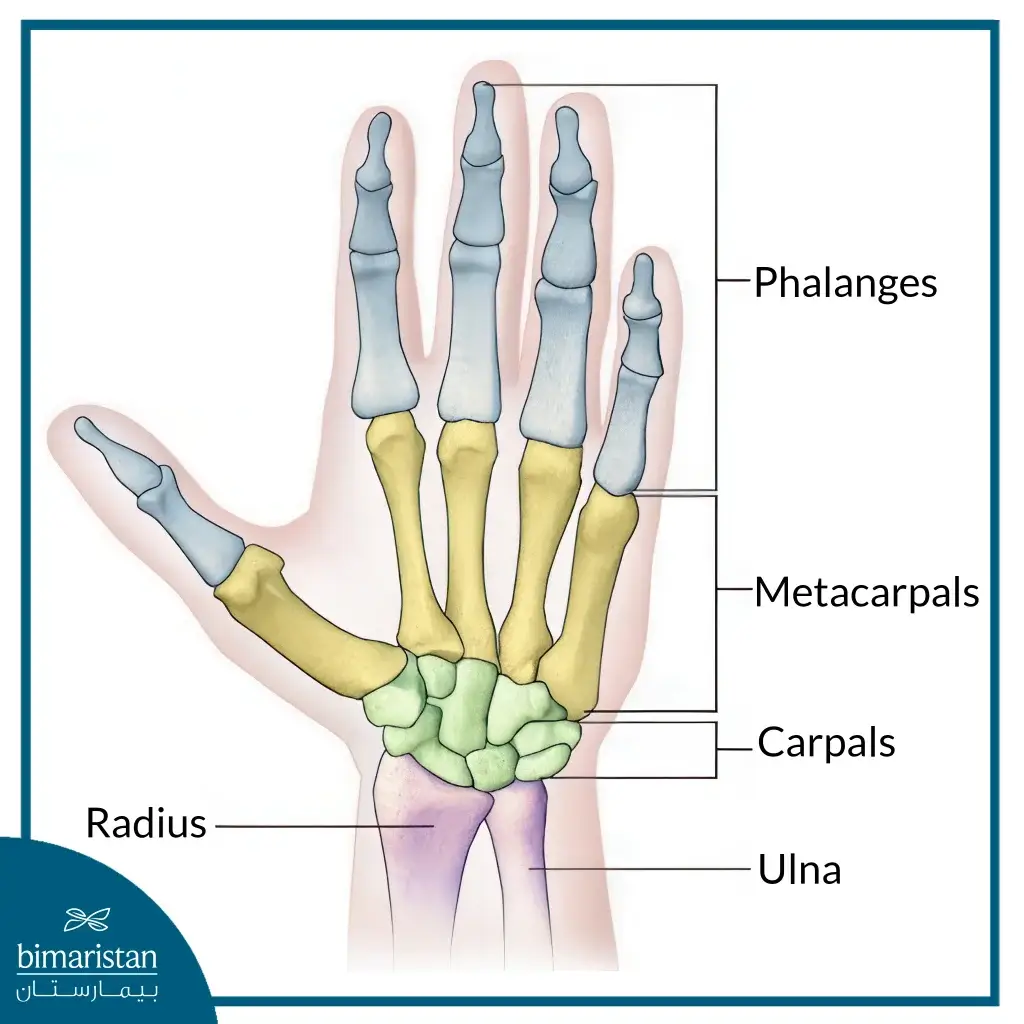
The human hand is composed of 27 bones, which are divided into three main groups: 8 carpal bones that form the wrist, five metacarpal bones that extend from the wrist to the base of the fingers, and 14 phalanges that make up the structure of the fingers. Each of the four fingers (index, middle, ring, and little finger) contains three phalanges: the proximal, middle, and distal phalanges. In contrast, due to its distinct anatomical and functional characteristics, the thumb consists of only two phalanges (proximal and distal).
Finger fractures occur when any of these bones or their joints are subjected to excessive mechanical stress or direct trauma, such as falling, being struck, or participating in sports activities. The fracture may involve the phalanges themselves or the joints between them, such as the proximal interphalangeal (PIP) joint, the distal interphalangeal (DIP) joint, or the metacarpophalangeal (MCP) joint, which connects the proximal phalanx to the metacarpal bone. Finger fractures are considered among the most common types of bone injuries, especially among adult and adolescent athletes.
Types of finger fractures
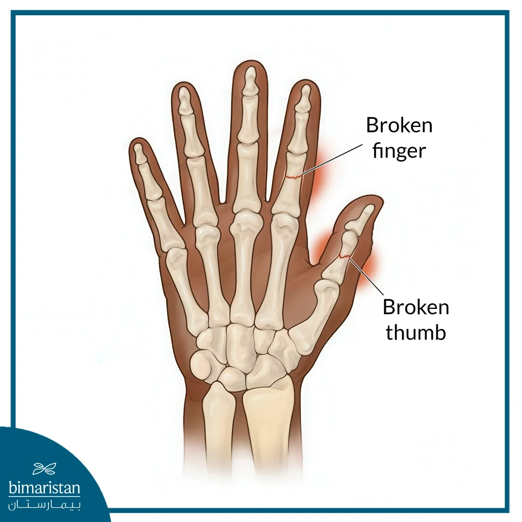
Common types of finger fractures include:
- Spiral fracture: Caused by a twisting force applied to the bone, creating a helical fracture line. Often results from violent finger torsion.
- Avulsion fracture: This occurs when a tendon or ligament pulls away a small bone fragment due to sudden tension. It is often associated with sports injuries or falls.
- Intra-articular fracture: Extends into the joint surface, potentially impairing motion and leading to post-traumatic arthritis. Usually caused by direct impact or falls.
Causes of finger fractures
Most finger fractures result from direct trauma or severe external force, commonly encountered in daily life, sports, or occupational settings. Fingers are particularly vulnerable due to their location at the front of the hand and frequent interaction with the environment. Key traumatic causes include:
- Direct impact from a hard or fast-moving object (e.g., baseball or basketball), especially in athletes
- Falling on an outstretched hand, transmitting force to the fingers
- Slamming a finger in a door or drawer, a frequent household injury, particularly in children
- Improper use of hand tools like hammers or power saws
- Motor vehicle accidents or other severe impacts to the hand
The fracture’s likelihood depends on the trauma’s severity and the bone’s strength at the moment of impact. Therefore, certain medical conditions may increase the risk of fractures even with minor injuries. Among the most prominent predisposing risk factors are:
- Osteoporosis: A common condition among older adults, it leads to weakened bone structure and makes bones more prone to fractures, even without severe trauma.
- Calcium deficiency or poor nutrition can weaken bone density, making bones more fragile and susceptible to fractures.
- Bone tumors or widespread neoplastic lesions affect the bone structure, reducing its ability to withstand pressure.
- Immunodeficiency or the use of immunosuppressive medications: These can impair the body’s ability to heal and increase the risk of complications following an injury.
Symptoms of a finger fracture
The clinical symptoms of a finger fracture vary depending on the location of the fracture, its type (simple, hairline, open, deformed, avulsion), and the severity of the injury. However, several common signs often appear immediately after the trauma and may indicate a fracture, requiring immediate medical evaluation.
The most common symptom is sharp, sudden pain that occurs right after the trauma or impact. This pain can range from mild and tolerable to severe, preventing the patient from using the affected hand. It’s important to note that the ability to move the finger does not rule out a fracture. Movement may still be possible in some mild or hairline fractures, but is usually accompanied by noticeable pain. Other symptoms that may accompany a finger fracture include:
- Localized swelling at the fracture site due to inflammation and internal bleeding.
- Bruising and discoloration (such as bluish or reddish tones), especially within the first 24–72 hours.
- Severe tenderness at the injury site when touched.
- Visible deformity of the finger’s natural shape; the finger may appear twisted or out of alignment.
- Discoloration of the nail or the appearance of bleeding spots under the nail may indicate trauma or a fracture in the distal phalanx.
- Numbness or tingling, caused by pressure on or injury to nerves near the fracture site.
- Limited range of motion in the finger joint and difficulty in bending or extending the finger.
In open fractures where the bone protrudes through the skin, the risk of infection is high, and urgent medical attention is required.
If a finger fracture is suspected, even if the symptoms are mild or tolerable, it is essential to seek medical attention at the nearest healthcare center. Starting treatment early increases the chances of a full recovery and helps prevent complications, such as finger deformity, a weak grip, or joint stiffness.
Diagnosis of a finger fracture
The diagnosis of finger fractures is divided into several stages:
Clinical examination
Diagnosis of a finger or thumb fracture is based on a thorough clinical evaluation and necessary radiographic investigations. The physician begins by taking a clinical history, asking about the details of the incident or mechanism of injury, such as a fall onto the hand or a direct blow, as well as the patient’s symptoms, including pain, swelling, or limited finger mobility. Next, the physician performs a physical examination of the affected area, focusing on any visible deformities such as misalignment, shortening of the finger compared to adjacent digits, or overlap when the hand is extended—signs that may indicate angular or rotational misalignment of the bone. The physician also assesses for localized swelling, bruising, or tenderness at the injury site and may extend the examination to include the wrist or elbow if a concurrent fracture in the arm is suspected due to significant trauma.
Radiographic imaging
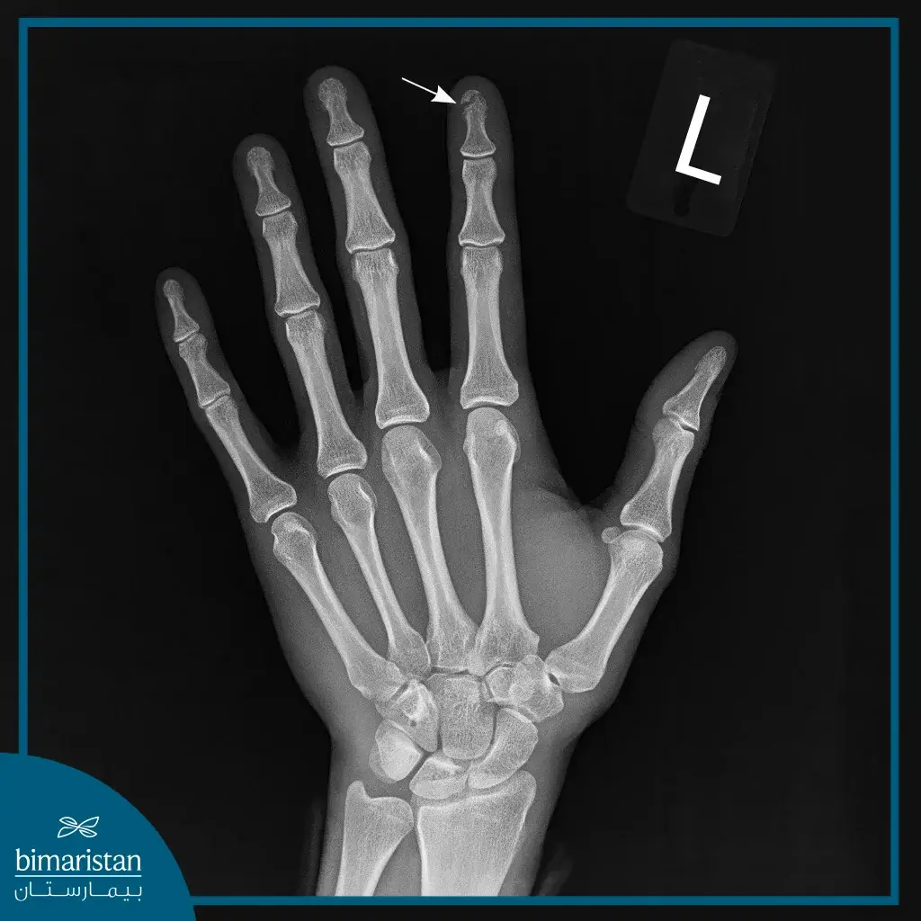
X-ray imaging is ordered following the clinical examination to confirm the fracture and accurately determine its type and location. Imaging helps reveal a fracture’s presence, assess any bone displacement or fragmentation, and determine its proximity to interphalangeal or metacarpophalangeal joints. Typically, X-rays are taken in multiple views—including anteroposterior, lateral, and oblique projections- to assess the bony structures comprehensively. When the fracture is unclear or complex, or if a subtle joint injury is suspected, a computed tomography (CT) scan may be requested for a more precise evaluation. These steps are crucial in distinguishing fractures from other injuries, such as sprains or dislocations, which may present similarly but require different treatment approaches.
Treatment of finger fractures
Treatment of a finger or thumb fracture depends on the type and location of the fracture, as well as the degree of stability and bone displacement. The treatment plan is divided into two main approaches: conservative (non-surgical) and surgical, with the most appropriate option determined based on clinical and radiographic assessment.
Conservative treatment
Initial management focuses on pain relief using analgesics to alleviate discomfort. If the fracture is displaced (unstable), the physician performs a closed reduction under local anesthesia to realign the bone. For non-displaced (stable) fractures or after successful realignment of a displaced fracture, a splint is applied to immobilize the affected finger and maintain proper alignment during the healing period. Resting the injured hand and relying on the unaffected hand for daily tasks is essential to reduce stress on the healing finger. Periodic X-rays are performed during follow-up visits to monitor healing progress and ensure proper bone union. After the fracture has healed, the patient undergoes physical therapy to regain normal finger motion. This helps prevent stiffness and provides full functional recovery of the digit.
Surgical management
Surgical intervention is required in cases that cannot be managed conservatively. These include failure of closed reduction in displaced fractures, ligament or tendon injuries, multiple fractures, or fractures involving the interphalangeal or metacarpophalangeal joints. Such injuries may lead to long-term complications such as osteoarthritis if not appropriately treated.
During surgery, local anesthesia is administered to minimize discomfort and pain. The surgeon makes an incision to access the fractured bone, realigns it, and secures it using metallic plates, wires, or screws. After the procedure, the finger is immobilized with a splint or cast for 4 to 6 weeks. The fixation devices are typically removed once the bone has healed, though in some cases, they may be left in place permanently if they cause no adverse effects, as determined by the treating physician.
In conclusion, finger fractures are common injuries that require accurate diagnosis and prompt management to prevent long-term complications, as they can directly impact the ability to perform fine motor tasks. Although diagnosis is often achieved through a simple hand X-ray, treatment options may range from conservative management with splinting to surgical fixation using plates and screws to ensure fracture stability and complete restoration of function.
In this regard, Bimaristan Medical Center in Turkey is leading in treating such injuries. The hospital is renowned for its highly skilled medical staff specializing in hand and orthopedic surgery, supported by state-of-the-art diagnostic and therapeutic technologies. Bimaristan is committed to providing comprehensive healthcare services—from precise diagnosis and safe treatment procedures to effective physical rehabilitation programs—ensuring that patients regain full hand function. The hospital’s high standards of care make it one of the most trusted medical destinations for treating finger fractures in Turkey.
References: