
A wrist fracture is medically defined as a break in one of the bones that form the wrist. Anatomically, the wrist consists of eight small bones known as the carpal bones, which connect to the distal ends of the radius and ulna— the two long bones of the forearm. Although a fracture can occur in any of these bones, the most common type of wrist fracture is a distal radius fracture.
Wrist fractures are common injuries, particularly in children due to falls on an outstretched hand, leading to fractures of the joint or the distal ends of the ulna or radius. These fractures also occur more frequently in women over the age of fifty and men over sixty-five, due to aging and the effects of osteoporosis, which make bones more prone to fractures even from minor falls.
Information about wrist fracture
The wrist joint is composed of a complex group of small bones known as the carpal bones, as well as the distal parts of the forearm bones. The carpal bones include seven bones: the scaphoid, lunate, triquetrum, trapezium, trapezoid, capitate, and hamate. The forearm consists of the radius and ulna, which connect proximally to the elbow joint and distally to the wrist joint. Notably, the radius plays a greater role in forming the wrist joint, while the ulna primarily contributes to the elbow joint.
The term “wrist fracture” refers to any fracture involving one of the carpal bones or the distal forearm bones, including the radius and ulna. The fracture may be displaced, where the bone fragments move out of their normal position, or non-displaced when the bone remains aligned despite the break. Fractures are also categorized as stable, where the bone pieces stay in place during healing, or unstable, which require surgical fixation due to movement of the fragments. Some fractures are complex, involving bone fragmentation or affecting the smooth surface of the joint, which increases the likelihood of joint misalignment and necessitates surgical intervention for realignment and fixation.
As we mentioned earlier, the most common fracture of the wrist is a distal radius fracture, typically resulting from falling onto an outstretched hand. The severity of the injury depends on the force of the fall and body weight, and it may sometimes be accompanied by additional fractures in the hand, such as fractures of the metacarpals, fingers, or even the forearm itself.
It is noteworthy that the radius is located on the thumb side, while the ulna lies along the side of the little finger. Near the elbow, the ulna is larger, whereas near the wrist, the radius becomes the larger bone and forms the primary component of the wrist joint.
In severe cases, an open fracture may occur—this is when the broken bone pierces the skin, exposing it to infection and requiring urgent surgical intervention and meticulous treatment to prevent complications.
Types of wrist fractures
Wrist fractures vary in pattern and severity and are classified according to several criteria, including the mechanism of injury, the extent of bone damage, and involvement of the joint or surrounding tissues. Understanding these types is crucial for selecting the appropriate treatment approach and planning a successful recovery. Based on general fracture structure, they are categorized as follows:
- Simple fracture: A closed fracture in which the bone splits into two parts without breaking the skin or tearing surrounding soft tissues.
- Compound (complex) fracture: Also known as an open fracture, where the bone shatters into multiple fragments and tears the skin and nearby tissues, causing bone fragments to protrude and increasing the risk of infection.
- Complete (displaced) fracture: The bone is completely separated into two or more parts with an apparent displacement.
- Partial (non-displaced) fracture: A cracked bone that remains partially intact, with the bone fragments staying in their normal position.
Based on joint involvement, fractures are further classified as:
- Intra-articular fracture: A fracture that extends into the joint surface, such as scaphoid fractures or distal radius fractures involving the joint cartilage.
- Extra-articular fracture: Does not extend into the joint surface and is generally less complex than intra-articular fractures, such as some distal radius or ulna fractures or metacarpal fractures.
Common clinical types also include:
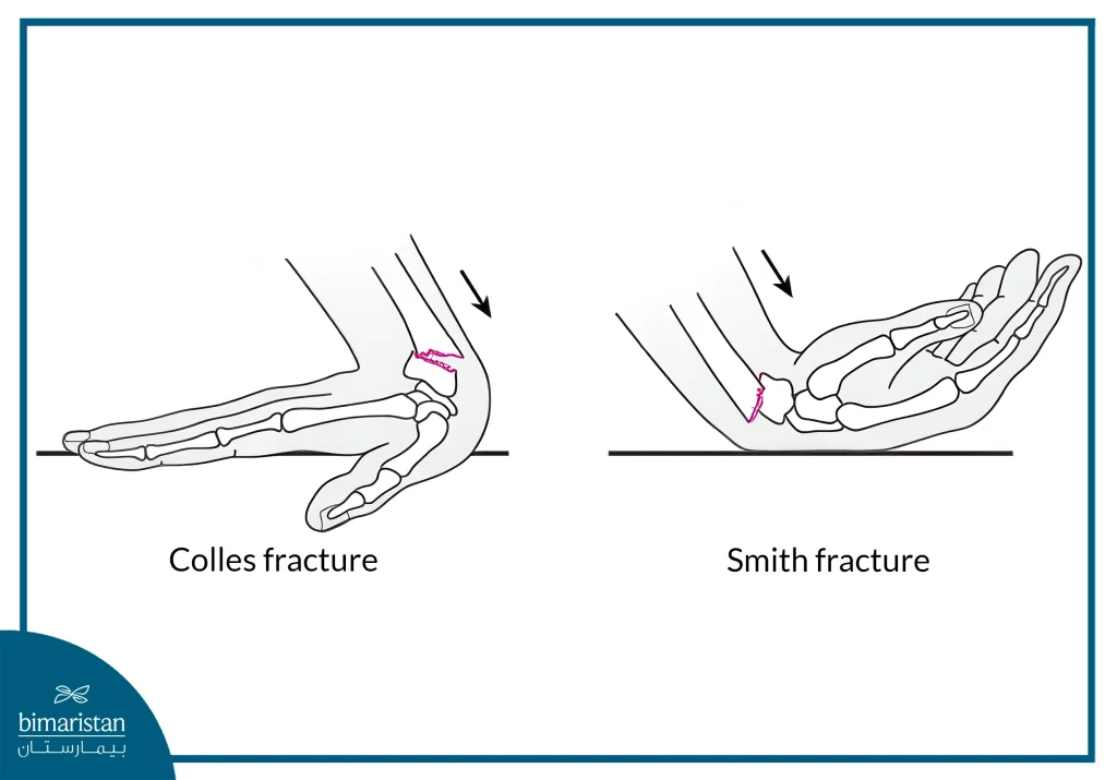
- Colles’ fracture: Occurs when falling on an outstretched hand, where the distal end of the radius tilts toward the back of the hand. It is one of the most common types, especially among older adults.
- Smith’s fracture: The opposite of a Colles’ fracture, it happens when falling on a flexed hand, causing the distal radius to tilt toward the palm.
Causes of wrist fracture
The causes of wrist fractures vary depending on age group, general health, and the individual’s level of physical or occupational activity. Understanding the mechanisms of injury is crucial for effective prevention, accurate diagnosis, and appropriate treatment.
- Falling on an outstretched hand: This is the most common cause of wrist fractures, particularly distal radius fractures. The injury occurs when the hand instinctively extends to protect the body during a fall, transferring the impact force to the wrist. It is especially common in children during play or running, and in older people due to balance loss or slipping indoors or outdoors.
- Sports injuries: Include sports with fall risks or direct impacts, such as basketball, football, skating, tennis, and mountain climbing. These can cause bone fractures, ligament tears, or joint injuries involving the wrist, fingers, and even the metacarpals.
- Osteoporosis: A common condition in adults over sixty, is one of the significant factors that increases bone fragility, making fractures more likely even from minor falls in a standing position.
Symptoms of a wrist fracture
Symptoms of a wrist fracture vary depending on the severity, type, and location of the broken bone. They may also differ among individuals based on age and bone structure. Clinical symptoms are important indicators for early diagnosis and treatment and include the following:
- Sharp, localized pain at the fracture site: Often one of the first signs, the pain is typically acute and centered directly over the break.
- Notable swelling: Appears quickly around the injured area due to inflammation and local bleeding around the fracture.
- Bruising and skin discoloration: Bruises result from subcutaneous bleeding and may appear blue or purple around the wrist and fingers. In some cases, finger color changes due to poor circulation or nerve compression.
- Visible deformity of the wrist or hand: In displaced or unstable fractures, the wrist may appear bent or misaligned.
- Numbness and tingling in the palm and fingers: Nerves may be affected due to pressure from the fracture, causing numbness or tingling, especially in the fingertips. The patient may lose the ability to move the wrist or use the hand normally.
Signs of wrist fracture
Signs of a wrist fracture are often clear and easily detectable by the physician during clinical examination. Key symptoms and signs include:
- Pain when using the wrist or hand: Whether during movement or activities requiring hand strength, such as writing or gripping objects.
- Pain with finger movement: Especially if the injury involves the fingers or bones surrounding the joint.
- Restricted range of motion in affected joints: This limitation can persist even after the fracture has healed if it was not treated correctly.
- Complete paralysis of the hand: May result from nerve or tendon damage surrounding the fracture.
- Pain when lifting objects with the injured hand.
Complications of wrist fracture
Although most wrist fractures heal without significant problems when treated appropriately, some complications can arise, especially in open, displaced, or severely fragmented fractures. These complications, though rare, may significantly affect joint function and include:
- Injury to blood vessels or nerves around the fracture site: This typically presents as numbness, tingling, or changes in color and temperature of the hand and fingers.
- Joint stiffness and chronic or persistent pain.
- Tendon and muscle tears around the joint: Can result from bone displacement or pressure from bone fragments.
- Osteomyelitis: One of the most serious complications of open fractures, where bacteria infect exposed bone tissue, potentially causing chronic, difficult-to-treat inflammation.
- Chronic arthritis or the triggering of rheumatoid arthritis.
- Carpal tunnel syndrome: Swelling due to the fracture or bone displacement compresses the median nerve within the carpal tunnel, causing numbness and pain in the fingers, especially at night.
Diagnosis of wrist fracture
Diagnosing a wrist fracture involves a detailed clinical evaluation, including a comprehensive physical examination and advanced imaging studies. The examining physician—whether an emergency doctor or hand surgeon—will begin with a physical assessment of the injured joint, palpating the wrist to identify points of tenderness and testing the range of motion by gently bending the joint in various directions. The patient is also asked about current symptoms, injury history, and the mechanism of trauma that caused the fracture.
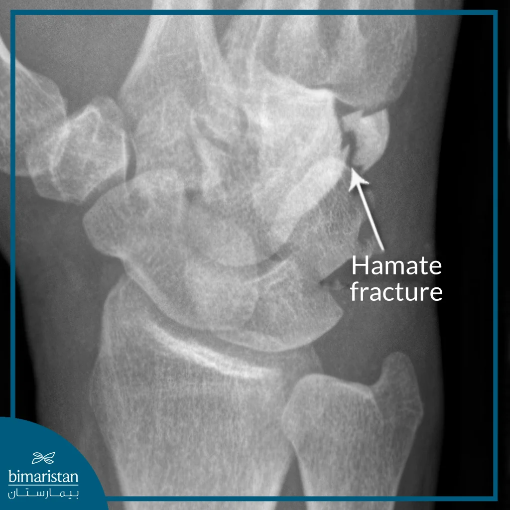
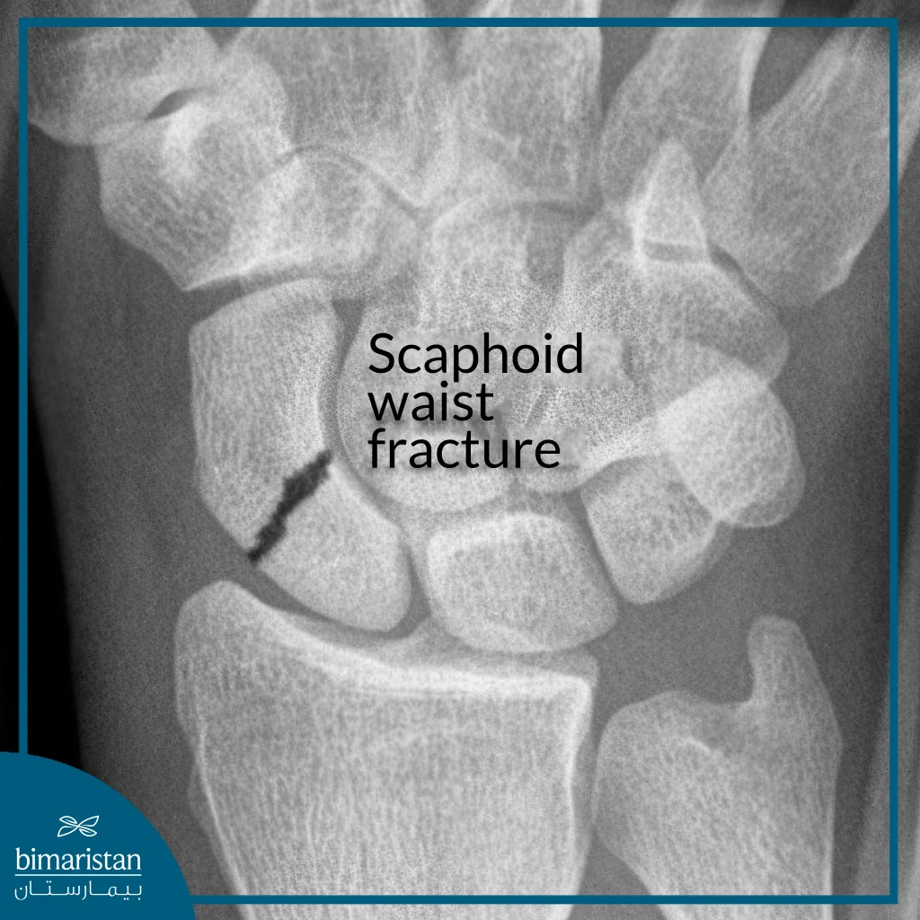
To confirm the diagnosis, X-rays of the wrist are ordered. These allow for visualization of the bone break, its location, whether it is displaced, and the number of fragments it has produced. In some cases, fractures may not be visible on early X-rays, particularly within the first few hours post-injury. Therefore, follow-up imaging may be recommended to monitor changes as healing begins.
If the injury is due to high-energy trauma or if there is suspicion of associated injuries to ligaments, tendons, muscles, or nerves, advanced imaging modalities are used:
- Computed Tomography (CT) scan: Provides high-resolution 3D images of broken bones, which help assess complex fractures that are not clearly visible on X-rays. It can also reveal associated soft tissue and vascular injuries.
- Magnetic Resonance Imaging (MRI): Used to detect very fine fractures that may not appear on X-rays and to evaluate injuries to ligaments and soft tissues around the joint. It is particularly effective at visualizing deep structures and early changes within the joint.
Treatment of wrist fracture
There are various types of treatments for wrist fractures depending on the type and severity of the fracture. Treatment is divided into two main categories:
Conservative treatment
Conservative treatment is the first and most common option for managing wrist fractures, especially in cases of simple and non-displaced fractures. This approach includes splinting, where the hand is positioned in a posture resembling the act of holding a cup. Then, gauze or cotton is applied from the middle of the palm to the middle of the forearm, and a cast is placed over it to immobilize the bones in a healing position. In the case of a displaced fracture, closed reduction under anesthesia is necessary, which is performed by an orthopedic surgeon. After casting, the patient is advised to avoid moving the hand for several days and to monitor for swelling or discoloration that may occur as a result of immobilization. If such symptoms occur, consult a doctor immediately. Emphasis is also placed on proper nutrition, rich in calcium and vitamin D, to enhance bone healing.
In the early stages of conservative treatment, a removable cast or splint is often used and adjusted as needed to accommodate the reduction in swelling. Hand immobilization typically lasts for five to six weeks, followed by a physical therapy program to restore strength and range of motion in the joint. During the healing period, regular X-ray imaging—usually weekly during the first three weeks and once again in the sixth week—is conducted to monitor bone healing and ensure proper alignment. If any displacement occurs during follow-up, surgical intervention may be required.
Surgical treatment
In cases where the fracture is severe or open, or when conservative treatment fails to maintain bone stability, surgical treatment becomes necessary. The surgical approaches vary depending on the type and severity of the fracture and include:
- Open reduction and internal fixation: This is the most common surgical option, in which metal plates and screws or pins are used to stabilize the bones after surgical realignment. This procedure is performed under general or local anesthesia and is considered safe and effective in maintaining accurate bone alignment.
- External fixation with pins and rods: This involves placing pins above and below the fractured wrist joint, connected externally by a rod that stabilizes the bone until healing is complete.
In general, the choice of treatment is based on several factors, including the type of fracture, its location, and the number of broken pieces, as well as the presence of ligament tears or other associated injuries. These factors are also influenced by the patient’s age, dominant hand, and daily activity level. Experts emphasize that the primary goal of all treatment options is to restore the joint’s full function and ensure that the patient returns to regular activity as quickly as possible.
Wrist fractures are among the most common injuries, particularly in children and the elderly, due to the complex and sensitive nature of this joint. Despite the wide range of fracture types from simple to complex, early diagnosis and appropriate treatment remain crucial in restoring full hand function and avoiding long-term complications. In this regard, advanced medical institutions play a vital role in providing optimal care, such as Bimaristan Hospital in Turkey.
Bimaristan Hospital is distinguished by its specialized medical staff and modern equipment, which ensure accurate diagnosis and a range of treatment options, from splinting and conservative management to precise surgical interventions utilizing the latest internal and external fixation technologies. The hospital utilizes advanced imaging methods, including MRI and CT scanning, to accurately assess each patient’s condition, thereby informing the most suitable treatment decision.
Moreover, Bimaristan Center in Turkey offers advanced rehabilitation services under the supervision of a trained physical therapy team to ensure full recovery of hand movement and strength after healing. Therefore, Bimaristan Hospital in Turkey is considered a leading model in the treatment of wrist fractures, thanks to its high medical expertise and integrated care that follows the latest global standards.
References:
- Wrist fracture, NHS Foundation Trust.
- A Patient’s Guide to Distal Radius Fractures (Broken Wrist)
- What is a wrist fracture? American Society for Surgery of the Hand
- Wrist Fracture. The Sports Medicine Patient Advisor
- An Overview of the Named Wrist Fractures Jenna A. Saxton, DO, Justin R. Montgomery, MD, and Paul J. Spicer, MD
- Distal radius fractures. Virtual Hand Fracture Clinic