Tetralogy of Fallot is one of the most recognized congenital heart diseases, causing health challenges in newborns, often marked by cyanosis and breathing difficulties within the first months of life. Despite its complexity, medical advancements have enabled early diagnosis and effective treatment strategies, significantly improving survival rates. In this article, we will explore Tetralogy of Fallot, its underlying causes, clinical symptoms, diagnostic approaches, and the most recent treatment options, along with post-surgical success rates.
What is Tetralogy of Fallot?
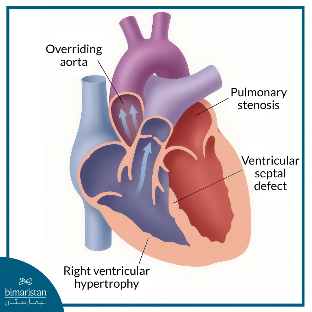
Tetralogy of Fallot is a rare congenital heart defect that is present from birth and is classified as a critical congenital heart disease. It is characterized by four concurrent structural issues that affect the way blood flows through the heart and to the rest of the body, leading to low oxygen levels in the blood and cyanotic skin.
Ventricular Septal Defect (Ventricular Septal Defect – VSD)
An opening in the wall between the left and right ventricles that allows oxygen-rich and oxygen-poor blood to mix, reducing the efficiency of oxygen distribution to the body’s organs.
Pulmonary valve or pulmonary artery stenosis
A narrowing of the right ventricular outlet or pulmonary valve and artery, which blocks blood from reaching the lungs for oxygen and increases pressure on the right ventricle.
Overriding Aorta
Abnormal positioning of the aorta so that it connects to both ventricles instead of just the left ventricle, allowing deoxygenated blood to pass into the general circulation.
Right Ventricular Hypertrophy
Thickening of the wall of the right ventricle as a result of the great effort to pump blood through the narrowed passage, which, over time, leads to impaired heart function.
These four defects combine to form what is known as tetralogy of Fallot, a condition that affects approximately one in 2,000 babies and is categorized as one of the most common complex congenital heart conditions.

Causes of Tetralogy of Fallot
The exact cause of tetralogy of Fallot remains unknown in most cases, as structural changes in the heart form during the first weeks of pregnancy. It is believed that genetic and environmental factors may play a synergistic role in the development of this heart defect. Factors associated with an increased risk include:
- Genetic factors: Children with genetic syndromes such as Down syndrome or DiGeorge syndrome are more likely to develop the condition, as well as if there is a family history of heart defects.
- The mother has a viral infection during pregnancy: Such as rubella or other viral diseases, that may affect the development of the fetal heart.
- Health issues in the mother: Uncontrolled diabetes during pregnancy.
- Environmental and lifestyle factors: Smoking, drinking alcohol, poor diet, or taking certain medications during pregnancy.
- Mother’s age: Pregnancy after age 35-40 may be associated with a slightly increased risk of having an affected child.
Despite these factors, most cases of tetralogy of Fallot do not have a clear cause. They are often caused by a combination of genetic factors and environmental conditions surrounding the mother during pregnancy.
Symptoms of Tetralogy of Fallot
Symptoms of tetralogy of Fallot vary depending on the degree of blockage of blood flow from the heart to the lungs, and may appear clearly from birth or later in the first months of the child’s life:
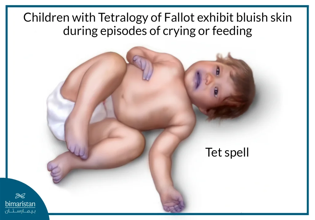
- Cyanosis: Blue or gray discoloration of the skin, lips, or nails due to low oxygen levels in the blood.
- Difficulty breathing or rapid breathing: Especially during feeding, crying, or exertion.
- Rapid fatigue and exhaustion: Appears when feeding, playing, or physical activity.
- Poor growth and slow weight gain: Due to insufficient oxygen supply to the tissues.
- Fainting or dizziness: The result of a sudden drop in oxygen.
- Cardiac murmur: The doctor hears it during a stethoscopic examination due to blood passing through an abnormal valve or septum.
- Changes in the shape of the fingers: Abnormal widening or rounding of the fingertips over time when hypoxia persists.
Tet Spells
Some babies may develop sudden episodes of cyanosis known as Tet spells, in which the skin, lips, and nails become very blue during crying, feeding, or excitement. These episodes often occur between the ages of two and four months, resulting from a rapid drop in blood oxygen levels, and may be accompanied by difficulty breathing or loss of consciousness in severe cases.

Diagnosis of Tetralogy of Fallot
Tetralogy of Fallot can be diagnosed before or shortly after birth. In some cases, during a pregnancy ultrasound (between weeks 18-22), the doctor may notice signs of an abnormal heart structure, which is later confirmed by a fetal echocardiogram.
After birth, the injury is usually suspected when the baby develops a blue or grayish color in the skin or when the doctor hears a heart murmur using a stethoscope. To confirm the diagnosis and determine the exact details, several tests are used, including:
- Pulse oximetry: A simple test used to determine the percentage of oxygen in the blood.
- Echocardiogram: An ultrasound scan that shows the shape of the heart and its valves and assesses blood flow.
- Electrocardiogram (ECG): Records the electrical activity of the heart and detects arrhythmias or right ventricular hypertrophy.
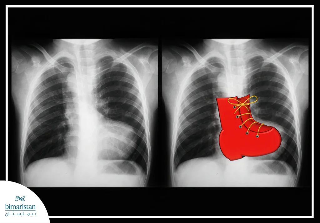
- Chest X-ray: Shows the shape of the heart and lungs, sometimes showing a shoe-shaped heart due to an enlarged right ventricle.
- Cardiac Catheterization: A thin tube inserted through a blood vessel into the heart to measure pressures and oxygen levels, used to plan surgery.
- Computed tomography (CT) or magnetic resonance imaging (MRI): To obtain detailed images that help in the accurate planning of surgical treatment.
With these tests, the doctor can assess the severity of the condition and develop a tailored treatment plan for each child.

Tetralogy of Fallot treatment
Tetralogy of Fallot is one of the most prominent congenital heart defects that requires early surgical intervention, and the primary solution in this case is pediatric heart surgery, which aims to repair structural defects within the heart and restore normal blood flow. The timing and type of surgery are determined based on the child’s age, health condition, and the severity of the heart defects. In some cases, medications or oxygen may be used temporarily to alleviate symptoms until the surgery is performed.
Palliative surgery
In some cases, the child is too small or weak to withstand complete surgery, and doctors resort to what is known as palliative surgery, which aims to temporarily improve blood flow to the lungs until the child is old enough to undergo a complete repair.
The most common procedure in this context is the shunt procedure, where the surgeon implants a small tube that connects the aorta to the pulmonary artery, creating an additional pathway that allows blood to pass to the lungs and obtain oxygen. In some cases, a stent may be placed inside the ductus arteriosus to keep it open for a longer period, which also facilitates the passage of blood to the lungs. These palliative solutions do not permanently cure the four malformations, but they alleviate the symptoms and give the child a chance to grow until they are ready for complete surgery.
Complete repair
This is the most common and efficient treatment, typically performed within the first few months or during the first year of life. It includes:
- Closing a ventricular septal defect (VSD) with a Patch
- Enlarging the pulmonary valve or pulmonary artery by removing enlarged tissue or replacing the valve if necessary
- Repair the overlay of the aorta and adjust its course so that it receives only oxygenated blood
- In some cases, the surgeon may need to place a conduit between the right ventricle and the pulmonary artery to improve blood flow
After this repair, the right ventricle gradually returns to its normal size. This happens because the surgery reduces the pressure and resistance to blood flow, relieving the strain on the right ventricle and allowing its muscle to relax and return to nearly normal size and function. The level of oxygen in the blood increases, resulting in a significant improvement in symptoms and a reduction in cyanotic episodes.
Surgery in adults
Tetralogy of Fallot rarely remains undiagnosed and untreated till adulthood. However, if it does, surgery is still possible, performed by surgeons who specialize in adult congenital heart disease, and is similar to a complete repair in children.
Thanks to these procedures, survival rates have improved dramatically, and most children who undergo complete repair can lead a normal life with regular medical follow-up.
Complications of Tetralogy of Fallot
Although surgical treatment has greatly improved the lives of most patients, tetralogy of Fallot can cause various complications, whether if treatment is neglected or even after surgery, and the following are the most important of these complications:
Complications of untreated disease
If left untreated, tetralogy of Fallot can lead to serious complications such as arrhythmias, heart failure, blood clots, stroke, endocarditis, delayed growth and development in children, and, in severe cases, premature death.
Complications of surgical treatment
Although surgery dramatically improves the lives of most patients, it may come with some immediate or delayed risks, such as:
- Electrical abnormalities in the heart may require a pacemaker
- Remaining partial ventricular septal defect or leakage around the patch
- An expansion of the ventricle or ascending aorta (Aneurysm)
- Shunt-related issues, such as stenosis or pulmonary hypertension
- Leaky or defective valves (especially the pulmonary valve) may require repair or replacement later
- Risk of endocarditis, which requires antibiotic prophylaxis and good oral and dental hygiene
Life after Tetralogy of Fallot surgery
Thanks to advances in modern medicine, the long-term outlook for patients with Tetralogy of Fallot has significantly improved after repair. Most infants who undergo corrective surgery within their first year of life go on to enjoy an active and healthy lifestyle. Research shows that the majority of those treated in childhood continue to live normal lives for decades, with survival rates exceeding 90% even 30 years after repair.
Nevertheless, individuals with Tetralogy of Fallot require lifelong cardiac monitoring, ideally under the care of specialists in congenital heart disease. Routine evaluations may include electrocardiograms, echocardiograms, cardiac MRIs, or stress tests to assess heart function and detect any late-onset complications.
During childhood, some patients may benefit from developmental support to ensure normal growth. Adults who underwent early surgery are encouraged to maintain regular follow-ups with their cardiologist. In some instances, additional medications or procedures, such as pulmonary valve replacement, may be necessary. Although there may be limitations on intense physical exertion or competitive sports, most patients can pursue education, careers, and even pregnancy with proper medical guidance.
In summary, Tetralogy of Fallot is a serious congenital heart condition that can impact life from birth, yet surgical innovations have transformed outcomes. With early intervention and consistent follow-up care, most children can thrive and lead full, active lives.
Sources:
- Centers for Disease Control and Prevention. (2024, October 21). About tetralogy of Fallot.
- American Heart Association. (n.d.). Tetralogy of Fallot.
- Johns Hopkins Medicine. (2025, February). Tetralogy of Fallot (TOF).

