Coronary artery disease is among the most common heart conditions. Coronary artery disease remains the leading cause of premature death in the developed world. In 2022, coronary artery disease accounted for 371,506 deaths in the United States, while in the United Kingdom, 1 in 2 men and 1 in 4 women die because of coronary artery disease.
The coronary arteries arise from the aorta, forming the first branches of the aortic root and originating from the corresponding sinuses of Valsalva. The left trunk divides into the anterior descending and circumflex branches, while the right trunk follows the right atrioventricular groove and continues as the posterior descending artery.
Causes of coronary artery disease
Coronary artery disease develops when the coronary arteries are unable to supply enough oxygen-rich blood to the heart muscle due to narrowing of the lumen, often caused by atherosclerosis. Other causes of coronary artery disease include functional, hematologic, or organic factors, such as:
- Coronary emboli: it occurs in conditions such as endocarditis, mitral stenosis, prosthetic valves, calcified aortic valves, and atrial myxoma.
- Aortic valve insufficiency: blood flows back into the left ventricle instead of reaching the coronary arteries, lowering diastolic pressure.
- Aortic valve stenosis causes Thickening of the heart muscle wall, which increases oxygen demand.
- Hematologic causes: Disseminated intravascular coagulation (DIC).
- Lumen narrowing by other mechanisms: Coronary artery spasm (e.g., Prinzmetal’s angina), aortic dissection, hypertrophic cardiomyopathy.
- Other contributing factors: Hyperthyroidism, carbon monoxide poisoning, and pulmonary hypertension.
- Metabolic diseases: Conditions like amyloidosis cause wall thickening.
- Coronary arteritis
The link between atherosclerosis and coronary artery disease
Coronary artery disease often stems from atherosclerosis, a progressive multifocal condition affecting large and medium-sized systemic arteries. This disease leads to arterial wall abnormalities and lesions, causing significant stenosis when fatty plaques accumulate. Stenosis becomes critical when it exceeds 70% of the luminal area, resulting in significant pressure differences. These plaques form when cholesterol-laden macrophages infiltrate the arterial lining, triggering cholesterol buildup and the gradual development of atheroma, which obstructs the lumen. Initially asymptomatic, but in severe cases, it can result in coronary artery disease, stroke, peripheral arterial disease, or kidney issues, with symptoms typically emerging in middle age.
What are the types of coronary artery disease?
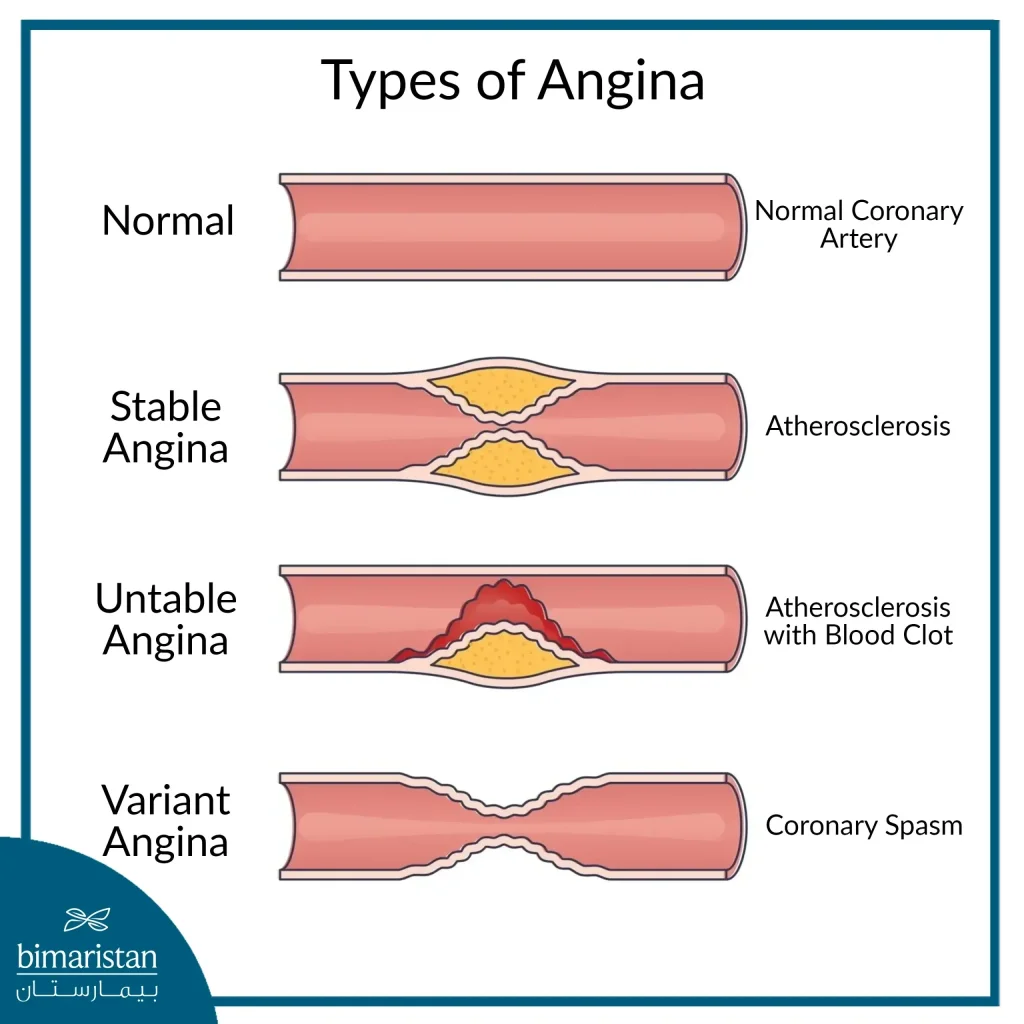
Coronary artery disease presents in various forms, determined by atherosclerosis status, whether plaques are stable or unstable. The complications of plaque rupture also influence classification, leading to transient ischemia or myocardial damage, which may be partial or widespread. The main manifestations of coronary artery disease include stable angina and acute coronary syndromes.
Stable chest angina
Stable angina, a chronic coronary syndrome, is a manifestation of coronary artery disease. It typically occurs during physical activity or emotional stress and diminishes with rest or appropriate medications. Symptoms include chest pressure or pain radiating to the shoulder, left arm, neck, or jaw, often described as a heaviness triggered by exertion or emotion. It is often described as a feeling of heaviness that occurs with exertion or emotion and does not usually occur with rest, which improves with rest or nitroglycerin and usually lasts 2 to 10 minutes.
Doctors diagnose stable angina by evaluating medical history and conducting a clinical examination. At rest, signs may not be evident, but exertion can cause pallor, cold sweating, increased heart rate, slight blood pressure elevation, and chest pain upon palpation. Electrocardiography (ECG) helps identify transient ST-segment depression or T-wave inversion during exertion.
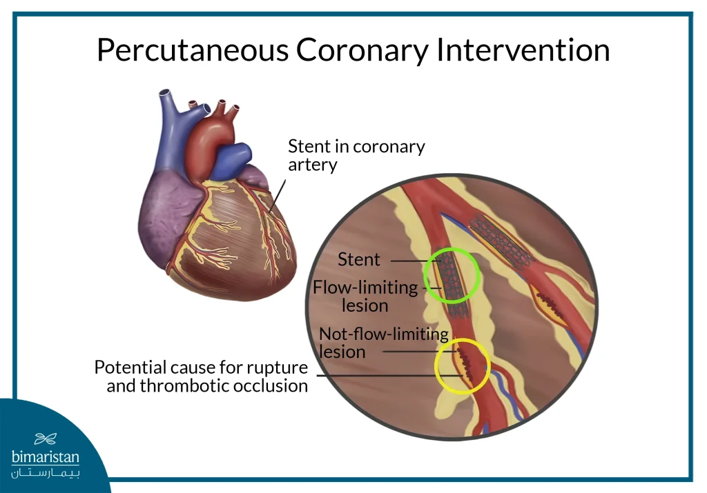
Additional tests like the Treadmill Test and Echocardiography (ECO) assess myocardial function, identify valvular disease, and rule out conditions like pericarditis. Computed tomography (CT) aids in detecting coronary artery abnormalities, while cardiac catheterization (coronary arteriography) measures pressure before and after stenosis to determine the need for reperfusion using a stent implant.

Acute Coronary Syndrome (ACS)
It is a manifestation of coronary artery disease resulting from the rupture of unstable atheroma. The condition is classified based on the rupture outcome and the extent of myocardial damage into unstable angina (UA), non-ST-segment elevation myocardial infarction (NSTEMI), and ST-segment elevation myocardial infarction (STEMI).
Unstable chest angina
Unstable angina is a manifestation of coronary artery disease caused by a partial or temporary blockage of the coronary artery, without myocardial infarction. It occurs due to the rupture of a fragile atheroma within the coronary artery, leading to platelet aggregation and the formation of a small thrombus. This thrombus dissolves quickly, reducing blood flow but not completely obstructing it. The condition presents as chest pain similar to stable angina, described as pressing or burning, with pain potentially radiating to the left arm, neck, jaw, or back. However, unlike stable angina, unstable angina is more severe, occurs at rest or with minimal exertion, lasts for more than 20 minutes, and does not improve with nitroglycerin.
Non-ST-segment elevation myocardial infarction (NON-STEMI)
Non-ST-segment elevation myocardial infarction (NSTEMI) is a type of coronary artery disease that occurs when blood flow to the heart muscle is reduced due to the formation of a large thrombus following the rupture of an atheroma. This leads to partial blockage in a coronary artery or the dissolution of the thrombus into smaller clots that obstruct the artery’s final branches, resulting in necrosis and damage to part of the heart muscle without affecting the entire thickness of the cardiac wall.
NSTEMI shares similarities with stable angina but presents with more severe symptoms, including persistent chest pain described as pressing or burning. The pain lasts for over 20 minutes, may radiate to the left arm, neck, jaw, or back, and does not improve with rest or nitroglycerin.
Diagnostic findings include elevated cardiac enzymes, particularly troponin, indicating heart muscle damage. Electrocardiogram (ECG) results may show non-specific changes, such as ST depression or T-wave inversion, but do not exhibit ST-segment elevation.
ST-segment elevation myocardial infarction (STEMI)
Coronary artery disease can lead to a complete blockage of the artery within minutes to hours. This happens due to the formation of a large clot following the rupture of an atheroma or arterial dissection without atheroma involvement, resulting in complete arterial blockage. As a result, heart cells in the affected region begin to die, causing myocardial infarction across the entire thickness of the myocardial wall. This life-threatening emergency demands immediate medical intervention to save the heart muscle and prevent fatal outcomes.
Symptoms include sudden, intensifying chest pain lasting from several minutes to half an hour, sometimes preceded by unstable angina. Additional manifestations include nausea, excessive sweating, and fever due to the infarction triggering an inflammatory response. Elevated cardiac enzyme levels, particularly troponin, indicate myocardial damage. Possible complications of the first manifestation include:
- Rhythm disorders (ventricular fibrillation)
- Heart failure
- Sudden death (within hours)
- Cardiac arrest
While chest pain and cardiac enzyme levels help diagnose an infarction, electrocardiography (ECG) is essential for confirmation and identifying the location of the infarct. In acute infarction, early signs include short-lived T-wave elevation, often within the first half hour. A concave downward ST-segment elevation appears in leads adjacent to the infarct site, while opposite leads may show ST depression, a phenomenon called the mirror effect. A first-degree branch block can also occur.
For an old myocardial infarction (after 24 hours), ECG may reveal a necrotic Q wave alongside wide T-wave inversion, suggesting a poor prognosis. Persistent ST-segment elevation despite treatment may indicate an ongoing arterial blockage or the development of a ventricular aneurysm due to infarction damage.
Ischemic cardiomyopathy
Ischemic cardiomyopathy is a type of coronary artery disease, a chronic condition caused by a chronic or recurrent lack of blood supply to the heart muscle due to narrowing or blockage of the coronary arteries, this lack of blood supply leads to weakness in the heart muscle and decreases its ability to contract and pump blood effectively, causing heart failure (Heart Failure).
Shortness of breath (especially with exertion or when lying down), general fatigue, poor exercise capacity, edema in the legs (fluid retention), palpitations, or irregular heartbeat, sometimes accompanied by chest pain. Echocardiography is an important investigation in case of cardiomyopathy as it shows impaired left ventricular function (low EF) and areas of the heart wall with little or no movement. Electrocardiography is also useful as it shows signs of old infarcts and arrhythmias.
Coronary artery disease treatment
The treatment of coronary artery disease, whether through medication or surgery, focuses on alleviating symptoms by reducing angina attacks and relieving exertional dyspnea. It aims to enhance long-term outcomes, including increased survival rates, improved quality of life, and a reduced need for re-interventions. Additionally, treatment helps prevent acute complications by lowering the risk of sudden cardiac arrest and acute coronary syndrome (ACS).
Lifestyle change
The foundation treatment of coronary artery disease, whether pharmaceutical or surgical, includes essential lifestyle modifications:
- Completely stop smoking: smoking damages the inner lining of blood vessels, accelerates cholesterol buildup, and increases the risk of blood clots.
- Eat a heart-healthy diet: reduce saturated fats, sugars, and excess salt while prioritizing fresh vegetables, fruits, whole grains, legumes, and fatty fish like salmon.
- Engage in regular physical activity: exercise enhances heart muscle efficiency, lowers blood pressure and LDL cholesterol, and improves circulation.
- Maintain a healthy weight: reducing excess weight alleviates strain on the heart.
- Control comorbidities: managing high blood pressure, diabetes, and elevated LDL cholesterol decreases the risk of coronary artery disease progression.
Pharmacological treatment of coronary artery disease
- Beta-blockers: relax the heart and alleviate symptoms (e.g., metoprolol, carvedilol). Caution is needed to prevent low blood pressure (shock).
- ACE inhibitors/ARBs: These drugs enhance heart function and lower the risk of future infarction (e.g., captopril, valsartan).
- Vasodilators: reduce blood pressure, decrease preload and afterload, and improve cardiac perfusion.
- Statins: lower LDL cholesterol and stabilize atherosclerotic plaques (e.g., atorvastatin, rosuvastatin).
- Anti-platelet agents: prevent platelet aggregation, reducing clot formation risk (e.g., aspirin, clopidogrel).
- Diuretics: administered post-episode rather than during; improve survival and support heart function (e.g., spironolactone).
Surgical treatment of coronary artery disease
Surgical treatment of coronary artery disease is typically considered when symptoms remain uncontrolled or the risk is high. Several approaches are used, including:
- Coronary artery catheterization: a procedure to widen narrowed or blocked coronary arteries. It is primarily used for acute coronary syndrome (ACS), cases where drug therapy fails, and patients with persistent ischemic symptoms.

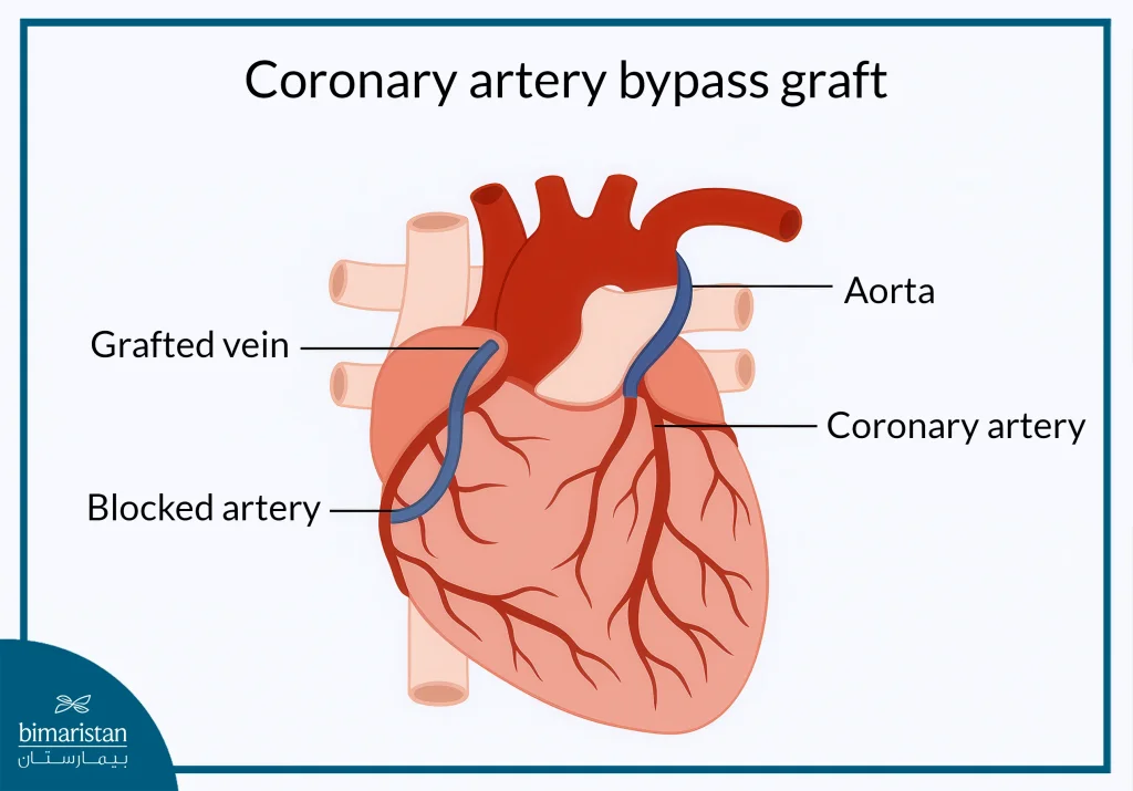
- Coronary artery bypass surgery (CABG): recommended when the left atrium is affected or when three arteries are narrowed, especially in the proximal section. The benefits are particularly significant for patients with diabetes.

Coronary artery disease: Follow-up and prevention
- Regular cardiologist visits to monitor heart health and treatment effectiveness
- Routine liver, kidney function, and cholesterol tests to ensure optimal metabolic balance
- Adjusting treatment based on response, ensuring the best possible outcomes
- Lifelong medication adherence, in most cases, to prevent complications and maintain stability
Finally, coronary artery disease is a crucial topic, as it remains a leading cause of death worldwide. This condition can impact lives in numerous ways, making awareness and education essential. Recognizing symptoms, understanding risk factors, and adopting a healthy lifestyle are key to both prevention and treatment. Early detection plays a vital role in reducing complications, so when experiencing symptoms or suspecting signs of coronary artery disease, seek immediate medical attention at the nearest health center or consult Bimaristan for expert guidance.
Sources:
- American Heart Association. (n.d.). Coronary artery disease. American Heart Association
- National Heart, Lung, and Blood Institute. (n.d.). What is coronary heart disease? National Institutes of Health

