Mitral valve repair surgery in Turkey is performed to treat most mitral valve diseases before the valvular issue progresses and causes systemic damage to the patient, resulting in death.
Overview of the mitral valve
The mitral valve, also known as the mitral valve, is one of the four heart valves that keep blood flowing in the right direction. It is a bicuspid valve consisting of two leaflets held together by tendons and papillary muscles that separate the two chambers of the heart, the left atrium and the left ventricle.
The back leaflet of the mitral valve has a C-shape, and the front leaflet has a semicircular shape to match the back leaflet. The name of the valve comes from the fact that the two leaflets of the valve have the shape of the crown worn by bishops.
The function of the mitral heart valve is to prevent the return of blood from the left ventricle to the left atrium. During each heart cycle, the walls of the ventricles expand to move blood from the left atrium to the left ventricle through the mitral valve, then the walls of the ventricles contract to move blood to the arteries, and the mitral valve closes to prevent blood from returning to the atria.
Congenitally or with age, the mitral valve may become damaged or diseased so that it does not function well and not enough blood reaches the left ventricle, making the heart unable to meet the body’s needs with oxygen-rich blood, and the patient develops symptoms such as fatigue and exertional dyspnea as the condition progresses.
Mitral valve repair surgery is often necessary when such damage occurs. Most people with mitral valve disease have no obvious symptoms, but if left untreated, the condition can become serious and lead to life-threatening complications such as heart failure and irregular heartbeat.
Types of mitral valve disease
Mitral valve issues are usually categorized into three types:
- Mitral valve stenosis: The mitral valve leaflets become stiff or calcified, and the valve doesn’t open enough, resulting in insufficient blood flow to the left ventricle
- Mitral valve insufficiency (mitral regurgitation): When the mitral valve closes, its leaflets don’t fit together, causing blood to leak from the left ventricle into the left atrium as the ventricles contract.
- Mitral valve prolapse: Mitral valve prolapse occurs when one or both mitral valve leaflets protrude into the left atrium during ventricular contraction, the valve does not close properly, and blood leaks into the left atrium, so it is considered by some to be a form of mitral insufficiency.
How are mitral valve diseases diagnosed?
It is important to have routine cardiac examinations, especially as we age, in order to diagnose mitral valve disease early and treat it as soon as possible before the condition progresses and we have to resort to mitral valve repair surgery.
When a doctor suspects mitral valve disease, he or she will listen to the patient’s heartbeat with a stethoscope and then perform additional tests and examinations to help confirm the diagnosis of mitral valve disease.
Some of the methods used to detect mitral valve disease:
- Echocardiography: This technique uses ultrasound to visualize the mitral valve and is considered the most important diagnostic tool in determining the cause and severity of valvular lesions.
- Plain chest X-ray: It is done by simple radiology and is important for detecting cardiac hypertrophy or heart failure.
- Cardiac catheterization: This procedure allows several tests, such as imaging the heart vessels and providing information about blood flow and heart health. It can also be used as a therapeutic tool in some cases.
- Electrocardiogram (ECG): Used to assess the heart’s electrical activity and detect many disorders caused by mitral valve issues.
- Exercise stress test: An ECG is performed during physical exertion to evaluate the heart’s response to stress.
These evaluations help determine whether mitral valve repair surgery is necessary or if the condition can still be managed with other medical options.
Mitral valve repair surgery in Turkey
When the mitral valve is not functioning well, the doctor may prescribe certain medications to increase the heart’s contractility, thereby increasing the blood flow to the body’s arteries without treating the underlying issue.
Mitral valve issues are mechanical issues that cannot be treated with medications alone. Surgery is required to repair or replace the damaged mitral valve, and leaving the condition untreated may reduce the options available to the patient.
Why should a mitral valve be repaired rather than replaced?
Repairing the diseased valve is better than replacing it if repair alone is able to restore normal heart function, because it avoids the complications that can occur in mitral valve replacement and also leads to a longer survival rate without the need for prolonged use of blood-thinning medications, as in mitral valve replacement with artificial valves. Therefore, mitral valve repair surgery is often preferred when feasible.
Looking for mitral valve repair surgery?
At Bimaristan Medical Center in Turkey, we treat mitral valve insufficiency and stenosis using advanced mitral valve repair surgery techniques.
Get expert care with the latest technology — contact us today.
More than half of mitral valve insufficiency cases can now be repaired using minimally invasive techniques that require 2 to 4 small incisions instead of open heart surgery, depending on preoperative tests that determine the optimal procedure for each patient.
Advantages of mitral valve repair
Mitral valve repair surgery is superior to replacement surgeries in several ways:
- Optimizes lifestyle in patients with mitral valve insufficiency
- Offers a longer survival rate compared to mitral valve replacement
- Helps maintain better heart function
- Carries a lower risk of stroke and infections
- Eliminates the need for blood-thinning medications
A study was conducted to compare the mortality rate of these two procedures in a large sample of mitral valve patients. The rate was 3.9% for mitral valve repair surgery and 8.9% for mitral valve replacement.
Mitral valve repair surgery is more technically challenging than replacement, and surgeons are often inclined to repair rather than replace the valve for the best long-term outcomes.
How is mitral valve repair surgery done in Turkey?
Before mitral valve repair surgery begins, the surgeon determines which leaflet will be repaired (anterior, posterior, or both), which technique is best suited for repair, and what surgical procedure will be used to access the mitral valve.
There are several techniques used to treat mitral valve stenosis or insufficiency, some of which require open-heart surgery and others that can be performed using minimally invasive robotic or catheter-based techniques.
In all mitral valve repair surgery procedures, the patient will be placed under general anesthesia and connected to a cardiopulmonary bypass machine (except for catheter-based techniques, which are performed without a bypass machine).
The procedure usually takes 4–6 hours, but this varies depending on the type of surgery and the technique used.
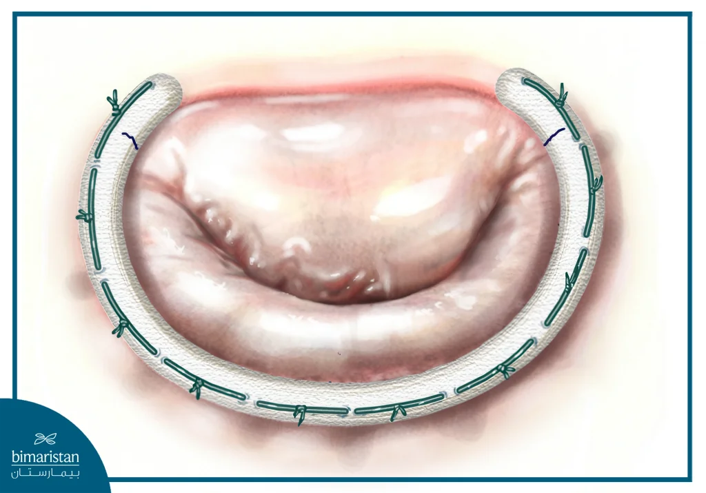
Annuloplasty
In this technique, the mitral valve ring on which the mitral valve leaflets rest is narrowed and strengthened by attaching a ring (or half ring) of tissue, fabric, or metal to it.
It should be noted that all mitral valve repair techniques may require annuloplasty to maintain the original size of the mitral valve and re-seal the valve leaflets, making the mitral valve repair last longer.
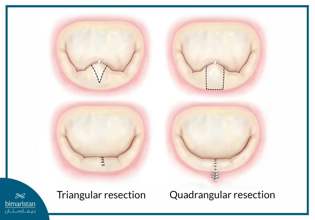
Triangular resection of a portion of the mitral valve leaflet
This technique involves cutting and removing the affected part of the valve leaflet in a triangular shape and then connecting the edges of the cut to the surgical sutures, This technique is usually used when posterior mitral valve leaflet prolapse occurs.
Mitral valve leaflet sliding repair
Similar to the previous method except that the resection of the posterior leaflet is wider (from the free edge of the valve to the mitral valve annulus), this method is used in special cases of posterior leaflet prolapse of the mitral valve.
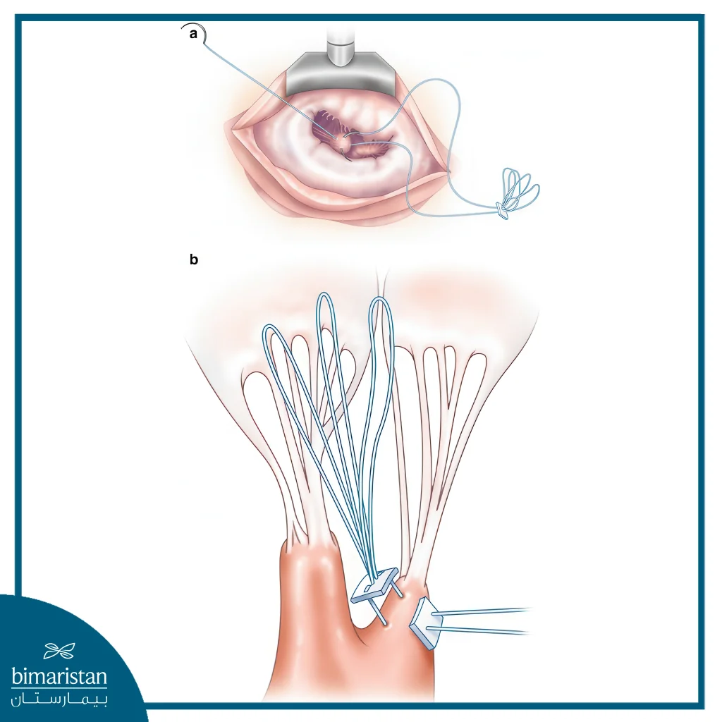
Chordal replacement
The tendons stabilize the valve leaflet against the inner walls of the heart and prevent it from drooping towards the atrium, and any stretch or break in these tendons leads to mitral valve prolapse and thus mitral regurgitation.
This technique involves replacing one or more of the stretched or severed tendons with artificial tendons made of a non-absorbable material such as Gore-Tex and is often used in mitral valve leaflet prolapse.
Another way to repair mitral valve leaflet prolapse is to transfer normal tendons with a portion of the associated posterior leaflet to the area containing the abnormal tendons in the anterior leaflet, and reattach the cutting edges of the posterior leaflet.
Both methods have excellent long-term results.
Mitral valve commissuroplasty
This technique is often used in cases of septic endocarditis affecting the mitral valve leaflets (the junction of the anterior and posterior leaflets) and adjacent portions of the leaflets, as one-third of the mitral valve can be removed without resulting in significant mitral stenosis.
In this technique, the surgeon removes the valve and neighboring septic parts from the two leaflets, and then joins the remaining parts of the valve at the cutting edge.
There have been studies that have shown the effectiveness of this technique in repairing mitral valve stenosis due to septic endocarditis.
Balloon valvuloplasty
This minimally invasive technique is used in cases of coronary stenosis and can be performed even before the onset of symptoms because it is easy to perform and has few complications.
In this procedure, a catheter with an inflatable balloon at the end is inserted through one of the body’s vessels into the heart, and when the balloon reaches the site of the mitral valve, it is inflated to enlarge the opening of the mitral valve.
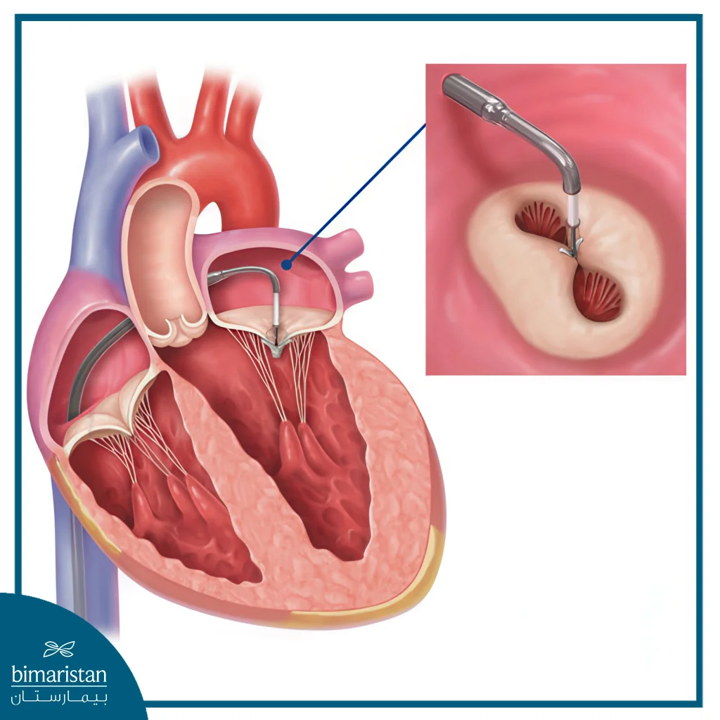
Mitral valve clip (MitraClip)
It is a modern catheter-based mitral valve repair technique that is usually used in patients with severe mitral valve regurgitation who cannot undergo surgery, and this minimally invasive technique is usually used in patients with severe mitral valve regurgitation who cannot undergo surgery.
In this technique, the doctor inserts a catheter with a clip on the end through a vessel until it reaches the site of the mitral valve, then uses the clip to join the two mitral valve leaflets together in the center, which is why it is also called transcatheter edge-to-edge repair (TEER).
By bringing the two valve leaflets closer together, the mitral valve is able to close tightly when the ventricles are contracting; however, the mitral valve remains open on both sides of the surgical suture and blood continues to flow from the left atrium to the left ventricle when the ventricles are diastolic.
Risks of mitral valve repair surgery
Like any other surgical procedure, mitral valve repair surgery can carry risks and complications that are more related to the surgical process than the repair itself, such as:
- Hemorrhage
- Infection
- Formation of blood clots
- Heart attack
- Cerebral hemorrhage
- Complications from anesthesia
- Death
These risks generally depend on the patient’s age at the time of the procedure, overall health condition, and cardiac function.
For asymptomatic patients who underwent TAVR, the risk of intraoperative death did not exceed one in one thousand, and for symptomatic patients, the risk did not exceed 1%. However, the presence of coronary artery disease or other health issues can increase the overall risk of mitral valve repair surgery.
Benefits of minimally invasive techniques
Minimally invasive surgery has several advantages over open-heart surgery:
- Faster healing
- Return to daily life faster
- Shorter hospital stays
- Smaller surgical scars
However, not all patients are suitable for minimally invasive surgery, and your doctor will tell you which procedure is best based on your medical history.
What happens after mitral valve repair surgery?
After the surgeon repairs the mitral valve, he removes the cardiopulmonary bypass device, restores a pulse to the heart, and closes the incision.
After completing the mitral valve repair surgery, the patient is transferred to the intensive care unit for a day or two, where family and friends can visit the patient.
The patient’s vital signs are closely monitored in the intensive care unit, after which they are transferred to a regular hospital room where they remain for about 4-7 days until discharge.
After discharge from the hospital, it is recommended to follow a healthy lifestyle and not to drive for at least 3 weeks and not to carry heavy weights for 6 weeks, after which you can do all the activities you want.
After the procedure, the patient is expected to experience an immediate improvement in symptoms that were present before the procedure, as well as an improvement in the patient’s quality of life and survival rate.
The recovery time after mitral valve repair surgery varies depending on the type of procedure used and the patient’s general health and adherence to the doctor’s instructions, the patient may recover in 4-6 weeks or it may take 3 months.
The patient must visit the doctor frequently to perform the necessary tests and examinations in order to ensure the healing process and the integrity of the mitral valve.
Studies have shown that when mitral valve repair is performed on the right patients at the right time, 95% of patients who undergo the procedure successfully repair the valve and do not require reoperation for 10 years and 90% do not require reoperation for 20 years.
Sources:

