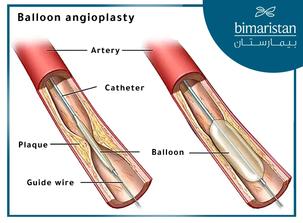
Endovascular therapy (balloon angioplasty) has recently become the first-line treatment option for diabetic ulcers. In 2024, endovascular treatment of diabetic foot is considered the latest treatment for diabetic foot in Turkey, as this therapy helps heal ulcers and reduce the risk of amputation.
Diabetic ulcers are one of the most severe complications of diabetes, with approximately 5% of all type II diabetes patients developing arterial diabetic ulcers. About 70% of lower limb amputations in diabetic patients are preceded by a diabetic ulcer, with 85% of these amputations occurring as a result of the ulcer. Therefore, it is well known that preventing ulcers is the best way to avoid amputation, but when an ulcer is present, the primary goal is to achieve its healing.
Causes of diabetic foot
Diabetic ulcers on the legs are complex lesions caused by multiple factors. The pathological basis of diabetic ulcers includes poor blood supply (ischemia), peripheral neuropathy, and bacterial foot infection, which usually coexist. Peripheral neuropathy and tissue ischemia are initiating factors in the disease, while bacterial infection is often secondary.
Peripheral neuropathy in diabetic foot
Diabetic peripheral neuropathy is common in clinical practice and is often associated with vascular disease. It includes motor, sensory, and autonomic neuropathy. Sensory neuropathy in its early stages can lead to altered sensation, increasing the risk of foot compression and mechanical or thermal injuries. In contrast, motor neuropathy alters the biomechanics of the foot, leading to anatomical changes, deformities, limited joint mobility, and changes in foot load capacity.
Diabetic foot ulcers
For every 1% increase in blood glucose levels, the risk of developing peripheral vascular disease increases by 25-28%. According to a European study, nearly half of diabetic foot cases arise from neuro ischemic (neuropathy + ischemia) or ischemic ulcers, with ischemia being one of the most significant factors preventing ulcer healing. As such, ischemia is the primary cause of diabetic ulcers and should be examined first unless there is clear evidence otherwise.
Neuroischemic ulcers result from the synergistic effects of peripheral neuropathy and tissue ischemia, which reduce oxygen delivery to the tissues. Both macrovascular and microvascular complications impair blood circulation in the diabetic foot. A key characteristic of diabetic macroangiopathy is that calcification of the lower limb arteries significantly reduces vascular elasticity. Clinically, neuro ischemic and ischemic ulcers share similar etiological factors, necessitating treatment through angioplasty (reopening of blocked vessels).

Diabetic foot infections – diabetic foot inflammation
Neuroischemic diabetic sores can easily become infected, although infections are rarely the cause of the ulcer. Additionally, the occurrence of an infection is closely associated with the likelihood of needing foot amputation, especially in patients with peripheral vascular disease. Deep infections are characterized by osteomyelitis or widespread soft tissue infection along the tendon, which is a direct factor associated with the risk of amputation and death. Patient outcomes are linked to the extent of the infection, comorbidities, and the presence or absence of peripheral vascular disease.
Symptoms of diabetic foot
Common symptoms of diabetic ulcers:
- Neuropathy:
- Numbness, tingling, or pain in the feet or legs, especially at night.
- Loss of sensation to heat or cold.
- Muscle weakness.
- Difficulty moving the feet.
- Foot deformities.
- Vascular Disease:
- Dry, pale, and cold skin on the feet.
- Slow-healing sores or wounds on the feet.
- Leg pain, especially when walking.
- Feet turning red or blue.
- Loss of hair on the feet.
- Other Complications:
- Foot infection.
- Foot gangrene (tissue death).
- Foot or leg amputation.
Evaluation and classification of diabetic ulcers
In cases of diabetic ulcers, consideration must be given to the ulcer’s location, depth of tissue involvement, associated infections, and overall tissue necrosis. Although no standardized evaluation criterion exists, the Wagner classification is commonly used to assess these factors. Diabetic foot infections can be diagnosed based on symptoms and signs of local inflammation, including discharge, localized redness, swelling, warmth, pain, systemic symptoms such as fever, increased white blood cell count, elevated erythrocyte sedimentation rate, and increased CRP levels in the blood.
Diabetic infections in the foot often occur secondary to ulcers and may not always accompany them. The extent and severity of the infection are crucial factors that influence the prognosis, and the risk of amputation and death is often predicted based on the presence of widespread infections and significant systemic inflammatory responses.
Diabetic ulcers treatment options in Turkey
There are four options for diabetic foot gangrene treatment: conservative treatment, primary amputation, surgical vascular bypass, and revascularization. The choice of treatment should be made by a multidisciplinary team, with decisions between surgical and endovascular treatments based on the anatomy of the affected area, the patient’s condition, and the expertise of the treatment team.
Endovascular treatment (angioplasty) for diabetic foot in Turkey
Endovascular treatment for neuro ischemic diabetic foot ulcers primarily focuses on the arteries below the knee. Ulcers may occasionally appear in the iliac and femoropopliteal regions in diabetic patients, but this is less common. For endovascular treatment, the focus should be on treating ulcers resulting from vascular injury below the knee. Diabetic foot ulcers often consist of long-segment lesions, while atherosclerotic lesions are often short.
Treating long-segment diabetic foot ulcers (endovascular therapy) requires a specialized center with extensive experience, as these treatments require personal expertise. The treatment plan is tailored according to the patient’s needs, clinical condition, and treatment feasibility. The goal of the treatment plan is to heal the ulcers while simultaneously saving the limb.
When is the endovascular treatment of diabetic foot needed?
Unhealed diabetic foot ulcers, whether accompanied by infection and gangrene, are reasons to opt for endovascular treatment, provided the limb is still viable, and the treatment will improve the quality of life. Therefore, a bedridden patient with dementia is not a candidate for endovascular treatment.
The decision between percutaneous angioplasty (endovascular treatment) and open surgery is usually the result of a discussion among the treatment team. The high risks associated with open surgery, the lack of good venous material, the absence of suitable surgical anastomosis sites, and poor blood flow are additional reasons to choose percutaneous angioplasty (endovascular treatment) as the therapeutic solution.
However, in many medical institutions, percutaneous angioplasty (endovascular treatment) is currently the first-line treatment option. The understanding that “time is tissue” in diabetic patients means that infected diabetic foot ulcers should be treated as an emergency procedure and preferably performed within 24 hours.
Method of performing endovascular treatment of diabetic foot in Turkey
Endovascular treatment in the leg, such as percutaneous angioplasty, is a specialized technique that should only be performed after extensive training and experience. This technique is applied through a skin puncture and should never be performed directly over vascular bifurcations.
For below-knee arteries, simple balloon angioplasty should be performed well, and stenting should only be considered in rare cases when prolonged low-pressure dilation does not result in adequate blood flow to the foot. Since the treatment is not intended to keep the blood vessels open in the long term, stenting should not be initiated as it may prevent repeat interventions on the vessels if needed. Additionally, stenting is less preferred for treating diabetic foot ulcers because diabetic foot lesions are often long and not focal compared to atherosclerosis.
The current trend is to perform atherectomy followed by drug-coated balloon angioplasty, with stenting only being used in cases with adequate anatomy or residual stenosis. Atherectomy can be performed using a laser, directional atherectomy device, or rotational atherectomy device, all of which remove and modify a certain amount of plaque, allowing for easier balloon angioplasty.
Most atherectomy procedures carry potential embolism risks, especially in long-segment lesions. However, using a filter is advised when treating long-segment lesions, even when using devices that perform simultaneous removal and aspiration.
Calcified vessels may impede balloon catheter passage, making angioplasty challenging. A small and short balloon of 2 × 40 mm size can be used for initial dilation of hard-to-cross arteries, followed by angioplasty with a larger balloon matching the size of the affected artery.
The balloon should be advanced with a “tapping” motion as static friction has a greater stopping force than kinetic friction. Crossing tight stenotic vessels requires strong balloon support, which can be achieved with a 4F sheath longer than the external 6-7 F sheath placed coaxially. This ensures strong balloon support for crossing the stenotic artery.
Two guidewires can be placed side by side through the narrow artery, and “swinging balloon dilation” with the balloon’s leading edge can help cross the calcified section of the artery. Other methods for dealing with calcified areas to allow balloon passage include gradual dilation with a low-profile tapered crossing catheter, rotational atherectomy, laser atherectomy, and lithotripsy devices.
Whittling is a technique where the back end of the wire is used to strike the calcified section to modify the plaque. Additionally, external puncture with a needle can modify the plaque and allow balloon passage, thereby enabling angioplasty of the occluded artery.
Some calcified arterial plaques may remain resistant to angioplasty despite the availability of modified plaque equipment and methods. In these cases, intentional plaque/artery rupture using an expandable balloon-covered stent and securing it with a SUPERA stent after achieving an adequate diameter may be necessary.
When should the doctors stop angioplasty and try another day?
Lower extremity arterial interventions can be complex procedures requiring several hours of surgery. The procedure should be considered for termination if the patient experiences discomfort under local anesthesia. An uncomfortable patient may move, leading to angiographic issues that make the procedure extremely challenging.
Angioplasty procedures may require the use of large amounts of contrast agents, which carry the risk of causing acute kidney injury. Foot arch intervention planning is best performed with careful study of anatomical variations before the procedure. Planning involves understanding not only the anatomy and location of arterial occlusion but also ensuring the availability of appropriate equipment. Patient comorbidities and how they may affect the outcome should be considered in the decision-making process, and clear treatment goals should be established before starting angioplasty.

Outcomes of the latest diabetic ulcer treatment via angioplasty
The outcomes of both open surgery and endovascular treatment for healing diabetic leg ulcers and limb salvage are widely discussed, with success rates ranging from 78-85%. However, post-bypass graft patency is generally better than that of endovascular treatment. This is not considered a major issue because the healing of diabetic leg ulcers and subsequent limb salvage almost always occurs within 6-9 months.
Typically, arterial patency is not needed beyond the healing of the diabetic ulcer because diabetic patients often do not experience rest pain. Endovascular interventions generally provide sufficient arterial patency to achieve the ultimate goal of diabetic ulcer healing. This is why reports on limb salvage after vascular treatment often indicate a 20% higher rate than actual vascular patency. As a result, angioplasty in acute ischemic foot is often called a “temporary bypass.”
Feel free to contact us to learn about the best hospitals for diabetic foot treatment in Turkey.
References:
-
- Li M. Guidelines and standards for comprehensive clinical diagnosis and interventional treatment for diabetic foot in China. Journal of Interventional Medicine 2021; 4: 117-129.
- Wong T.Y. Endovascular treatment of diabetic foot ischemic ulcer – Technical review. Journal of Interventional Medicine 2020; 3: 17-26.
- Current endovascular management of the ischaemic diabetic foot, HIPPOKRATIA
- Endovascular treatment of diabetic foot ischemic ulcer, Journal of Interventional Medicine.
- Guidelines and standards for comprehensive clinical diagnosis and interventional treatment for diabetic foot in China, Journal of Interventional Medicine.

