Astigmatism is a vision disorder that is highly prevalent in the world, and the diagnosis and treatment of this disorder have accelerated significantly in Turkey.
Overview of the eye and refractive media
To understand more about astigmatism, we need to understand the photorefractor.
Vision occurs when light enters the eye through the pupil. Other important structures in the eye, such as the iris and cornea, help direct the right amount of light to the lens, which directs the light onto the retina. The iris controls the amount of light entering the eye by narrowing or widening the pupil.
The light-refracting structures of the eye are the cornea and the lens, both of which have anterior and posterior surfaces. The cornea is the transparent, circular outermost part of the front of the eyeball, which is devoid of vessels, highly neurovascularized, and sensitive to pain.
The lens is a transparent structure behind the pupil inside a thin capsule. Just like the lens in a camera, the lens in the eye refracts (bends) incoming light to fall optimally on the retina.
The retina consists of millions of specialized visual cells known as rods and cones. These cells work together to convert the image or light falling on the retina into electrical energy. This energy is sent to the optic disc and then transmitted via electrical impulses along the optic nerve to be processed by the brain, and we get the image of the object in its correct position and size.
What is Astigmatism?
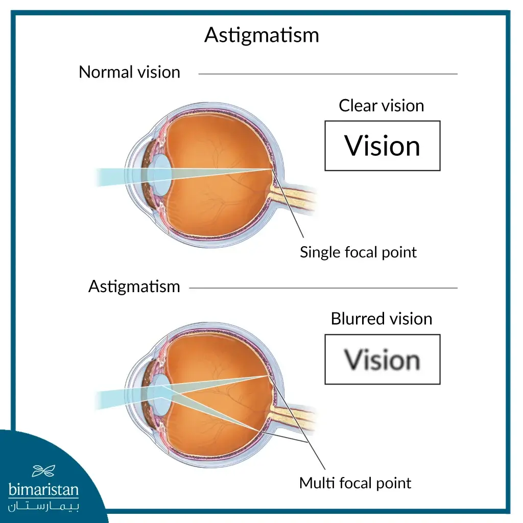
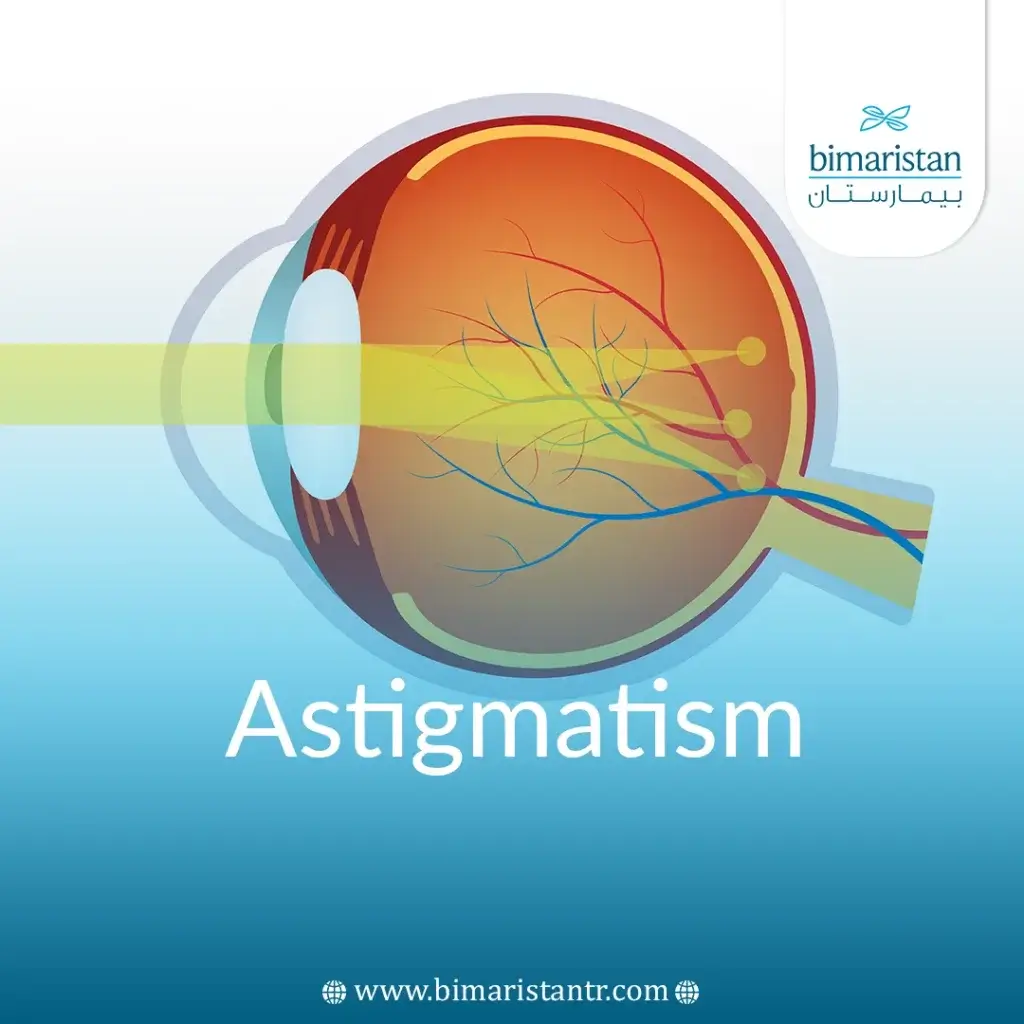
Astigmatism is a common eye disorder or error in the curvature of any of the refractive surfaces of the eye that results in blurry, hazy, or misdirected vision.
Astigmatism occurs when the cornea isn’t perfectly round, resulting in a non-spherical surface. Many people have mild conditions without noticeable vision issues.

There are several ways to categorize this condition based on several factors:
Depending on the structure responsible for the defect
- Corneal astigmatism: The defect is in one of the surfaces of the cornea.
- Lens astigmatism: The defect is in one of the surfaces of the lens.
The defect can be in both structures and in their front or back surfaces.
Based on longitudes or axes
To explain further, we assume that the front of the eye is a clock face or circle with the center of the pupil, each meridian connects two different numbers (like the diagonals in a circle), and that the main meridians are two lines: One between 3 o’clock and 9 o’clock and the other between 6 o’clock and 12 o’clock.
In astigmatism, the principal meridians are the flattest and most convex (sharpest) meridians.
Astigmatism is classified by understanding corneal meridians, which helps define its different types. Based on these meridians, astigmatism is divided into:
Regular astigmatism
In this case, the principal meridians are perpendicular and the corneal refractive force is constant in each of the perpendicular axes, which is the most common type of astigmatism.
Regular astigmatism can be further subdivided into:
- With-the-rule astigmatism: Common in children in which the vertical meridian’s refractive power is greater, is the most convex (sharp), and stays close to 90 degrees.
- Against-the-rule astigmatism: The horizontal meridian remains close to 180 degrees and its refractive power is greater than the vertical axis, Against-the-rule astigmatism is common in the elderly.
- Oblique astigmatism: Oblique astigmatism is diagnosed if the major meridians are not at 90 degrees or 180 degrees. Here, the shape of the eyeball resembles an oblique American football, with the major meridians falling between 30 degrees, 60 degrees, 120 degrees, and 150 degrees.
Regular astigmatism can also be subdivided based on the dysphoria that occurs (which is caused by different breaking forces in the orthogonal axes rather than in the axis itself):
- Simple regular astigmatism: This means that one axis of the cornea brings the focus of the image onto the retina while the other axis brings it in front of or behind the retina, meaning that this type of astigmatism is neither nearsighted nor farsighted.
- Compound regular astigmatism: In this type, the patient suffers from hyperopia or myopia, where both meridians focus the focus of the image in front (myopia) or behind (hyperopia) the retina.
- Mixed regular astigmatism: In this case, one axis focuses the image in front of the retina and the other behind the retina.
Irregular astigmatism
The principal meridians are not perpendicular and the corneal breaking force varies along the same axis. It is uncommon compared to the regular type and is most commonly seen in external eye injuries due to trauma or surgery.
The classification of astigmatism is complex, and each patient has his or her own classification. However, these classifications are designed to make things easier, lay the groundwork for the treatment plan, and then modify it.
Astigmatism Causes
Doctors don’t yet know why the cornea or lens is shaped differently and convex in normal people. The cause of astigmatism is not clearly known. The condition can be present at birth, indicating a significant role for genetics in this disorder.
Astigmatism can also be caused by an eye injury, eye surgery (such as an artificial lens implant), or a condition known as keratoconus, which causes the cornea to not only thin but to become cone-shaped and lead to a refractive error.
This condition leads to a severe degree of this disease, causing the patient’s vision to be impaired. This impairment cannot be fully corrected with glasses, and in many cases, a corneal transplant is required.
Astigmatism can be caused by the eyelids pressing against the cornea or constantly rubbing the eyes (preferably not) and is not caused, as many people think, by holding a book close to the eyes while reading or reading in dim light.
Astigmatism is usually associated with nearsightedness or farsightedness, and together, they are called refractive disorders.
Symptoms of astigmatism
Symptoms of astigmatism vary depending on the severity and types, and symptoms often include:
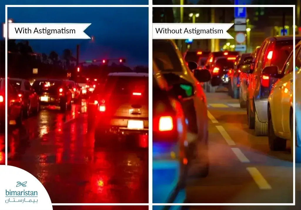
- Blurred or distorted vision at all distances.

- Headache.
- Difficulty seeing at night Night Vision (difficulty driving at night).
- Eyestrain and irritation.
- Stare for a long time until the vision is clear.
- Astigmatism affects the depth of vision, the definition of distance, and the clarity of the true shape of objects and can make the patient feel unbalanced.
Astigmatism in children can cause what is known as lazy eye.
Methods of diagnosing astigmatism in Turkey
Methods of diagnosing astigmatism in Turkey have evolved considerably, and regular check-ups have helped many people solve issues that they have overlooked.
This is especially important for children who do not complain about vision defects either because they can’t or because they think it is normal. Periodic examinations are necessary and even imperative because astigmatism affects their degree of productivity, participation and interaction at school and home, and academic achievement.
Adolescents should also be screened for keratoconus. Different tests will determine the degree of astigmatism and what treatment or lenses should be used.
The doctor first performs a routine eye examination; if there is an abnormality, more precise diagnostic methods can be used. Of the tests or devices used:
Visual acuity test
In this test, your doctor will ask you to read or locate letters from a chart at a specific distance to determine how well you can see the letters.
Refraction test (how the eye refracts light)
A visual test that uses a machine called a phoropter. The device has multiple corrective glass lenses with different strengths.
The doctor will ask you to read a chart as you look at the lenses that the doctor changes to fit your vision. Ultimately, which lens best corrects your vision and astigmatism will be determined.
Keratometer
It measures the degree of curvature of the cornea, in which light is focused on the cornea, and measures the reflected light and how it is reflected to know the curvature of the cornea.
This test is important to get the right lenses and to differentiate between normal astigmatism and the one caused by disease.
Corneal Topography
Astigmatism can be better understood through detailed information on corneal topography, curvature, and shape, aiding in selecting the right treatment.
To inquire about more diagnostic methods or astigmatism symptoms, do not hesitate to contact us, your family bimaristan center in Turkey.
Astigmatism Treatment in Turkey
Astigmatism treatment in Turkey has advanced recently, with varied devices and methods tailored to different types and degrees of vision defects and astigmatism severity.
The condition can be mild and asymptomatic and not require any treatment, or the symptoms can be bothersome and affect the patient’s daily life and productivity. In this case, the patient must see a doctor to determine whether or not he has astigmatism and how to treat it.
The main treatment for astigmatism is corrective lenses, whether eyeglasses or contacts, which correct most cases of astigmatism.
Contact lenses used to treat astigmatism were thought to be rigid contacts, but now another type of lens has been developed, called toric contacts, which are small soft lenses.
Toric lenses differ from normal lenses by having two powers—one for distance vision and one specifically for astigmatism—while rigid contact lenses are the best choice for severe astigmatism.
Contact lenses have a wider field of vision compared to glasses and are more convenient in working life, but they have several disadvantages, such as infections due to neglect of hygiene and health rules, and may not be suitable for all patients.
Of other treatment methods:
Orthokeratology
In ortho-k, the doctor prescribes a rigid contact lens that the astigmatic wears at bedtime that corrects the shape of the cornea.
This procedure is not intended to cure astigmatism, but it is a temporary treatment, and the issue may return if you stop using these lenses.
Surgery
There are several surgical methods of treating astigmatism, mostly using lasers, which are mostly definitive treatments but may have some issues and complications:
LASIK
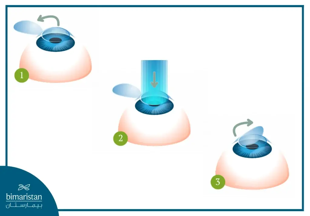
In laser in situ keratomileusis, a small flap is created in the cornea using small, rapid pulses of laser. The flap is then bent back, and a guided laser is used to reshape the cornea. The flap is then returned to its place, where it adheres naturally.
Laser treatment of ocular disorders has developed significantly in Turkey and has achieved high rates of cure and success.

PRK Corrective Epithelial Keratectomy
Photorefractive keratectomy removes the superficial epithelial layer of the cornea in the patient’s eye with a brush or medical instrument and then directs a laser to reshape the cornea.
After the procedure is completed, the doctor places a contact lens over the cornea while the epithelium grows back.
RK Corneal Finishing
Previous procedures have superseded this procedure due to its many complications and inaccurate results.
If the cornea is severely damaged, a new one may need to be transplanted, which is why corneal transplants in Turkey are performed. These operations can be performed on patients who are in good health and eligible for surgery.
Everyone should have regular check-ups to ensure the health of their most important sensory organ and correct any abnormalities. See a specialist at Bimaristan and communicate with them, as you may have stigmata and not even realize it.
Sources:
