
Meningiomas are among the most common tumors of the central nervous system, accounting for about 30–40% of primary brain tumors in adults. These tumors arise from the meninges surrounding the brain and spinal cord and are often benign and slow-growing. Meningiomas are diagnosed more frequently in women than in men, especially after the age of 40. Although they grow slowly, their location can lead to a variety of neurological symptoms that require careful medical evaluation.
What are meningiomas?
Meningiomas are tumors that originate from the cells lining the meninges that surround the brain and spinal cord, specifically from the cells of the arachnoid membrane. These tumors do not originate from the brain tissue itself, but from the protective layers surrounding it, so they are classified as extra-axial tumors.
Meningiomas are among the most common central nervous system tumors in adults, accounting for approximately 30–40% of primary brain tumors. In most cases, they are benign and slow-growing; however, their location may lead to gradual compression of nearby brain tissue or nerves, causing symptoms that vary depending on the tumor’s location and size.
Meningiomas are histologically classified into three main grades: Grade I (benign), Grade II (atypical), and Grade III (malignant and relatively rare). This classification determines the tumor’s expected behavior and the appropriate treatment plan.
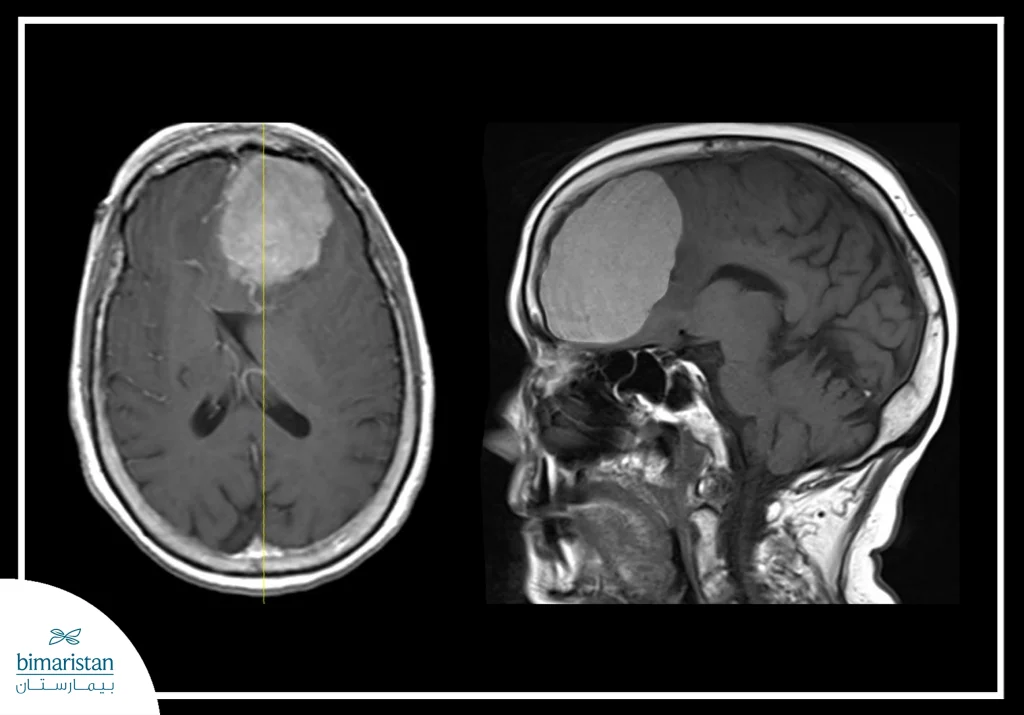
Are meningiomas dangerous?
The severity of meningiomas depends on three main factors: size, location, and histological grade. In most cases, these tumors are benign and slow-growing, and may remain for years without obvious symptoms, especially if they are small and do not press on sensitive nerve structures.
However, meningiomas can become dangerous when they grow in size or are located in vital areas such as the base of the skull, near the optic nerves, or in the brainstem. The danger here stems not from the nature of the tumor itself, but from its compressive effect on brain tissue, which can cause chronic headaches, seizures, vision problems, motor weakness, or cognitive changes.
From a classification standpoint, Grade I tumors often have a good prognosis following appropriate treatment, while Grade II tumors carry a higher risk of recurrence. Grade III tumors, on the other hand, are rare but more aggressive and may grow more rapidly. Therefore, meningiomas cannot be considered absolutely dangerous; rather, the risk is assessed on a case-by-case basis according to the tumor’s characteristics and the patient’s clinical condition.
Causes of meningiomas and risk factors
The danger of meningiomas depends more on the characteristics of the tumor and its local impact than on the tumor itself. Most of these tumors are benign and slow-growing, and may be discovered by chance during brain imaging for another reason without causing obvious symptoms. In these cases, periodic monitoring may be recommended without immediate intervention.
The risk increases when the tumor grows larger or is located in sensitive areas of the brain; gradual pressure on the nervous tissue may lead to neurological symptoms whose severity varies depending on the location of the tumor. Furthermore, some atypical or malignant types may tend to grow rapidly or recur after treatment. The factors that determine the severity of meningiomas can be summarized as follows:
- Tumor size: The larger the tumor, the greater the likelihood of pressure on the brain.
- Anatomical location: Tumors at the base of the skull or near the optic nerves are more sensitive.
- Histological grade: Grades II and III carry a higher risk of recurrence or rapid growth.
- The presence of concomitant cerebral edema: This may exacerbate symptoms and complications.
- The patient’s overall health: This affects their ability to tolerate surgical or radiation therapy.
Accordingly, meningiomas are not considered serious in all cases; rather, they are evaluated on a case-by-case basis using precise clinical and imaging criteria to determine the need for follow-up or treatment.
Symptoms of meningiomas by location
The symptoms of meningiomas vary depending on their location within the skull or along the spinal cord, as their primary effect results from localized pressure on adjacent neural structures; therefore, some cases may remain asymptomatic for a long time, while others exhibit clear neurological signs depending on the affected area.
Meningiomas in the frontal lobe
When the tumor is located in the frontal lobe, gradual changes in behavior and personality may occur, such as impaired concentration, apathy, or impaired judgment; In advanced cases, motor weakness in one of the limbs may occur, especially if the tumor is close to the motor cortex, and the patient may also complain of chronic headaches that gradually worsen.
Meningiomas near the optic nerve or the occipital lobe
If the tumor is near the optic nerve or in the areas responsible for vision, visual symptoms such as blurred vision or loss of part of the visual field may appear, and sometimes a gradual weakening of visual acuity occurs in one eye. Diagnosis may be delayed if the symptoms are slow to develop.
Meningiomas in the temporal lobe
A tumor located in the temporal lobe may lead to partial seizures or memory disturbances, and some patients may have difficulty understanding or expressing speech if the tumor is in the dominant hemisphere of the brain. Auditory symptoms may also occur if the auditory nerve pathways are affected.
Meningiomas in the parietal lobe
When a tumor affects the parietal lobe, it may cause sensory disturbances such as numbness or loss of sensation on one side of the body. In some cases, the patient may have difficulty orienting themselves or performing fine motor movements, especially if the areas responsible for sensorimotor integration are affected.
Meningiomas in the posterior fossa or near the brainstem
This area is more sensitive, as pressure on the cerebellum may lead to balance and gait disturbances, along with dizziness and unsteadiness. If the brainstem is affected, more serious symptoms may appear, such as difficulty swallowing, weakness of the facial muscles, or breathing disturbances in severe cases.
Spinal meningiomas
When meningiomas develop in the spinal cord, the symptoms differ from those of brain meningiomas; the patient may experience localized back pain that gradually worsens, along with weakness or numbness in the lower extremities. In advanced stages, bladder or bowel control may be impaired as a result of pressure on the spinal nerve pathways.
How are meningiomas diagnosed?
Diagnosis of meningiomas relies on a combination of clinical assessment and advanced imaging tests, with histological analysis sometimes used to confirm the tumor grade. Because these tumors often grow slowly, they may be discovered incidentally during brain imaging for another reason.
Clinical evaluation
Diagnosis begins with taking a detailed medical history, focusing on progressive neurological symptoms such as chronic headaches, seizures, visual disturbances, or motor weakness. This is followed by a comprehensive neurological examination to assess sensory and motor functions and reflexes, which helps pinpoint the suspected cause.
Magnetic Resonance Imaging (MRI)
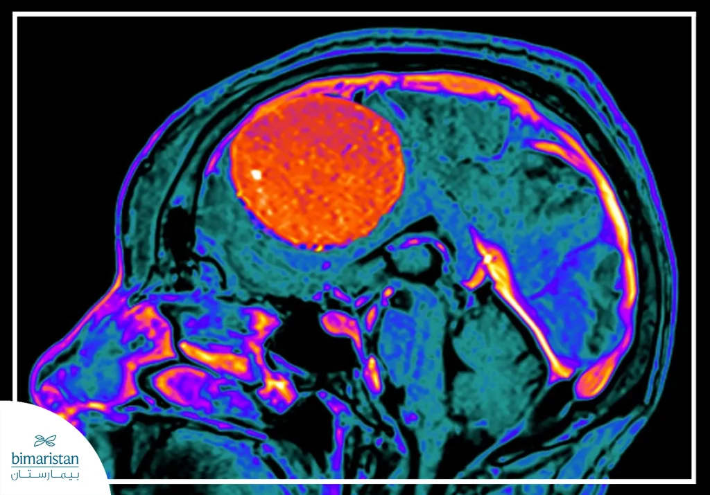
Magnetic resonance imaging with contrast injection is the most accurate examination for diagnosing meningiomas, where the tumor usually appears as a well-defined mass connected to the dura mater, and a characteristic sign known as the extension of the dura tail is often seen. This examination allows for determining the size of the tumor, its precise location, and the extent of its impact on the surrounding tissues.
Computed Tomography (CT)
Computed tomography is used when there is a need to evaluate calcifications within a tumor or bony changes in the skull. It is also useful in emergency situations. Meningiomas may appear in this examination as a dense mass attached to the inner surface of the bone.
Biopsy and histological examination
In some cases, particularly when an atypical or malignant tumor is suspected, a tissue sample is obtained during surgery for microscopic analysis; the histological examination determines the tumor grade (stage I, II, or III), which is a key factor in developing a treatment plan and assessing the likelihood of recurrence.
Treatment of meningiomas
The treatment of meningiomas depends on several factors, including the tumor’s size, location, growth rate, and histological grade, as well as the patient’s age and overall health. Not all cases require immediate intervention; in some patients, watchful waiting may be an appropriate option, while other cases require surgical or radiation therapy to control the tumor and prevent complications.
Periodic monitoring
If the meningiomas are small and asymptomatic, and slow-growing, regular monitoring via MRI every few months or annually may be recommended. This approach aims to track any changes in size or the emergence of new symptoms before a decision to intervene is made. This option is particularly suitable for older adults or patients at high surgical risk.
Surgery
Surgery is considered the primary treatment option when there are clear symptoms or noticeable tumor growth. The goal is to completely remove the tumor along with the portion attached to the dura mater, if possible, thereby reducing the likelihood of recurrence. The success rate depends on the tumor’s location; Superficial tumors are easier to remove than those located at the base of the skull or near sensitive neural structures. In some cases, complete resection may not be possible in order to preserve neural function.
Radiation therapy
Radiation therapy is used in several treatment settings, whether following surgery when tumor residue remains, or as a primary option when surgery is not possible. One common technique is stereotactic radiation therapy, which precisely directs high doses toward the tumor while minimizing the impact on healthy tissue; this option is particularly effective for small or medium-sized tumors.
Drug therapy
Drug therapy is not a primary treatment option in most cases of benign meningiomas; however, it may be used for recurrent or malignant tumors that do not respond to surgery or radiation. Treatment options include targeted or experimental drugs administered as part of research protocols, but their role remains limited compared to surgical and radiation options.
Follow-up after treatment for meningiomas
Even after complete resection, regular follow-up remains essential to monitor for any potential tumor recurrence, especially in atypical grades. The follow-up schedule depends on the tumor grade and the results of the surgery, and typically includes periodic MRI scans and regular neurological examinations.
In conclusion, meningiomas vary in their clinical behavior depending on their size, location, and histological characteristics, making a specialized evaluation crucial for determining the most appropriate treatment plan. Advances in microsurgical techniques and targeted radiation therapy have contributed to improved tumor control rates and reduced complications. Regular follow-up is essential even in benign cases to ensure that any early changes are detected. Bimaristan Medical Center is committed to providing comprehensive care based on a multidisciplinary team and modern diagnostic and treatment techniques to ensure the best therapeutic outcomes.
Sources: