Skull base surgery is a highly delicate procedure used to treat tumors and deformities in the lower part of the skull, the critical area separating the brain from the face and neck. This region is extremely sensitive, as it houses major cranial nerves and vital blood vessels that supply the brain, making skull base surgery one of the most intricate and demanding operations in modern medicine.
In the past, even minor errors in this area could lead to complications affecting vision, hearing, or motor function. However, with recent advances in endoscopic and robotic-assisted techniques, skull base surgery has become significantly safer, more precise, and minimally invasive, resulting in better outcomes and faster recovery times for patients.
What is skull base surgery?
Skull base surgery targets the lower part of the skull, which is made up of five bones forming the eye socket, parts of the sinuses, the roof of the nasal cavity, and the bones surrounding the inner ear. This area is highly sensitive, as it contains openings that allow the spinal cord, cranial nerves, and blood vessels to connect the brain to the rest of the body.
Surgical intervention in this region is performed to treat cancerous and non-cancerous tumors, congenital malformations, or abnormal growths located beneath the brain or at the top of the spine. In the past, skull base surgery required open procedures involving direct access through the skull, which increased both surgical risks and recovery time.
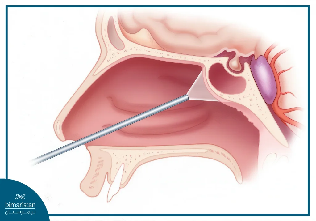
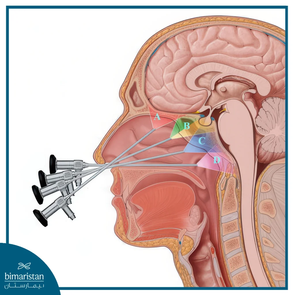
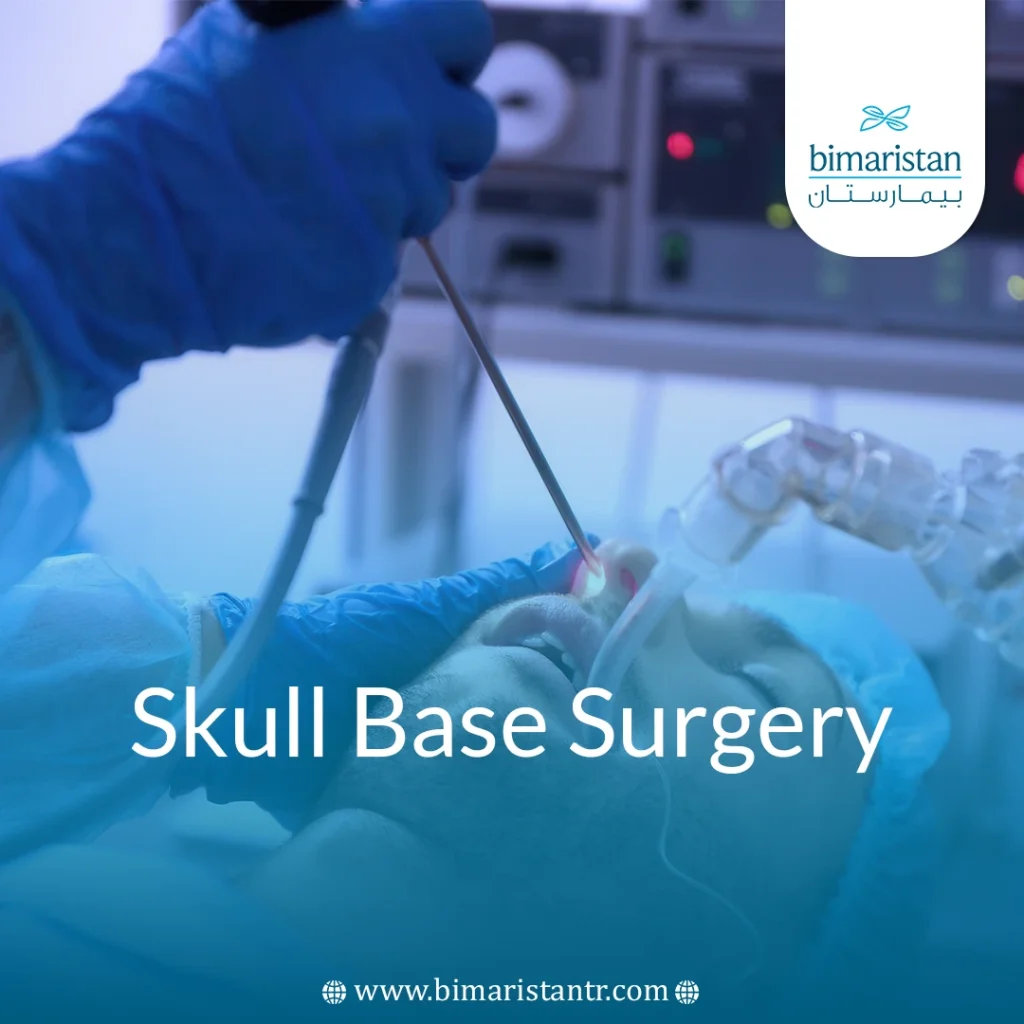
Today, with advances in medical technology, endoscopic skull base surgery is increasingly used. This minimally invasive technique is performed through the nose or mouth using an endoscope, offering greater precision, fewer complications, and faster recovery. Due to the complexity of the area, this surgery necessitates a multidisciplinary team to ensure maximum safety and optimal outcomes.

Causes and conditions that require skull base surgery
Conditions that require skull base surgery include a variety of diseases and tumors, including
Benign and malignant tumors
- Meningioma
- Acoustic neuroma (vestibular schwannoma)
- Chordoma
- Schwannomas of the cranial nerves
- Basal cell carcinoma
- Squamous cell carcinoma
- Sensory neuroblastoma
- Inverse papilloma
- Nasopharyngeal angiofibromas
- Pituitary cancer and tumors
- Eye and brain tumors (benign or malignant)
Vascular diseases and vascular malformations
- Cerebral aneurysm
- Arteriovenous malformations
- Brain bubbles
- Tangled blood vessels
Injuries and bruises
- Complex fractures of the skull base
- Traumatic lesions resulting from accidents
- Brain hernia
Inflammation and chronic issues
- Chronic or fungal infections of the skull base
- Cerebrospinal fluid (CSF) leak
- Infection-induced growth
- Congenital cysts
Congenital and acquired abnormalities
- Early craniosynostosis
- Craniofacial dysostosis
- Rathke’s incision cysts
- Third nerve pain
Types of skull base surgery
A clear comparison chart between the three types of skull base surgery you mentioned:
| Type of surgery | Description of the procedure | Key features/notes |
|---|---|---|
| Nasal endoscopic surgery | Accessing the base of the skull through the nostril using an endoscope with a camera | Minimally invasive procedure, no external scars, faster recovery |
| Transcranial surgery (conventional surgery) | Opening the skull with an incision in the scalp for direct access to the base of the skull | Used for large or complex tumors, it provides a direct view |
| Transoral surgery | Accessing the base of the skull through the mouth, especially for tumors near the throat or jaw | Less invasive than transcranial surgery, but provides limited and deep access |
Modern techniques in skull base surgery
Endoscopic Skull Base Surgery
Endoscopic or minimally invasive skull base surgery typically does not require a large incision. Instead, the surgeon may create a small opening (keyhole) inside the nose or in another part of the skull. Through this access point, the neurosurgeon removes the tumor using a thin, lighted instrument called an endoscope.
To support precision during skull base surgery, an MRI scan may be used. This imaging technique captures detailed views of the skull base using magnets, coils, and a computer. In some cases, a radiologist performs real-time MRI during the procedure to help ensure the complete removal of the tumor and improve surgical accuracy.
Surgical navigation integrates preoperative imaging with real-time data during the procedure to enhance surgical precision and improve patient outcomes. In skull base surgery, this technology is especially valuable, as it helps guide access to the complex three-dimensional anatomy of the skull base and directs tumor removal near critical structures such as the eye socket, carotid artery, and brain. This approach enhances the surgeon’s ability to preserve healthy anatomy while ensuring tumors are removed with adequate margins.
Microsurgery
Microsurgery plays a crucial role in skull base surgery, particularly in cases that demand high precision around cranial nerves and small blood vessels. Using a surgical microscope, the surgeon can magnify the operative field, enabling detailed visualization and precise manipulation of delicate structures. This technique is especially important when removing tumors near the optic or facial nerves, as it helps reduce the risk of nerve damage and enhances the likelihood of functional recovery following skull base surgery.
Artificial intelligence and 3D printing: The next revolution in surgical planning
AI and 3D printing are revolutionizing the way skull base surgeries are planned and performed. AI algorithms are used to analyze radiological images (such as MRI or CT scans) to accurately detect tumor boundaries and map the complex relationships between the tumor and surrounding neural structures.
3D printing provides tangible models of the skull or tumor based on real patient data, allowing surgeons to simulate the surgical process in advance, determine the best surgical access route, and minimize risks during the actual surgery. The integration of AI and 3D modeling enhances clinical outcomes, reduces complication rates, and accelerates surgery and recovery times.

Details about the skull base surgery
Preoperative preparation
Skull base surgery begins with careful planning that includes several key steps:
- Comprehensive clinical assessment: A complete medical history is collected, and the patient’s general condition and neurological function are assessed.
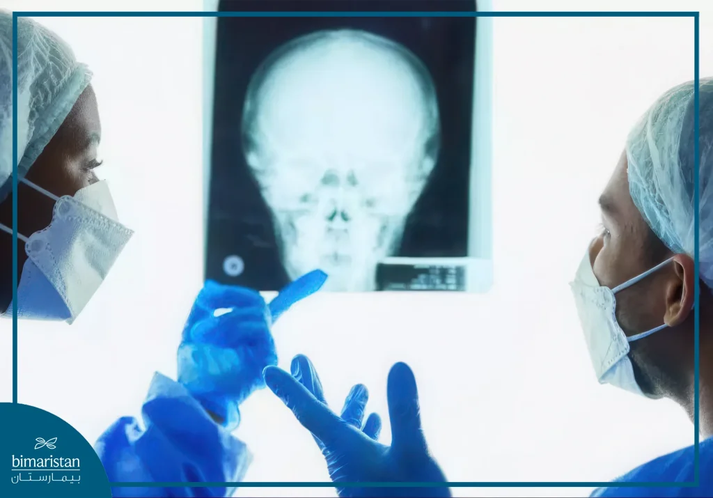
- Advanced imaging tests: Magnetic resonance imaging (MRI), computed tomography (CT), and sometimes angiography are used to determine the exact location of the tumor and its relationship to sensitive structures, including nerves and vessels.
- 3D surgical planning (when available): Artificial intelligence or 3D printing technologies can be used to simulate surgery and determine the optimal route.
- Anesthetic preparation: The patient is evaluated by an anesthesiologist to determine a safe plan during the procedure.
- Psychological preparation and family support: Education is provided to the patient and their family about the course of the surgery, the expected risks, and the support needed.

Performing skull base surgery (steps in order):
The surgery is performed in a structured and systematic manner based on the technique used: endoscopic, open, or combined.
- Patient positioning and stabilization: The patient is positioned on the operating table in a manner that provides optimal access to the surgical site.
- Surgical access is initiated: The incision is made according to the chosen route (nasal, oral, or transcranial).
- Navigating through deep structures with precision: Using a surgical microscope or endoscope, the surgeon carefully advances through the tissue to reach the tumor or lesion.
- Removing the tumor or treating the condition: The tumor or lesion is removed using the most precise instruments while preserving nearby nerves and arteries as intact as possible.
- Intraoperative neuromonitoring (when available): Neural activity is monitored during the procedure to avoid injury to vital structures.
- Closing the wound and reconstructing the area (if necessary): Tissue grafts or stents may be placed to reconstruct the base of the skull.
- Transfer the patient to the neurointensive care unit: Where the patient’s condition is continuously monitored after surgery.
Recovery and post-surgery
Length of hospitalization
The length of hospitalization after surgery depends on the type and complexity of the surgical intervention. In cases of endoscopic surgery, patients can typically leave the hospital in 2 to 4 days. In contrast, open surgeries or those involving sensitive areas, such as the cranial nerves or brain stem, may require a recovery period of one week or more, allowing for close monitoring and management of potential complications.
The importance of physical therapy and neurological follow-up
Neurological and physical rehabilitation therapy plays a crucial role in enhancing the quality of life after surgery, particularly when motor function, speech, or vision are impaired. It includes:
- Balance and mobility training to prevent falls
- Speech and swallowing therapy if the facial or cranial nerve is affected
- Exercises to improve muscle function in the face or neck. It is also recommended that you follow up with your neurologist periodically to assess improvement and detect any early signs of complications
Post-operative instructions and return to normal activity
After leaving the hospital, the patient receives careful instructions, including:
- Rest at home for a sufficient amount of time and avoid excessive exertion
- Watch for any unusual symptoms, such as severe headaches, nasal discharge, or a change in vision
- Avoid bending or lifting heavy weights for the first two to four weeks
- Consult your doctor to monitor your healing progress and to have stitches removed or bandages changed as needed
- Gradual return to work or study as assessed by your doctor
Possible need for radiation or chemotherapy in cases of cancer
If the tumor is malignant or the surgeon is unable to remove it completely, complementary procedures may be recommended, such as
- Conventional radiation therapy or stereotactic radiosurgery (SRS) to target tumor remnants.
- Chemotherapy in selected rare cases (e.g. in glioblastomas or tumors with high metastatic potential).
- Proton therapy is a precise option for tumors near sensitive nerve structures.
What are the risks of skull base surgery?
Although skull base surgery is a very effective treatment option, it does come with some risks, which include:
- Infection: As with any surgery, there is a risk of infection.
- Bleeding: Bleeding can occur in or around the brain, especially in delicate surgeries.
- Nerve damage: Damage to the nerves that control vision, hearing, speech, or motor skills may occur despite efforts to minimize it.
- Cerebrospinal fluid leakage: In rare cases, cerebrospinal fluid leakage may occur after surgery.
Generally, the risks are low, especially when surgery is performed by experienced neurosurgeons who have specialized training in skull base surgery.
In conclusion, skull base surgery represents a complicated integration of medical precision, advanced technology, and surgical expertise. Despite the anatomical and technical complexities of this critical region, innovations in imaging, microsurgical techniques, and minimally invasive approaches, such as nasal endoscopy, have enabled safer and more effective treatment for a wide range of benign and malignant conditions. The success of skull base surgery relies not only on the surgeon’s skill but also on multidisciplinary collaboration, comprehensive rehabilitative care, and careful postoperative monitoring. With the continued advancement of artificial intelligence and 3D modeling, the future of skull base surgery holds promise for even greater accuracy in surgical planning and improved patient outcomes.
Sources:
- NPİSTANBUL. (n.d.). Skull base surgery
- UCSF Brain Tumor Center. (n.d.). Minimally invasive skull base surgery

