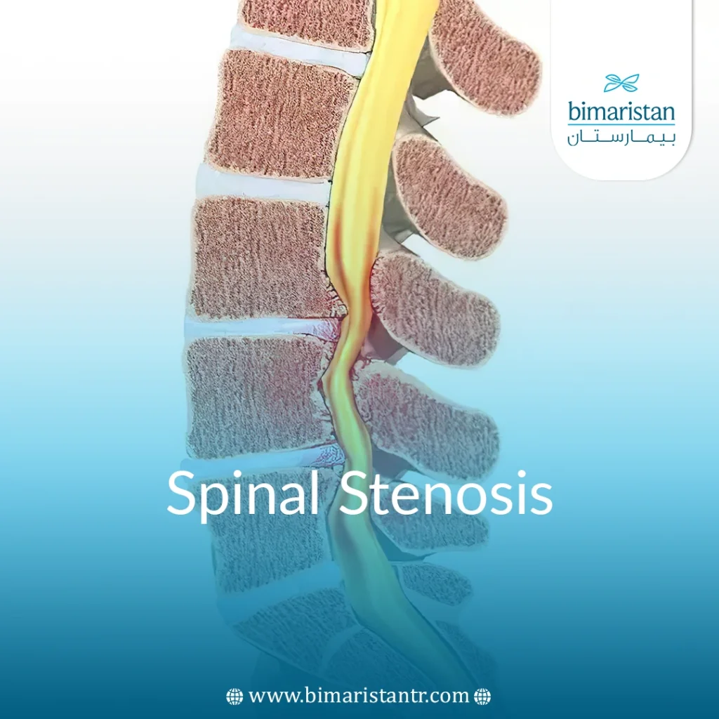
Spinal stenosis is one of the most common spinal diseases. Types of spinal stenosis are classified according to where they occur. Lumbar stenosis is a narrowing of the lumbar spinal canal at the bottom of the spine. In this article, you will learn about spinal stenosis, its types, symptoms, and treatment in Turkey.
Spinal stenosis (a narrowing of the space through which the spinal cord passes through the spine) puts pressure on the spinal cord and nerve roots that emerge from each vertebra.
Symptoms caused by spinal stenosis include pain in the arms or legs/tingling and numbness and, muscle weakness / lower back and neck pain.
Many treatments are available for this condition, including physical therapy, medication, and surgery.
What is spinal stenosis?
Spinal stenosis is narrowing one or more vertebral foramina within the spine. This narrowing puts pressure on the spinal cord (spinal cord) and nerves.
The most common causes of spinal stenosis are osteoporosis and vertebrae degeneration that occurs naturally with age.
Types of stenosis: Spinal stenosis can occur anywhere along the spine but is most common in the lower back (lumbar) or cervical spine.
The condition usually develops slowly over time, which is why many people may not experience symptoms of spinal stenosis despite changes visible on X-rays.
Depending on the location and severity of the spinal stenosis, neurological symptoms can range from pain, numbness, tingling, and muscle weakness in the neck and/or lower back to weakness in the arms and legs.
What are the symptoms of spinal stenosis?
Spinal stenosis is diagnosed by MRI or CT scan, and spinal stenosis may be diagnosed despite the absence of symptoms.
Although any part of the spine can be affected, spinal stenosis in the lower back and neck is the most common.
The type of stenosis, the location of the stenosis, and many other factors determine the appearance of symptoms.
Symptoms of spinal stenosis in the neck (cervical spine)
- Numbness or tingling in the hand, arm, foot, or leg (anywhere below the point of stenosis).
- Weakness in the hand (clumsiness), arm, foot, or leg
- Loss of function (weakness) in the hands, such as having trouble writing or buttoning shirts.
- Walking and balance problems
- Neck pain
- In severe cases, inability to control the bowels and bladder.
- Some may experience dizziness and balance problems
Symptoms of lumbar spinal stenosis
- Numbness or tingling in the buttocks, foot, or leg
- Weakness (weakness) in the foot or lower leg (heavy legs).
- Feeling pain, cramps, or spasms in one or both legs when standing for long periods of time or when walking, which is relieved by bending forward, sitting, or walking uphill.
- Lower back pain: It is described as bouts of mild pain such as an electric or burning sensation.
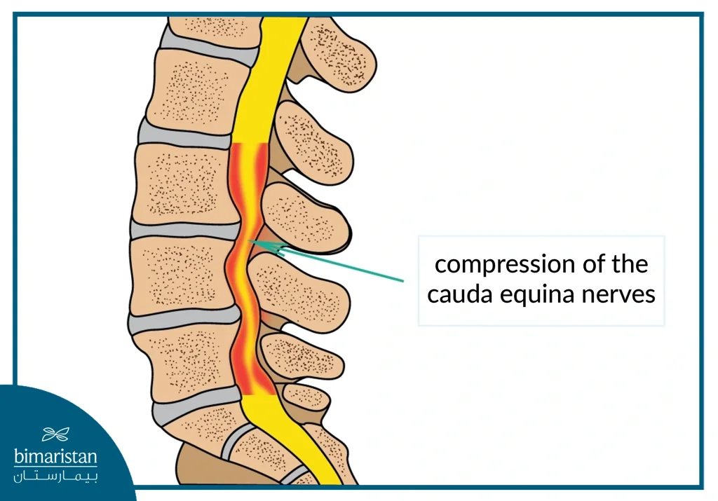
- Sciatica (sciatic nerve compression): Pressure on the sciatic nerve causes pain that starts in the buttocks and extends to the lower leg and may extend to the feet.
- Loss of bladder or bowel control in severe cases.
Spinal stenosis in the thoracic spine
- Numbness or tingling in the arm, hand, leg, or foot.
- Balance problems.
What are the causes of spinal stenosis?
The spine extends from the neck to the lower back.
The spinal cord is located inside the spinal canal in the spine, as the spine’s job is to protect the spinal cord and nerves from any shocks or bruises.
Spinal stenosis may be congenital, but it is often acquired.
Spinal stenosis has many causes, but the common denominator is the change it causes in the vertebrae. This change narrows the space around the spinal cord and nerves, putting pressure on them.
Some causes:
- Congenital spinal stenosis:
This is a condition in which a person is born with a small and narrow spinal canal. Some diseases of the spine may cause narrowing of the spinal canal, such as scoliosis.
- Bone overgrowth (joint spurs):
In chronic inflammatory conditions, erosion occurs in the articular cartilage that covers the bony surfaces in the joints.
Cartilage erosion causes the bones to rub against each other, and the body responds by forming a new bony protrusion.
The protrusions commonly occur, extending from the vertebrae into the spinal canal and thus pressing on the spinal cord.
Paget’s disease, which also affects bones, can cause an increase in bony growth in the spine and narrowing of the nerves.
Between the vertebrae are flat, round cushions (intervertebral discs) that absorb shocks from the spine.
As we age, the outer edge of these discs may tear, and a gel-like substance may come out of them, pressing on the spinal cord (spinal cord) and narrowing the spinal canal.
- Thick ligaments:
These are fibrous bands that hold the spine together. Arthritis leads to the thickening of the ligaments and the narrowing of the spinal canal.
- Meningioma or spinal cord tumors:
This is an uncommon condition diagnosed by radiography. As it grows, it presses on the spinal cord and nerves, causing narrowing of the spinal canal.
- Vertebral injuries and fractures:
Traumas or accidents can cause dislocations or fractures of the vertebrae, changing their position and pressing on the spinal cord and nerve roots, narrowing the spinal canal.
Risk factors for spinal stenosis
- Spinal stenosis can affect anyone, but it is most common at the age of fifty.
- Scoliosis or vertebral fractures.
Complications of spinal stenosis
If the condition worsens and is neglected, spinal stenosis may lead to the following:
- Numbness and muscle weakness
- Imbalance
- Urinary incontinence
- Paralyze
Diagnosis of spinal stenosis
To diagnose stenosis, the doctor asks about the symptoms, details the patient’s medical history, and performs a complete neurological examination. An accurate diagnosis may require several X-rays.
Imaging tests to diagnose spinal stenosis may include:
- X-rays: X-rays show bone spurs that may be pressing on the spinal cord and nerve roots.
- Magnetic resonance imaging (MRI): An MRI produces detailed images of the spinal cord and nerve roots and can detect any tumors.
- Computed tomography (CT) scan or myelography: Your doctor may recommend a CT scan, which is a procedure that images the spinal cord and nerves and may show herniated discs, bone spurs, and tumors.
Spinal stenosis treatments
The choice of treatment for stenosis depends on the cause of the symptoms, the location of the stenosis, and the severity of the signs and symptoms. If your symptoms are mild, your doctor may recommend home remedies first. If these treatments don’t work and your condition worsens, your neurologist may recommend physical therapy, medications, and finally surgery.
Home treatments for spinal stenosis
Heat
Heat is usually the best option for treating stenosis caused by osteoporosis. It increases blood flow, relaxes muscles, and relieves joint pain. However, be careful when using heat—don’t set the settings too high, or you may get burned.
Cold compresses
If heat doesn’t relieve symptoms, try cold compresses (an ice pack, a frozen gel pack, or a frozen bag of peas or corn). Ice is usually applied for 20 minutes on and 20 minutes off. Ice reduces swelling and inflammation.
Exercises
Exercise helps relieve pain and strengthen muscles to support the spine and improve flexibility and balance. These exercises are done under the supervision of a specialist.
Non-surgical treatments for spinal stenosis
Medications
Non-steroidal anti-inflammatory drugs (NSAIDs) such as ibuprofen and naproxen can help reduce inflammation and relieve pain caused by spinal stenosis. Do not take these medications randomly. Talk to your doctor about them. Random use may put you at risk for stomach ulcers.
Pain-relieving medications such as tricyclic antidepressants such as amitriptyline are recommended. Opioids, such as oxycodone (Oxycontin®) or hydrocodone (Vicodin®), may be prescribed for short-term pain relief. However, they are usually prescribed with caution because they can be addictive. Muscle relaxants can relieve muscle tension and spasms.
Physiotherapy
A patient with spinal stenosis may avoid movement to relieve pain, which, in the long term, leads to muscle atrophy and increased pain. Physical therapists will give each patient a special exercise program for the back to help the patient gain strength and improve balance, flexibility, endurance, and spinal stability. Strengthening the back and abdominal muscles will make the spine more flexible.
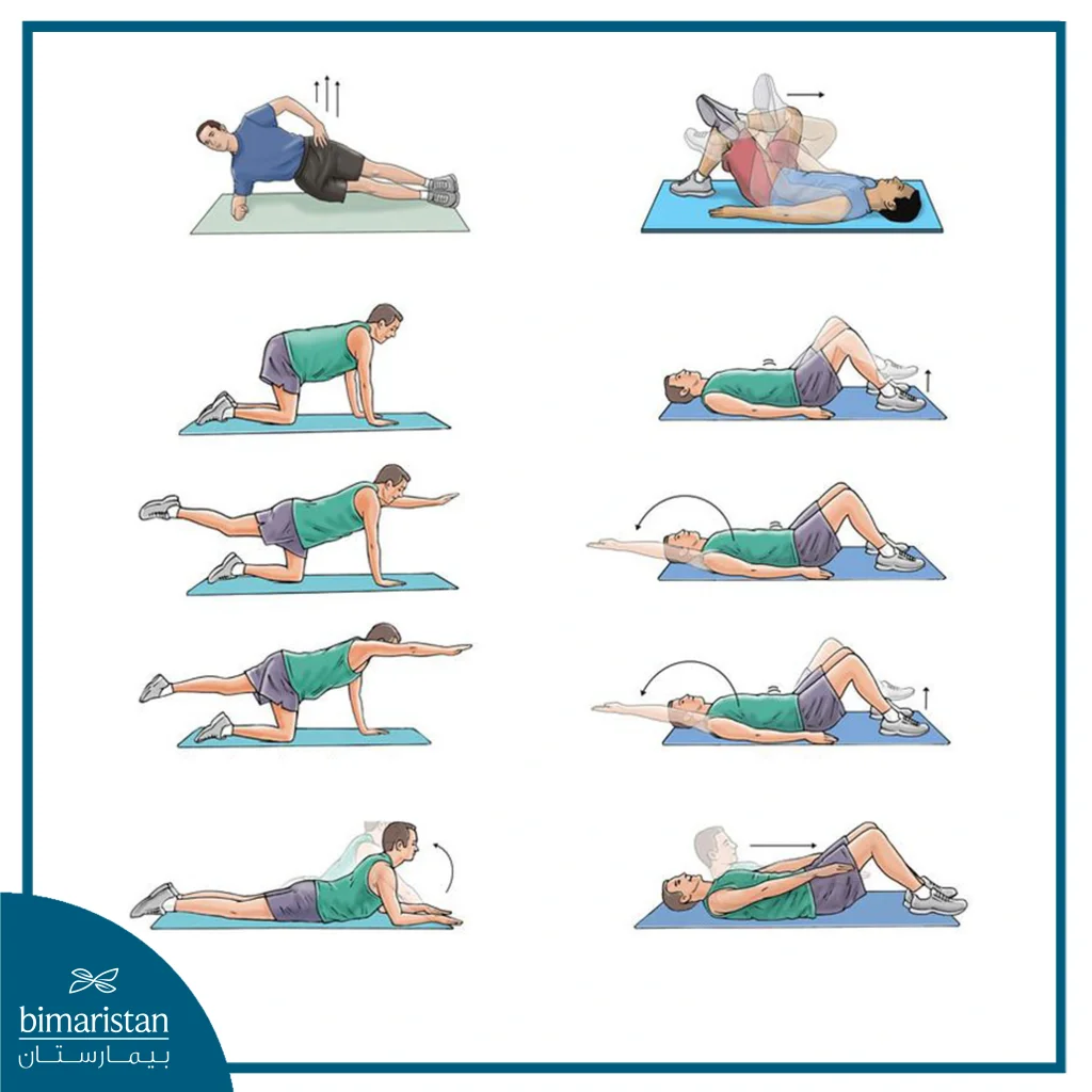
Physical therapists can teach you how to walk in a way that relieves spinal stenosis, which in turn relieves pressure on the nerves.
Steroid injections
Injecting corticosteroids near the narrowed spinal canal can help reduce inflammation and swelling of the surrounding nerve roots. However, because corticosteroids can cause damage, they are not recommended more than three or four times a year.
Decompression procedure
This procedure, also known as image-guided lumbar decompression (PILD), specifically treats lumbar spinal stenosis caused by the thickening of the ligaments (ligamentum flavum) in the back of the spine. It is performed through a small incision and does not require general anesthesia or stitches. X-rays guide the procedure. The surgeon uses special tools to remove part of the enlarged ligament and widen the narrowed spinal canal, which expands the space within the spinal canal and reduces pressure on the nerve roots.
The advantages of this procedure are that the bone structure of the spine is left intact, it is minimally invasive, and patients recover quickly. Patients usually go home within a few hours of the procedure and begin walking and/or physical therapy shortly thereafter. Compared to before the procedure, they will be able to walk and stand for longer periods of time and will have less numbness or tingling sensations and muscle weakness.
Surgery
Surgery is used when all other treatment options have failed. Fortunately, most people with spinal stenosis do not need surgery. However, your doctor may recommend surgery if your signs and symptoms are very severe and are interfering with daily life, such as difficulty with balance and walking due to pressure on the spinal cord and nerves. Surgery aims to relieve the narrowing of the spinal canal and free up the nerves.
Surgery options include removing parts of the vertebrae, spurs, or discs that are narrowing the spinal canal and compressing the spinal cord (spinal cord). Spinal stenosis surgeries include:
Laminectomy
The most common type of spinal stenosis surgery involves removing the lamina, the back portion of the vertebra. The bone spurs and surrounding tissue can also be removed, which relieves the narrowing of the spinal canal and makes room for the spinal cord and nerves. Doctors may need to connect the vertebra to adjacent vertebrae with bone grafts or metal hardware.
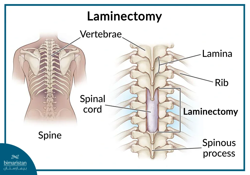
Laminotomy (partial laminectomy)
In laminotomy, only the portions of the lamina that are pressing on the spinal cord are removed.
Laminoplasty (for cervical spinal stenosis)
Laminoplasty is performed to treat stenosis in the neck only. The spinal canal is widened by removing the lamina of several vertebrae in a row and using metal plates and screws as a hinged bridge across the vertebrae.
Vertebral puncture
The foramen is the space inside the vertebrae where nerve roots exit. Vertebrae removal involves removing bone and tissue protrusions to create space and relieve pressure on the spinal cord and nerves.
Minimally invasive surgery for spinal stenosis
The protrusions or lamina are removed conservatively, minimizing harm to nearby healthy structures and tissues. This procedure is performed under local anesthesia. It is considered one of the most important types of stenosis surgeries for people with chronic diseases who cannot tolerate general anesthesia.
Vertebral fusion
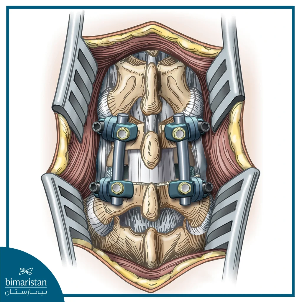
This is a complex procedure based on the principle of permanently fusing (connecting) two consecutive vertebrae together. Laminectomy is usually performed first, and bone is removed during this procedure to create a bridge between two vertebrae.
The vertebrae are held together with screws or wires until they heal and grow together. The healing process takes six months to one year. It helps stabilize the spine and relieve pain.
What are the risks of spinal stenosis surgery?
Surgery carries a number of risks, including infection, bleeding, blood clots, and reactions to anesthesia. Some complications from spinal stenosis surgery include:
- Nerve injury
- Tearing of the meninges (the membrane that covers the spinal cord)
- Failure of the bone to heal after surgery
- Failure of metal plates, screws, and other fixators
- Need for additional surgery
- Relapse of symptoms
How long does it take to recover from spinal stenosis surgery?
Full recovery from spinal stenosis surgery and return to normal activities usually takes three months. It may take longer for spinal fusion, depending in part on the complexity of the surgery and the progress of your rehabilitation.
If you had a laminectomy, you will likely be able to return to work in an office job within a few days of returning home.
If you had spinal fusion surgery, you will likely be able to return to work a few weeks after surgery.
Can spinal stenosis be prevented?
Spinal stenosis cannot be prevented 100%. However, you can take some measures to reduce the risk or slow down the progression, including Maintaining an ideal body weight, avoiding smoking, and adhering to the exercises prescribed by the physical therapist, which is an important condition to relieve pressure on the spine.
Bimaristan Medical Center is your first choice for treatment in Turkey. We direct you to the best neurologists in Turkey. Do not hesitate to contact us; Bimaristan Center is your family in Turkey.
Sources:
- NHS
- Johns Hopkins Medicine
- NIH
