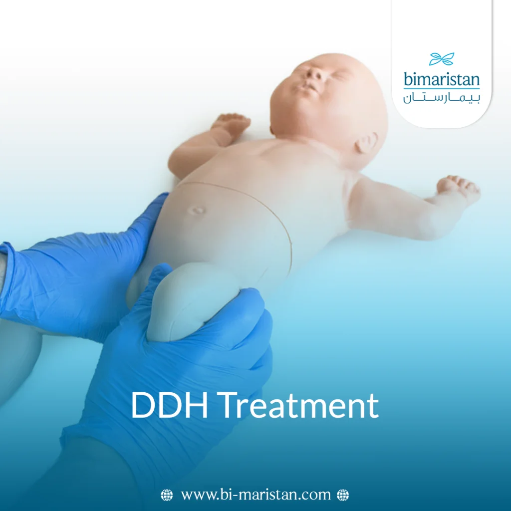
Congenital hip dislocation is a relatively common condition in pediatric orthopedics, a congenital disorder affecting the hip joint from birth, in which the head of the femur does not sit properly within the acetabulum of the pelvis, leading to joint instability and subsequently affecting the child’s motor development. The severity of the condition ranges from mild instability to complete dislocation; therefore, early diagnosis is crucial for improving outcomes and minimizing future complications that could permanently affect gait and joint function. It should be noted that this condition may not be apparent in the first days of life, making regular medical follow-up essential for early detection and optimal DDH treatment.
Turkey is gaining significant attention in DDH treatment as it has become a preferred medical destination for many patients thanks to major advancements in pediatric orthopedics and medical rehabilitation, as well as the availability of specialized centers and high-level medical expertise. Many parents are seeking safe and effective treatment options in Turkey because of the modern techniques and advanced medical care it offers, which ensure better outcomes for affected children, with a focus on improving quality of life and gradually and safely restoring the joint’s normal function.
What is congenital hip dislocation?
Congenital hip dislocation is a birth defect affecting the hip joint in newborns, in which the head of the femur is not properly seated within the acetabulum. This condition leads to varying degrees of joint instability, ranging from mild ligament laxity to partial or complete dislocation. It often does not cause obvious pain in the first few months of a child’s life, so it may not be detected until a routine examination or when a delay in motor development is noticed later on.
The importance of understanding this condition lies in the fact that, at this age, the hip is still growing and developing, making early intervention essential to ensuring the joint’s normal development. If the condition is not addressed in a timely manner, it may lead to gait abnormalities, shortening of the affected limb, and future functional problems. Therefore, early recognition of congenital hip dislocation is an important step in avoiding complications and achieving the best possible treatment outcomes.
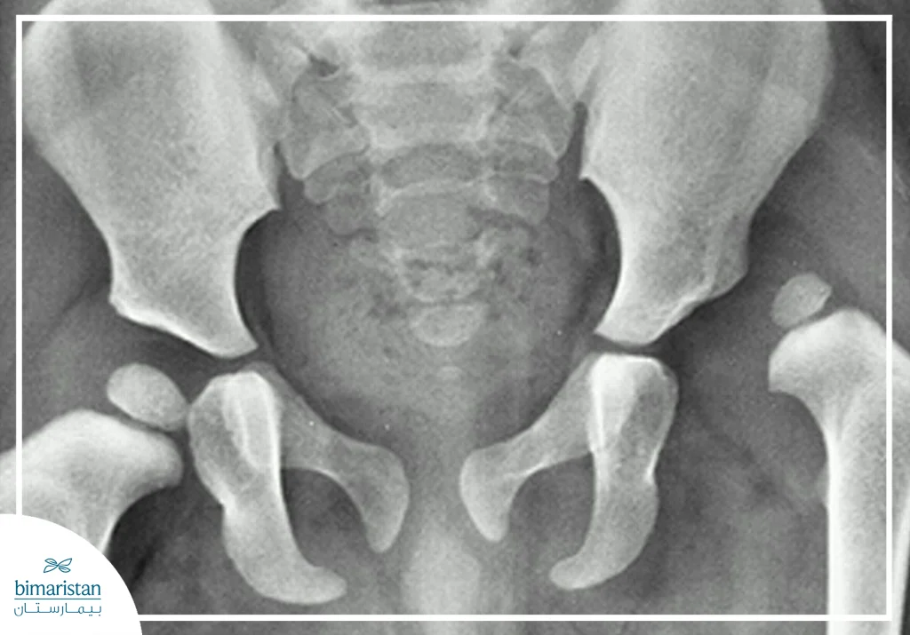
Grades of Congenital Hip Dislocation
Congenital hip dislocation is classified into grades based on the severity of hip joint instability and the extent of femoral head displacement from the acetabulum. These grades include:
- Incomplete formation: The joint cavity is insufficiently developed, and the femoral head is not firmly stabilized within the joint, although there is no obvious dislocation.
- Partial dislocation: The head of the femur is loosely situated within the acetabulum and can easily move partially out of its normal position.
- Complete dislocation: The head of the femur is completely outside the acetabulum, with no normal contact between the bones; this is the most severe form of the condition and requires early medical intervention.
Causes of Congenital Hip Dislocation
The causes of congenital hip dislocation vary between prenatal factors and those related to postnatal conditions, and include:
- Genetic factors: A family history of the condition increases the likelihood that a newborn will develop it.
- Breech presentation: When the fetus is in a breech position inside the uterus, putting pressure on the hip joint.
- Female gender: Women are more susceptible because their ligaments become more flexible under the influence of hormones.
- Low amniotic fluid: Reduces fetal movement in the uterus and affects normal joint development.
- Postnatal factors: such as wrapping the legs too tightly or adopting incorrect postures that restrict hip movement and affect hip stability
Symptoms of Congenital Hip Dislocation
The symptoms of congenital hip dislocation vary depending on the child’s age and the degree of joint instability, and include:
- Leg length discrepancy: where one leg appears shorter than the other.
- Asymmetry of skin folds: A noticeable difference in the thigh folds between the two sides.
- Difficulty moving the legs: The child resists or cries when the thighs are spread apart during a diaper change.
- A clicking sound in the joint: This may be heard when moving the hip during the examination.
- Delayed motor development: such as a delay in sitting, crawling, or walking in an older child.
- Limping while walking: This occurs in children after they begin walking and involves the body leaning to one side.

Diagnosis of Congenital Hip Dislocation
The diagnosis of congenital hip dislocation relies on a thorough clinical examination, along with imaging studies to confirm the condition and determine its severity. Diagnostic methods include:
- Clinical examination: The doctor moves the hip joints to assess for instability or dislocation, while noting external signs such as leg-length discrepancy or skin folds.
- Ortolani test: Used to reduce the head of the femur back into the acetabulum in cases of dislocation; the doctor feels a characteristic click.
- Echo: Used in young infants to assess joint structure and femoral head stability within the acetabulum.
- X-rays: These are typically obtained after 4–6 months of age to more clearly assess hip bone development.
DDH Treatment Methods
Treatment for congenital hip dislocation is essential to restoring the hip joint to its normal position and ensuring proper growth in the child. The treatment plan depends on the child’s age, the degree of dislocation, and when the condition was detected. The earlier the diagnosis, the higher the chances of success and the better the prospects for avoiding future complications. In general, treatment aims to stabilize the femoral head within the acetabulum, allowing for normal joint development and achieving natural movement throughout the various stages of growth.
The treatment method is carefully selected by a specialist physician on a case-by-case basis to ensure the best possible outcome, with a focus on restoring the natural balance between the hip bones during early growth stages, thereby reducing the need for surgical interventions later and improving long-term functional outcomes. The treatment methods are as follows:
From birth to 6 months of age
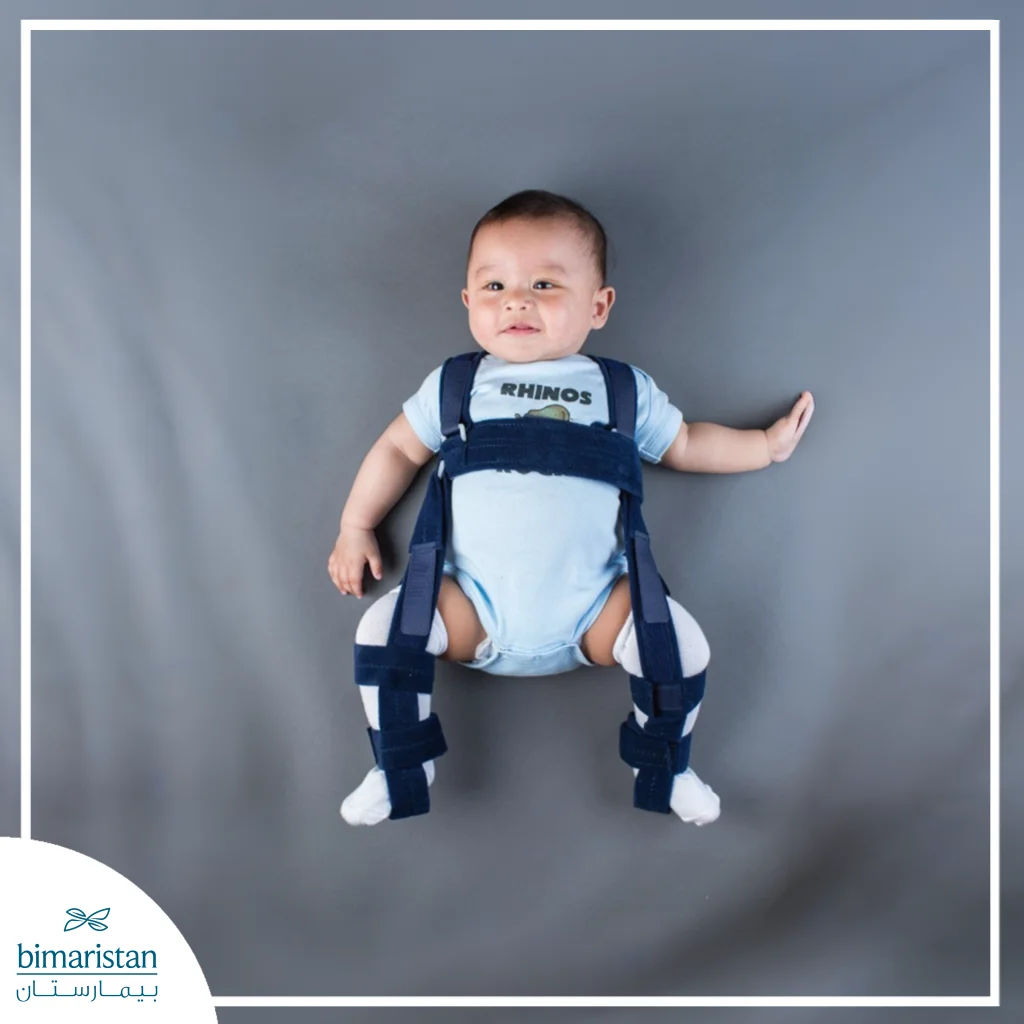
At this age, the hip joint is still highly flexible and malleable, so treatment often relies on a non-surgical approach using the Pavlik harness, a medical brace that stabilizes the hips in a position that helps keep the head of the femur consistently within the acetabulum, allowing the joint to develop normally over time.
This treatment is considered one of the most successful methods in early-stage cases, as the joint responds very quickly if medical instructions are followed. The device is used continuously throughout the day at first to ensure complete stabilization, then the number of hours is gradually reduced as the condition improves and the joint stabilizes, with ongoing medical follow-up required to ensure normal growth.
From 6 months to 1 year
If diagnosis is delayed or the condition does not respond to treatment with a brace, a more invasive treatment approach is used under general anesthesia, in which the doctor carefully repositions the head of the femur back into the socket without the need for open surgery, thereby helping to correct the joint’s anatomical alignment in a relatively safe manner.
The joint is then immobilized with a specialized cast designed to maintain proper bone alignment for an extended period, allowing for complete healing and stabilization. The immobilization period lasts several months, with the cast changed periodically according to the treatment plan to strengthen the joint and gradually improve the shape of the hip.
One year of age and older
At this stage, congenital hip dislocation is often more severe and persistent; therefore, surgical intervention becomes the primary treatment option, with the goal of restoring the hip joint to its normal position and achieving the greatest possible functional stability, while attempting to correct deformities resulting from delayed treatment.
The surgical procedure begins with the removal of any tissue or obstructions inside the joint that might prevent the femoral head from returning to the acetabulum. The joint is then immobilized using a special cast after the procedure. In some advanced cases, modifications may be made to the pelvic bones to improve coverage of the femoral head and increase the joint’s long-term stability.
Follow-up After Treatment for Congenital Hip Dislocation
Follow-up care after treatment for congenital hip dislocation is just as important as the treatment itself, as it aims to ensure that the hip joint remains stable and continues to develop normally. The doctor schedules periodic visits for the child to monitor joint movement and check the stability of the femoral head within the acetabulum, as well as to assess the gradual development of bone growth.
X-rays or ultrasounds are also used as needed to ensure the joint is intact and that there has been no recurrence of the dislocation. Follow-up typically continues for long periods, which may extend for several years depending on the severity of the condition, with the aim of early detection of any potential complications, such as poor acetabular coverage or limb length discrepancy, which allows for prompt and timely intervention and ensures the best long-term functional outcome for the child.
Why is Turkey a preferred choice for treating congenital hip dislocation?
Turkey is considered one of the leading medical destinations for the treatment of congenital hip dislocation thanks to several important factors, the most notable of which are:
- It offers state-of-the-art medical centers that utilize the latest technologies in diagnosis and treatment
- Highly experienced physicians specializing in pediatric orthopedic surgery
- Thorough post-treatment medical follow-up to ensure the best outcomes
- Lower costs compared to Europe and the United States, combined with high-quality medical care
- Provides comprehensive rehabilitation and physical therapy programs for children following treatment
- Ease of access to medical services and the speed with which appointments are scheduled compared to other countries
In conclusion, congenital hip dislocation is a condition that requires a high level of awareness on the part of parents and close medical follow-up from the first months of a child’s life, because early diagnosis is considered the most important factor in the success of treatment and in avoiding complications that may affect walking and the joint’s normal development. The earlier the condition is detected, the greater the chances of successfully repositioning the femoral head into the acetabulum, which supports healthy hip joint development and reduces the need for complex therapeutic interventions later on.
Furthermore, significant medical advances in pediatric orthopedic surgery have clearly improved treatment outcomes and increased recovery rates, especially in specialized centers that rely on modern techniques and precise treatment plans tailored to the child’s age and the severity of the condition. Turkey is considered a leading medical destination in this field due to the availability of medical expertise and advanced equipment that contribute to providing comprehensive and effective care. Therefore, committing to regular follow-up visits with the doctor and strictly adhering to treatment instructions remains the most important foundation for ensuring the normal and healthy development of the hip joint and achieving the best long-term outcomes.
Sources: