
Congenital Pneumothorax Surgery is an important medical procedure when air accumulates around a newborn’s lung, causing it to collapse and making it difficult for the lung to expand normally. Pneumothorax may occur immediately after birth, especially in premature infants or those requiring respiratory support, and may cause shortness of breath, low oxygen levels, and a rapid heart rate if severe.
The term “surgery” here does not always refer to a major operation; treatment often begins with observation, needle aspiration, or chest tube insertion, depending on the severity of the condition. The goal of treatment is to remove the accumulated air, re-expand the lung, and improve the child’s breathing as quickly as possible, with thoracoscopic or open surgery used only in rare cases where air leakage persists, or there is a concomitant lung problem.
What is congenital pneumothorax?
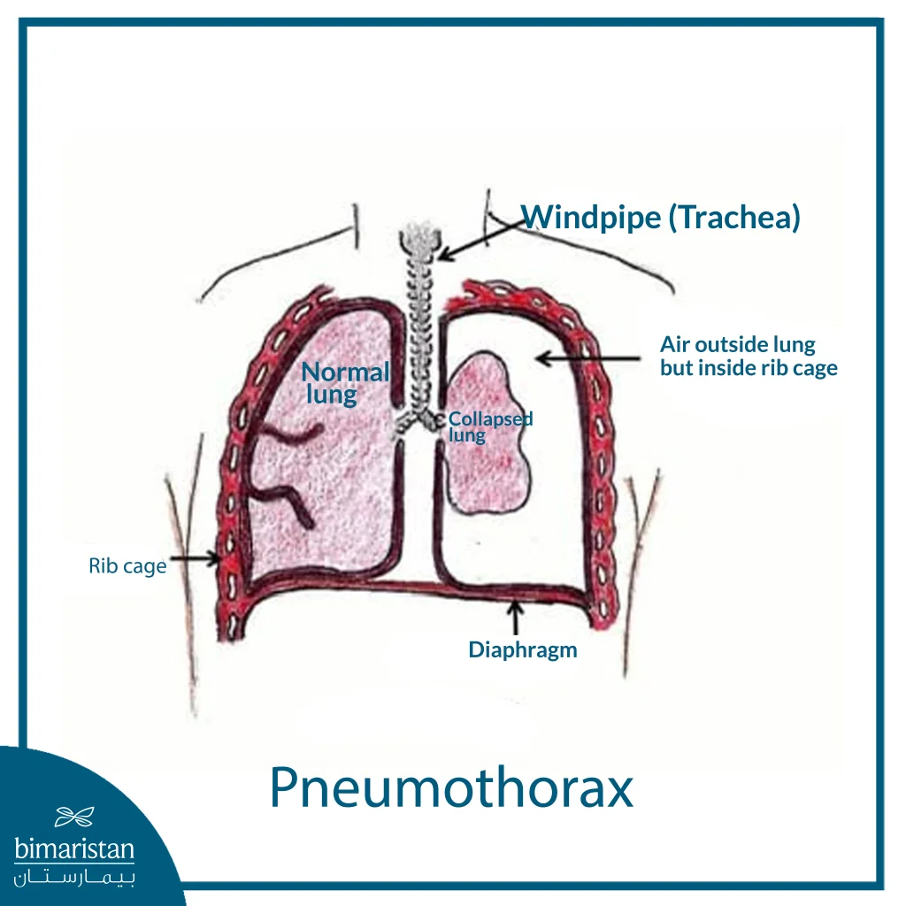
Congenital pneumothorax is the accumulation of air in the space between the lung and the chest wall at birth, which puts pressure on the lung and prevents it from expanding normally during breathing. It may occur immediately after birth or within the first few hours of life and is more common in premature infants or in infants who require mechanical ventilation or other respiratory support.
The severity of the condition varies from child to child; the pneumothorax may be small and cause no obvious symptoms, or it may be severe and lead to difficulty breathing, cyanosis, low oxygen levels, and a rapid heart rate. In severe cases, it may progress to tension pneumothorax, a medical emergency that requires rapid air removal to relieve pressure on the lungs and heart.
When does a newborn need surgical intervention?
A newborn requires intervention when chest retractions are severe or interfere with breathing, especially if signs such as severe difficulty breathing, low oxygen levels, cyanosis, a rapid heart rate, or reduced lung movement on the affected side. In these cases, monitoring alone is not sufficient, and air must be removed to relieve pressure on the lung and help it re-expand.
Intervention is most urgent in cases of tension pneumothorax, a medical emergency that can affect the heart and circulatory system if not treated promptly. Treatment usually begins with needle aspiration or the insertion of a chest tube, while endoscopic or open surgery is rarely used when air leakage persists, the chest tube fails, or there is a congenital lung defect that requires repair.
Diagnosis of Pneumothorax in Newborns
The diagnosis of pneumothorax in newborns relies on a combination of clinical signs and imaging studies, as the symptoms may resemble those of other respiratory disorders such as respiratory distress syndrome or neonatal pneumonia. The goal of diagnosis is to confirm the presence of air around the lung, estimate its volume, and determine whether it is causing dangerous pressure that requires immediate intervention.
Clinical Examination and Respiratory Monitoring
The doctor begins by immediately assessing the child’s condition, monitoring breathing patterns, skin color, oxygen levels, and heart rate. The doctor may notice rapid breathing, retractions between the ribs, bluish discoloration around the mouth or extremities, or weaker movement on one side of the chest compared to the other.
When listening to the chest with a stethoscope, breath sounds may be weaker on the affected side due to lung compression. In severe cases, signs of instability may appear, such as low oxygen levels despite respiratory support or sudden deterioration in a child on a ventilator.
Chest X-ray
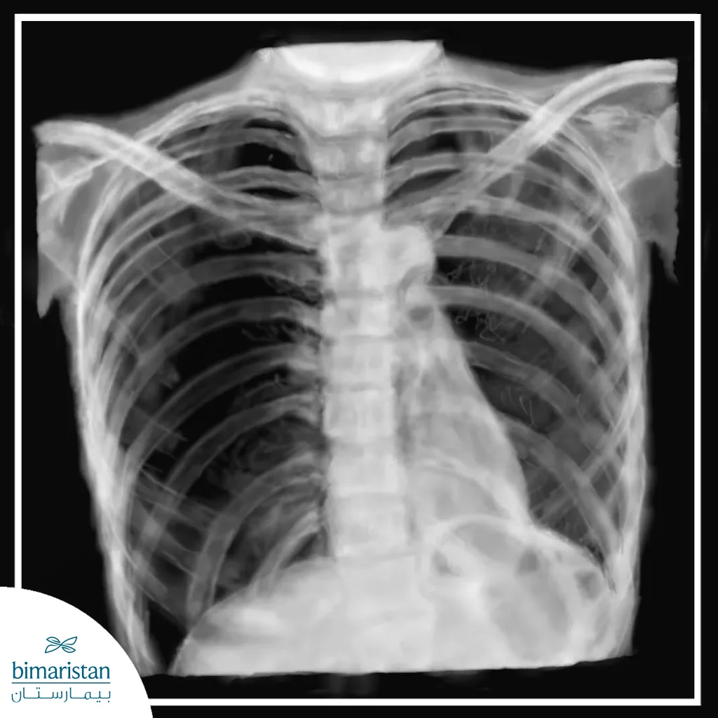
A plain chest X-ray is one of the most important tools for confirming the diagnosis, as it shows air outside the lungs within the pleural space. The X-ray also helps determine the affected side and the size of the pneumothorax, as well as whether the lung is mildly or severely compressed.
In some cases, the image may show displacement of the heart and midline toward the opposite side, which indicates a compressive pneumothorax requiring emergency treatment. A chest X-ray also helps rule out other causes of breathing difficulties in newborns.
Trans-thoracic Illumination Test
A doctor may use a chest transillumination test on some newborns, especially in intensive care units. This is done by shining a bright light on the chest wall; the affected side may appear brighter due to air around the lung.
This test is quick and useful in some cases, but it is no substitute for a chest X-ray once the child’s condition is stable enough to undergo imaging. Its primary benefit is that it provides a rapid assessment of the likelihood of a pneumothorax before deciding on a course of action.
Ultrasound
The use of lung ultrasound has become more common in neonatal units because it can be performed at the infant’s bedside without moving the baby. The ultrasound helps detect the presence of air around the lungs through specific signs that the doctor observes during the examination. This test has the advantage of not involving radiation, and it can be repeated as needed to monitor improvement following treatment. However, it requires considerable expertise in imaging newborns’ lungs and correctly interpreting the results.
Assessment of Disease Severity Before Treatment
After confirming the diagnosis, the doctor does not simply note the presence of a pneumothorax; rather, he or she assesses the extent to which it affects the child’s breathing and circulatory stability. A small pneumothorax may require only observation, while a large or compressive pneumothorax requires rapid air drainage. This assessment helps determine the appropriate course of action, whether it involves monitoring, administering oxygen, needle aspiration, inserting a chest tube, or considering a rare surgical intervention if the air leak persists.
Steps for Treating Congenital Pneumothorax
The treatment steps for congenital pneumothorax vary depending on the extent of air accumulation around the lung, the infant’s respiratory status, and the presence of signs of pressure on the heart or lungs. Therefore, not all cases are treated the same way; in mild cases, the doctor may simply monitor the child, while severe pneumothorax requires rapid air drainage or the insertion of a chest tube.
Monitoring and Oxygen in Mild Cases
In mild cases where the pneumothorax is minor and does not cause a noticeable deterioration in breathing or oxygenation, the doctor may choose to monitor the infant in the neonatal unit without immediate intervention. Monitoring includes measuring oxygen levels, tracking respiratory rate, observing skin color, and assessing the baby’s effort during breathing.
The child may be given oxygen to help breathing and improve blood oxygen saturation, with repeated clinical examinations or imaging as needed to ensure the pneumothorax does not increase in size. If the child’s condition remains stable and the air is gradually absorbed, no further intervention is necessary.
Fine Needle Aspiration in Emergency Situations
Fine needle aspiration is used when the pneumothorax is severe or constricting, especially if there is a rapid deterioration in the child’s breathing or a marked drop in oxygen levels. In this case, the accumulated air is putting pressure on the lung and may also affect the heart and circulation, so the child needs immediate intervention.
The doctor inserts a needle or a thin catheter through the chest wall on the affected side to allow trapped air to escape. This procedure helps quickly relieve pressure and improve lung expansion, but it may be a temporary measure before a chest tube is inserted if the air leak continues or the pneumothorax remains large.
Insertion of a chest tube for air drainage
The insertion of a chest tube is one of the most important steps in treating neonatal pneumothorax when observation or needle aspiration is insufficient. A thin tube is inserted between the ribs into the pleural space and then connected to a drainage device that allows air to escape and prevents it from re-entering the chest.
The chest tube helps maintain airflow until the lung returns to its normal expanded state and the leak stops. During this time, the medical team monitors the child’s breathing, the volume of air exiting the tube, the oxygen level, and the chest X-ray. Once the condition improves and no new leaks are detected, the tube can be removed while continuing to monitor the child for an appropriate period of time.
Laparoscopic or open surgery in rare cases
Endoscopic or open surgery is not routinely used to treat congenital pneumothorax, as most cases improve with observation, air drainage, or the placement of a chest tube. However, the child may need surgery if the air leak persists for several days, if the lung does not expand despite adequate drainage, or if a congenital lung problem is found, such as a pulmonary cyst or a malformation that causes recurrent pneumothorax.
In thoracoscopic surgery, the surgeon attempts to reach the source of the air leak through small incisions in the chest, while open surgery may be used in complex cases or when thoracoscopy is not appropriate. The goal of the surgery is to stop the air leak, treat the underlying cause, and help the lung return to normal function as safely as possible.
Recovery After Treatment for Congenital Pneumothorax
Recovery after treatment for neonatal pneumothorax depends on the severity of the condition and the treatment method used. Infants with a small pneumothorax may improve within a short period with observation and oxygen alone, while infants who have undergone air evacuation or chest tube placement require closer monitoring in the neonatal unit until the lungs stabilize and the air leak resolves. After treatment, the medical team monitors several key factors to ensure the infant’s improvement, the most important of which are:
- Stabilization of the oxygen level in the blood
- Improvement in the child’s breathing rate and effort while breathing
- Restoration of lung expansion in the chest X-ray
- Air is no longer escaping through the chest tube
- A child’s ability to breastfeed or be fed in a manner appropriate for their age
- No signs of return of spontaneous breathing after tube removal
If a chest tube is inserted, it is usually removed only after confirming that the air leak has stopped and the chest X-ray has improved. After the tube is removed, the child continues to be monitored for a period of time to ensure that air does not accumulate again. However, if the child has a concomitant lung disease or is a preterm infant, the hospital stay may be prolonged because recovery depends not only on the drainage of the air but also on the overall condition of the lungs and the child’s ability to breathe without significant support.
Congenital pneumothorax surgery is a rare therapeutic procedure, as most cases improve with observation, oxygen therapy, air drainage, or chest tube placement, depending on the severity of the condition. The success of treatment depends on rapid diagnosis, assessment of lung and respiratory function, and selection of the appropriate intervention before pressure on the lungs and heart worsens. The Bimaristan Medical Center helps guide families toward appropriate treatment options for newborns in collaboration with specialized medical teams.
Sources: