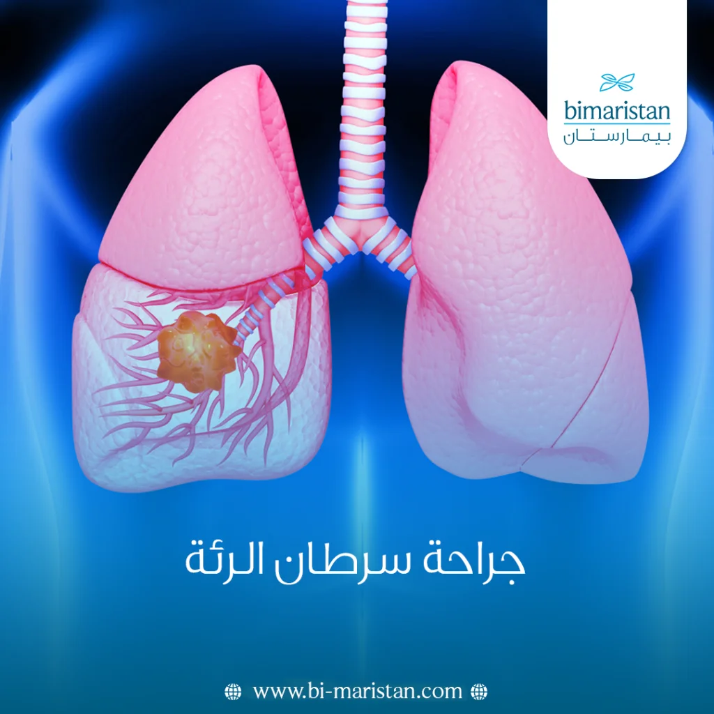
Lung cancer surgery, or surgical resection of lung cancer, is considered the optimal treatment option for lung cancer in its first three stages, when the tumor is of an appropriate size, has not deeply invaded other tissues, and there are no distant metastases. Unfortunately, however, most patients diagnosed with lung cancer are in the late stages of the disease, making it difficult to completely remove the tumor.
The percentage of lung cancers that can be resected at diagnosis is only about 25 to 30 percent in non-small-cell lung cancer, while this percentage is much lower in small-cell lung cancer due to its central location around the major arteries.
How is lung cancer surgery performed?
There are two main methods for removing lung cancer, and the choice of method depends on several factors, including the tumor’s location, size, and stage, as well as the surgeon’s experience and their awareness of the latest medical studies and developments.
Open-chest surgery (Thoracotomy)
Also known as open or conventional surgery, this procedure is performed through a transverse incision on the side of the rib cage. This method requires cutting through some of the chest wall muscles and using a special instrument to create a space between two ribs to allow the surgeon access to the lung. When the incision is closed, the muscles that were cut during the procedure are sutured back together.
This procedure is performed on very large masses that cannot be removed through small incisions; it is also still occasionally used in some countries that are not scientifically advanced.
Minimally invasive surgery
This surgery is usually performed through several small incisions, ranging from 1 to 4, to access the lung. It is divided into two parts:
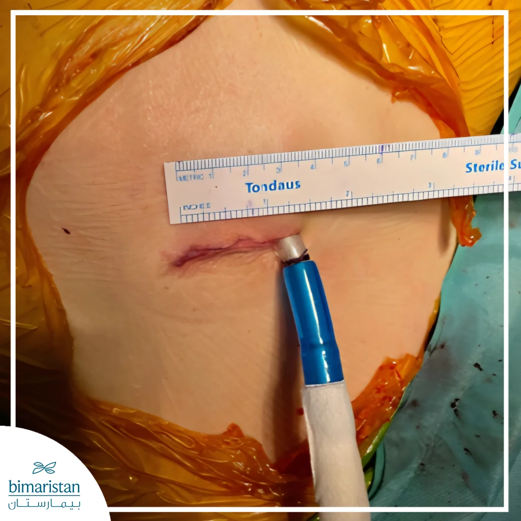
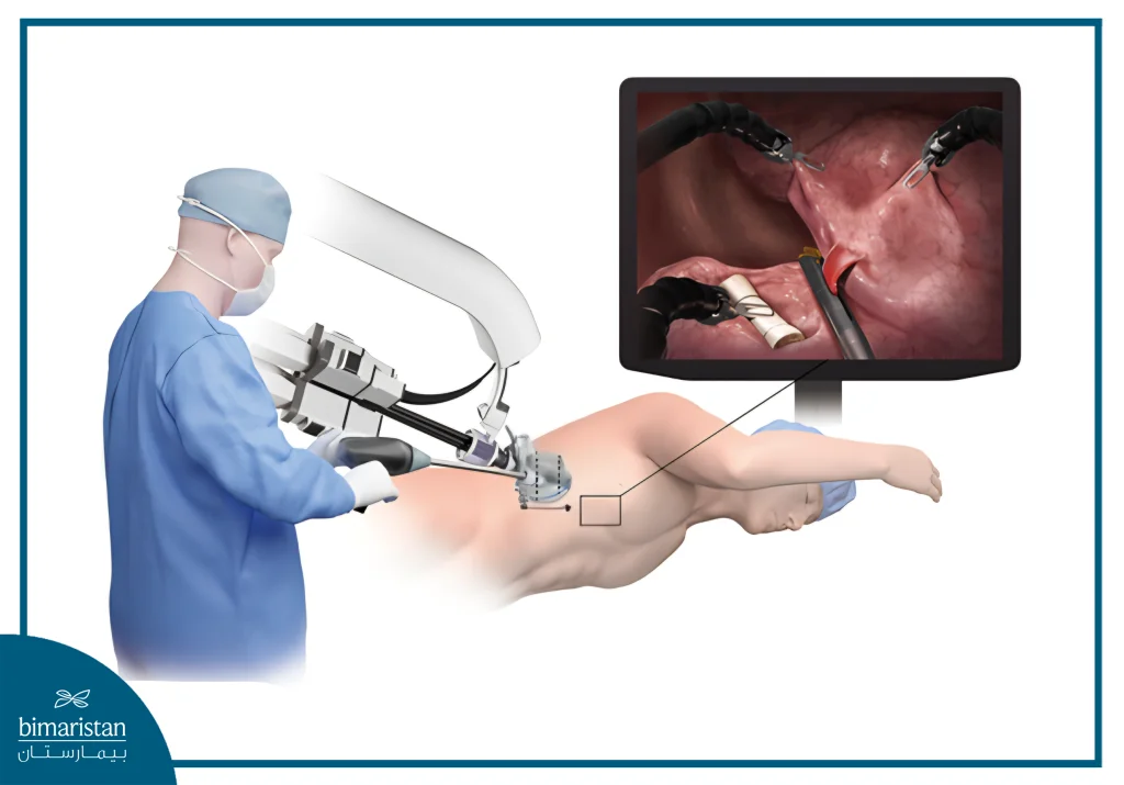
- Video-ِAssisted Thoracoscopic Surgery (VATS): The surgeon uses a camera to view the lung and special instruments to perform the surgery. Currently, this procedure is performed through a single surgical incision measuring only 3 to 4 cm in length.
- Robotic thoracic surgery: The first robotic lobectomy was performed in 2001, and the number has since increased significantly as the robot has developed and surgeons have adapted to it2.
Benefits
- Less surgical intervention: Because the incisions are very small, there is less damage to the patient.
- Faster recovery: Patients are often discharged from the hospital sooner than with open surgery.
- Less pain: Smaller incisions result in less postoperative pain. Larger incisions in thoracotomy involve cutting more types of tissue, such as muscles, nerves, and bones.
- Fewer complications: Because the camera magnifies the view, there is significantly less bleeding, and the smaller incision leads to less infection and therefore fewer complications.
Special technique: Single-port thoracoscopy
Single-port (uniportal) Video-ِAssisted Thoracoscopic Surgery (VATS) is performed through a single small incision rather than the multiple incisions used in conventional VATS. By minimizing surgical trauma, this minimally invasive approach may reduce postoperative pain, improve patient comfort, speed recovery, and shorten hospital stay in appropriately selected patients.
Types of lung cancer resection surgeries
The type of lung cancer resection varies depending on several factors, including the size, location, and type of the tumor, as well as the patient’s overall condition, tolerance, lung capacity, and ability to withstand lung resection. Therefore, the decision is often made collectively by a team of doctors. The types of surgery are as follows:
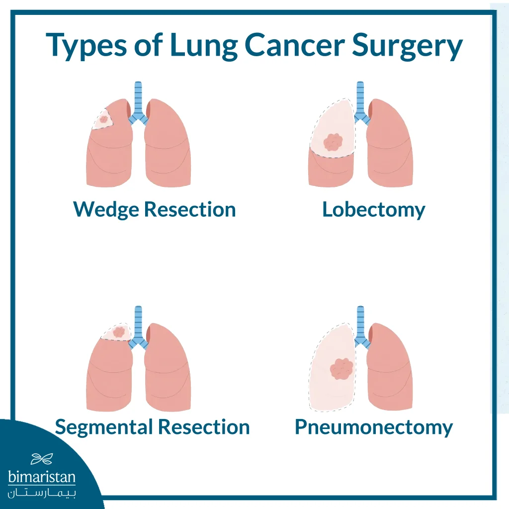
Lobectomy
In a healthy human body, the right lung has three lobes (the upper, middle, and lower lobes), while the left lung has two lobes (the upper and lower lobes).
Resection of the lung lobe affected by cancer is the most common method of treating lung cancer.
- Bilobectomy: Performed only in the right lung if the tumor has invaded the adjacent lobe (e.g., upper and middle lobe resection or middle and lower lobe resection).
- Sleeve lobectomy: Performed when the tumor invades the bronchus connecting to the affected lobe. The procedure involves removing the affected lobe along with a portion of the main bronchus, after which the remaining bronchus is reconnected to the unaffected lobe(s), preserving as much healthy lung tissue as possible.
Segmentectomy
Each lung lobe is divided into 2 to 5 bronchopulmonary segments. When the tumor is smaller than 2 cm, or the patient’s overall condition limits the amount of lung tissue that can be safely removed, the surgeon may perform a segmentectomy, removing one or more segments of the affected lobe while preserving as much healthy lung tissue as possible.
The goal of this procedure is to remove the smallest possible amount of healthy lung tissue in a manner consistent with the lung’s anatomical structure.
Wedge Resection
A wedge resection involves removing a small, wedge-shaped portion of lung tissue containing the tumor. Unlike segmentectomy, it does not follow the natural anatomical boundaries of the lung, making it a technically simpler procedure but generally associated with higher local recurrence rates and lower oncologic effectiveness than anatomical resections.
It is typically performed when the patient is not fit for a more extensive operation or when the lung nodule is very small or has not yet been definitively diagnosed.
Pneumonectomy
A pneumonectomy is the complete removal of one lung. It is performed when the tumor is centrally located and cannot be completely removed by lobectomy alone, typically in patients with more locally advanced lung cancer. Because it removes an entire lung, pneumonectomy is one of the most complex and highest-risk operations in thoracic surgery.
When is lung cancer surgery performed?
Surgical removal of cancerous tissue is the most effective treatment for early-stage lung cancer, with a success rate exceeding 90 percent in some stages.
A patient’s eligibility for surgery is a valuable opportunity because, unfortunately, most patients are not suitable for surgery since they are diagnosed at a late stage.
Lung cancer surgery is performed only if the cancer is detected in its early stages.
Lung cancer surgery is performed only if certain of the following conditions are met:
- Lack of cancer spread to other organs: This is determined through imaging, particularly PET-CT scans of the body and MRI scans of the brain. In very rare cases, lung cancer resection is performed even when metastases are present in another part of the body.
- The possibility of completely removing the tumor: Surgery is only performed if the tumor can be completely removed. If the cancer has spread to nearby important organs or blood vessels so that it cannot be safely removed, surgery is not appropriate in most cases, and other treatments such as chemotherapy, radiation, or immunotherapy may be used.
- Good heart and lung function: Respiratory and cardiac tests are performed to ensure the patient can tolerate partial lung resection.
Preparation before lung cancer surgery
Before surgery, certain tests must be performed to assess your overall health and lung function.
These tests may include the following:
- Electrocardiogram (ECG): To rule out any underlying heart conditions.
- Pulmonary function test (spirometry): To assess the extent to which the patient is affected by the removal of part of the lung.
- Exercise test: To assess the body’s ability to tolerate physical exertion before surgery.
Patients who smoke should also quit as soon as possible, because the sooner they stop smoking, the fewer complications they will experience after surgery.
It is best to begin physical therapy and pulmonary rehabilitation if the surgery is going to be delayed.
Follow-up after lung cancer surgery
Immediately after surgery, the patient is transferred to the intensive care unit (ICU) for close monitoring during the first postoperative day. If no complications occur, the patient is usually transferred to the regular hospital ward the following morning.
It is highly recommended that the patient has a healthy companion available to assist with walking, breathing exercises, and physical therapy, all of which help reduce the risk of postoperative complications.
Limiting the number of visitors during the first few days is also advisable to reduce the risk of respiratory infections, including pneumonia.
At the end of the operation, a chest tube is placed into the chest cavity to drain air and fluid (including blood) from around the lung. The tube is removed once the air leak has stopped and the daily drainage decreases to less than 250 mL. Chest tube removal is generally not painful and typically occurs between the first and fourth postoperative day. In some patients, the tube may remain in place for several additional days if the air leak persists. Once the chest tube is removed, most patients are ready to be discharged home.
How long does lung cancer surgery take?
The duration of lung cancer surgery depends largely on the experience of the surgical and anesthesia teams. Some tumors are larger, located in technically challenging areas, or occur in patients with previous lung diseases that cause scar tissue and adhesions, such as tuberculosis, pneumonia, or pleuritis, which can make the operation more complex and time-consuming. Open surgery also requires additional time to open and close the chest compared with minimally invasive procedures
The total operating room time includes anesthesia preparation, which usually takes 30 to 45 minutes.
On average:
- Open lung cancer surgery (thoracotomy): approximately 90 minutes
- Minimally invasive surgery (VATS or robotic surgery): approximately 60 minutes
Overall, the total time from anesthesia preparation until the patient awakens is typically 2 to 2.5 hours.
Chest tube (chest drain)
At the end of surgery, a chest tube is inserted into the chest cavity to drain air and fluid. It is removed once the air leak has resolved and daily drainage falls below 250 mL. Chest tube removal is generally painless and usually occurs between postoperative days 1 and 4. In some cases, the tube may remain in place for several additional days if air leakage continues.
Risks of lung cancer surgery
The risks associated with lung cancer surgery have decreased substantially over the past decade due to advances in minimally invasive and robotic thoracic surgery. However, as with any major operation, complications can still occur. The key to minimizing these risks is careful preoperative evaluation and appropriate patient selection. For example, patients with severely impaired lung function or significant heart disease are considered high-risk and may not be suitable candidates for surgery.
The likelihood of complications varies depending on the patient’s overall health, the type of operation, and the extent of lung resection. Overall, postoperative complications occur in approximately 27% of patients and continue to decline as minimally invasive techniques become more widely adopted. Potential complications include:
- Bleeding during surgery (more common after pneumonectomy)
- Persistent air leak lasting more than one week (approximately 17%; usually not serious)
- Postoperative infections, including pneumonia or wound infection
- Cardiac arrhythmias (abnormal heart rhythms)
- Respiratory failure (particularly if the remaining lung function is insufficient or if the patient continues smoking)
- Stroke or heart attack caused by blood clots or emboli (preventive blood thinners are routinely administered before and after surgery)
- Prolonged stay in the intensive care unit
- Chylothorax (leakage of lymphatic fluid into the chest due to injury of the thoracic duct)
- Drooping of the eyelid (Horner syndrome) caused by injury to the sympathetic nerves, particularly when tumors involve the chest wall
- Hoarseness resulting from injury to the recurrent laryngeal nerve
- Prolonged chest tube drainage
- Pain (generally less severe after minimally invasive surgery and well controlled with modern pain management)
- Psychological complications, including delirium and depression
- Other rare complications
Risk of death in lung cancer surgery
The risk of death during the procedure is extremely low. It is also important to note that the mortality rate is calculated based on deaths occurring either during the surgery or within 30 days after the procedure.
Due to the recent shift from open surgery to thoracoscopic surgery, the rates of complications and the risk of postoperative death have decreased significantly; In lung lobe resection surgeries, the 30-day mortality rate ranges from 0.6% to 1.3%, which, of course, depends on the quality of medical equipment, the surgeon’s experience, and the presence of comorbidities such as heart disease, diabetes, or high blood pressure6.
In total lung resection procedures, the 30-day mortality rate can be as high as 10 percent. Therefore, when planning a total lung resection, cardiac and pulmonary function must be evaluated more carefully than in standard7 lobectomy procedures.
Lung cancer surgery is a cornerstone treatment for early-stage lung cancer and offers the best chance for long-term control or cure in suitable patients. Advances in minimally invasive techniques, such as VATS and robotic surgery, have improved safety, reduced recovery time, and enhanced patient outcomes. Careful evaluation and proper patient selection remain essential to achieving the best results.
Sources:
1- Noy J, Chang A, Chow NP, De Jesus Fernandez J, Dureja R, Cotamo LM, Alnajar A, Nguyen DM, Villamizar N. Outcomes of Resectable Locally Advanced Non-Small Cell Lung Cancer After Neoadjuvant Chemoimmunotherapy: A Single-Institution Experience. Journal of Clinical Medicine. 2025; 14(3):988. https://doi.org/10.3390/jcm14030988
2- Ashley L. Deeb, Luis De Leon, Emanuele Mazzola, Suden Kucukak, Anupama Singh, Miles McAllister, Matthew Garrity, Michael T. Jaklitsch, Jon O. Wee, Matthew M. Rochefort, “Early Adoption of Robotic Lung Resection in an Established Video-Assisted Thoracic Surgery Practice,”
Surgery Open Science, Volume 20, 2024, pp. 189–193, ISSN 2589-8450,
https://doi.org/10.1016/j.sopen.2024.07.004.
3- Gopaldas RR, Bakaeen FG, Dao TK, Walsh GL, Swisher SG, Chu D. Video-assisted thoracoscopic versus open thoracotomy lobectomy in a cohort of 13,619 patients. Ann Thorac Surg. 2010;89(5):1563-1570. doi:10.1016/j.athoracsur.2010.02.026
4- Jing Sun, Shuzhao Ji, Bo Li, “Incidence and Risk Factor Analysis of Postoperative Complications After Lung Cancer Surgery Based on CT Follow-up,”
Current Problems in Surgery, 2025, 101887, ISSN 0011-3840, https://doi.org/10.1016/j.cpsurg.2025.101887.
5- Shinohara S, Kobayashi K, Kasahara C, et al. Long-term impact of complications following lung resections in non-small cell lung cancer. J Thorac Dis. 2019;11(5):2024-2033. doi:10.21037/jtd.2019.04.91
5- Sandri A, Papagiannopoulos K, Milton R, et al. Major morbidity following video-assisted thoracic surgery lung resections: a comparison between the European Society of Thoracic Surgeons definition and the Thoracic Morbidity and Mortality system. J Thorac Dis. 2015;7(7):1174-1180. doi:10.3978/j.issn.2072-1439.2015.06.07
6. Time-varying analysis of readmission and mortality during the first year after pneumonectomy. Jones, Gregory D. et al. The Journal of Thoracic and Cardiovascular Surgery, Volume 160, Issue 1, 247–255.e5. doi: 10.1016/j.jtcvs.2020.02.086