
Lung cancers are among the most complex malignant tumors in terms of symptoms and diagnosis, as they do not all present in the same way or exhibit the same pattern of clinical symptoms. While some tumors begin with obvious respiratory symptoms, others may present with unexpected signs related to the nervous system or localized pain. In this context, a Pancoast tumor stands out as a form of lung cancer characterized by its specific location at the apex of the lung, which influences the nature of its symptoms and the course of its diagnosis and treatment.
What is a Pancoast tumor?
A Pancoast tumor is a form of non-small-cell lung cancer in most cases. It is located at the apex of the lung rather than in the usual central regions. This tumor is characterized by its early spread to structures adjacent to the apex of the chest, such as the upper chest wall, the upper ribs, and the neural plexus. It may extend as far as the cervical or upper thoracic vertebrae, as well as the subclavian vessels.
A tumor located at the apex of the lung is not automatically classified as a Pancoast tumor; it must be accompanied by local invasion of adjacent structures, leading to characteristic clinical symptoms, particularly neurological symptoms or severe shoulder and arm pain.
Why is a Pancoast tumor different from other types of lung cancer?
A Pancoast tumor is no different from other lung cancers in terms of histological type; in most cases, it is considered a form of non-small-cell lung cancer. However, what sets it apart is its anatomical location at the apex of the lung. This location places the tumor in direct contact with nerves, blood vessels, and sensitive thoracic structures, leading to a clinical presentation that differs from that of other lung tumors. As a result, the initial symptoms may be non-respiratory. Diagnosis and surgical intervention also face particular challenges due to the complexity of the affected area compared to the rest of the lung.
Causes and risk factors for Pancoast tumor
The causes and risk factors for a Pancoast tumor are similar to those associated with lung cancer in general; this tumor is not considered a distinct entity in terms of its causes, but differs primarily in its anatomical location.
Smoking is the most important factor, as it is the leading cause of most lung cancers, including Pancoast tumors. Exposure to secondhand smoke also significantly increases the risk of lung cancer, especially when living for long periods with smokers. The use of electronic cigarettes (vaping) may also contribute to an increased risk of lung cancer, although the precise mechanisms are not fully understood, and there is not yet sufficient data to accurately determine the level of risk. The potential danger lies in the fact that these products contain new substances, some of which have been shown to have carcinogenic properties.
In addition, environmental and occupational factors play an important role in increasing the risk of developing lung cancer, particularly chronic exposure to carcinogens such as asbestos, radioactive gases such as radon, and some heavy metals. Air pollution, especially fine particulate matter from industrial emissions and vehicle exhaust, is also associated with an increased risk of developing lung cancer.
Symptoms of a Pancoast tumor
The symptoms of a Pancoast tumor differ from those of typical lung cancer. This is due to the tumor’s location at the apex of the lung and its proximity to nerves, blood vessels, and sensitive structures in the chest. Symptoms often begin as localized or neurological, while respiratory symptoms may appear later or be absent in the early stages, leading to delayed diagnosis in many cases.
1. Shoulder and arm pain
Shoulder pain is the most common symptom of Pancoast tumors. It is often severe and persistent, and may radiate to the arm, neck, or shoulder blade. This pain results from the tumor invading the upper ribs or chest wall and is characterized by its poor response to conventional analgesics.
2. Neurological symptoms
Invasion of the nerve plexus may lead to neurological symptoms in the upper extremities, such as weakness, numbness, or loss of reflexes, and, in some cases, even result in muscle atrophy of the hand. These symptoms are often mistaken for degenerative cervical conditions such as cervical disc herniation, which delays diagnosis and treatment.

3. Pancoast–Tobias syndrome
This syndrome presents with characteristic signs, including severe pain along the course of the cervical and upper thoracic nerves and the development of Horner’s syndrome, which involves ptosis, miosis, and loss of sweating on the affected side of the face.
4. Vascular symptoms
If the tumor invades the vessels below the clavicle, particularly the subclavian vein, swelling may appear in the arm and hand due to venous obstruction and may be accompanied by a feeling of heaviness or localized skin changes.
5. Respiratory symptoms
Classic respiratory symptoms, such as coughing, hemoptysis, or shortness of breath, are less common in the early stages of a Pancoast tumor, due to its peripheral location away from the large bronchi, and often appear in later stages of the disease.
Diagnosis of a Pancoast tumor
The diagnosis of a Pancoast tumor is a clinical challenge, due to its location at the apex of the lung and the predominance of non-respiratory symptoms in the early stages, which in many cases leads to a delay in suspecting this condition. Therefore, diagnosis relies on a combination of advanced radiological examinations and histological confirmation.
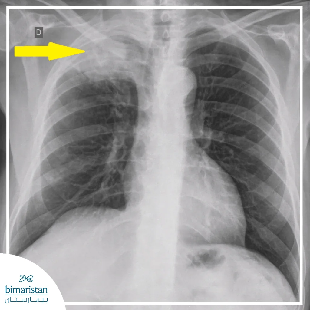
Chest X-ray
Chest X-ray is an initial step in the evaluation; however, it may not reveal the tumor in its early stages because it is obscured by the clavicle or adjacent bony structures. Therefore, it cannot be relied upon alone to rule out the diagnosis.
Chest Computed Tomography (CT) Scan
Computed tomography (CT) is considered the primary imaging modality for diagnosing a Pancoast tumor, as it determines the tumor’s size and the extent of its invasion into the chest wall, ribs, and vertebrae, in addition to assessing the lymph nodes and the condition of the surrounding lung tissue.
Magnetic Resonance Imaging (MRI)
Magnetic resonance imaging (MRI) is considered the most accurate method for evaluating invasion of the brachial plexus, the subclavian vessels, and adjacent neural and soft tissue structures, and is primarily used when planning surgical interventions.
Positron Emission Tomography-Computed Tomography (PET-CT)
This test is used to detect distant metastases and accurately determine the stage of the disease; it also helps plan radiation therapy and evaluate the response to treatment.
Biopsy and histological confirmation
Histological confirmation is an essential step in diagnosing a Pancoast tumor, and a biopsy can be obtained in several ways depending on the tumor’s location, extent of spread, and the patient’s condition. Common methods include Computed Tomography (CT)-guided needle biopsy, which is the primary option in many cases. A biopsy can also be obtained via Video-Assisted Thoracoscopic Surgery (VATS), particularly when needle biopsy is insufficient or not feasible. In some cases, a biopsy may need to be taken through a small incision in the chest wall, particularly when direct access to the tumor is required to obtain an adequate tissue sample.
Mediastinoscopy and bronchoscopy
Mediastinoscopy is crucial for assessing enlarged mediastinal lymph nodes, as it provides an accurate determination of the disease stage. In contrast, bronchoscopy has limited utility for diagnosing Pancoast tumor because this type of tumor is located at the apical periphery of the lung, far from the larger airways. As a result, Pancoast tumor is typically not visible through a bronchoscope.
Stages of Pancoast tumor
As with other types of lung cancer, a Pancoast tumor is classified according to the established cancer staging system, which uses Roman numerals from Stage I to Stage IV, along with subclassifications. The TNM system is also used to determine the extent of the disease, where the letter T refers to the size and local spread of the tumor, N indicates the presence of metastases to the lymph nodes, and M indicates the presence of distant metastases.
Because Pancoast tumors are difficult to detect in the early stages due to their location at the apex of the lung, most cases are diagnosed at locally advanced stages and are often classified as T3 or T4 at the time of diagnosis. A T3 classification indicates that the tumor has invaded the chest wall or the sympathetic nerves, while a T4 classification indicates that it has extended to deeper structures, such as the brachial plexus, the vertebrae, or the spinal cord.
Determining the tumor stage is an essential step in identifying the most appropriate treatment plan and assessing the feasibility of surgery, as the extent of local invasion and the spread of the disease are directly related to the selection of the appropriate treatment strategy for each case.
Treatment of a Pancoast tumor
The treatment of a Pancoast tumor relies on a multidisciplinary approach, given the complexity of the tumor’s location and its proximity to sensitive anatomical structures. The treatment plan is determined by the disease stage, the extent of local invasion, the feasibility of surgical resection, and the patient’s overall condition.
Preoperative chemotherapy and radiation therapy
In resectable cases, chemotherapy is administered in combination with radiation therapy as a first-line treatment, with the goal of reducing tumor size and minimizing its invasion of adjacent structures. This approach improves the likelihood of complete surgical resection and reduces the risk of local recurrence.
Surgical treatment
The goal of the surgical intervention is to completely remove the tumor while ensuring that the surgical margins remain intact. This often involves resection of the affected lung lobe and any nearby affected tissue whenever possible. The tumor’s location at the apex of the lung makes the surgery technically challenging, especially if there is invasion into the chest wall, ribs, or surrounding neural structures. However, advancements in surgical techniques now allow for the use of thoracoscopic or robotic methods in certain cases. These innovative approaches help reduce the size of the surgical incision, minimize postoperative pain, and lower the risk of complications compared to conventional open surgery.
Radiation therapy in non-surgical cases
In cases where surgical resection is not possible, radiation therapy is used as the primary treatment, often in combination with chemotherapy, with the goal of achieving local control of the tumor and slowing its progression.
Palliative care
When a Pancoast tumor is diagnosed in advanced stages with distant metastases, treatment focuses on palliative care, which aims to relieve pain, improve quality of life, and control symptoms resulting from tumor pressure on nerves or blood vessels through appropriate analgesics, local radiation therapy, and comprehensive medical support.
In conclusion, a Pancoast tumor is a specific form of lung cancer, and its primary distinguishing feature is its anatomical location at the apex of the lung and the resulting atypical symptoms that may lead to a delayed diagnosis. Early recognition of the tumor’s characteristic signs is a pivotal step in guiding the patient toward appropriate evaluation. With advances in diagnostic methods and multidisciplinary treatment, the chances of controlling the disease and the possibility of effective therapeutic intervention in appropriate cases have improved. Accurate diagnosis and the development of an appropriate treatment plan for each case remain essential factors in improving patient outcomes and quality of life.
Sources:
- Gundepalli SG, Tadi P. Lung Pancoast Tumor. . In: StatPearls . Treasure Island (FL): StatPearls Publishing; Jan. 2025. Available from: https://www.ncbi.nlm.nih.gov/books/NBK556109/
- Panagopoulos, N., Leivaditis, V., Koletsis, E., Prokakis, C., Alexopoulos, P., Baltayiannis, N., Hatzimichalis, A., Tsakiridis, K., Zarogoulidis, P., Zarogoulidis, K., Katsikogiannis, N., Kougioumtzi, I., Machairiotis, N., Tsiouda, T., Kesisis, G., Siminelakis, S., Madesis, A., & Dougenis, D. (2014). Pancoast tumors: characteristics and preoperative assessment. Journal of Thoracic Disease, 6 Suppl. 1 (Suppl. 1), S108–S115. https://doi.org/10.3978/j.issn.2072-1439.2013.12.29