Keratoconus is a condition that gradually affects the eye, reducing the sharpness of vision, It often affects the middle age group, emphasizing the importance of early diagnosis.
Estimates indicate that keratoconus affects one in every 500 to 2,000 people worldwide and is more common among certain populations.
The cornea is the clear window at the front of the eye chamber, focusing light and visible rays onto the appropriate spot on the retina. Due to keratoconus, the focus changes, causing blurred and distorted vision, which impairs daily tasks like reading and driving. Follow along in this interview to learn about the main symptoms of keratoconus and its various treatments.
What is Keratoconus?
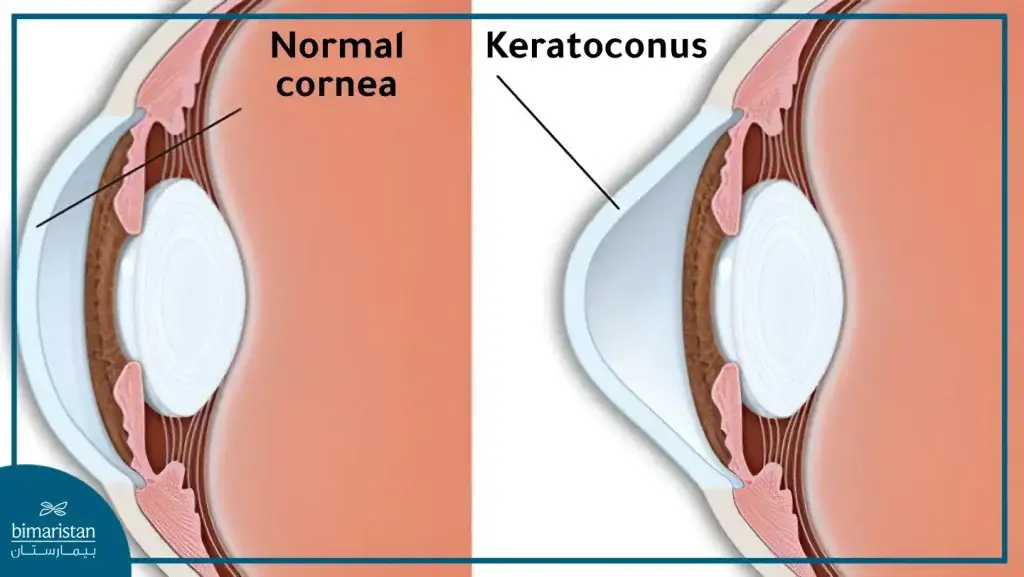
The cornea is a dome-shaped layer of tissue covering the iris and pupil, with its main function being to help focus light onto the lens and pupil.
Keratoconus is a progressive vision disorder that occurs when the round cornea thins and becomes cone-shaped. This deformation prevents incoming light from focusing correctly on the retina, causing blurred vision.

Causes of Keratoconus
Most cases develop without a clear cause, but researchers believe that a combination of environmental and genetic factors contribute to its development. Some predisposing factors include:
- Genetics: Genes play a role in some patients, with one in ten affected individuals having a parent with the disorder.
- Connective Tissue Disorders: Conditions such as Marfan syndrome, Ehlers-Danlos syndrome, Leber congenital amaurosis (a congenital retinal disorder), brittle cornea syndrome, and osteogenesis imperfecta.
- Chronic Eye Inflammation: Persistent inflammation from allergies or irritants can damage corneal tissue, leading to keratoconus.
- Underlying Disorders: The disease may occur with certain conditions like sleep apnea syndrome, Down syndrome, and asthma.
- Continuous Eye Rubbing: Considered a factor in disease progression.
- Eyelid Drooping
Symptoms of Keratoconus
Keratoconus often affects individuals in their late teens to early twenties, with vision symptoms worsening slowly over 10 to 20 years.
Symptoms may begin in one eye but about 96% of cases eventually affect both eyes to varying degrees, with symptoms differing in each eye over time.
Early Stages:
- Slightly blurred vision
- Slightly distorted vision, with straight lines appearing curved or wavy
- Increased sensitivity to light or glare
- Redness or swelling of the eye

Later Stages:
- More blurred or distorted vision
- Increased nearsightedness or astigmatism, requiring frequent changes in eyeglass prescriptionsIn rare cases, corneal blisters that can cause scarring or swelling
Complications of Keratoconus
Potential complications of untreated keratoconus include:
- Fleischer Ring: Iron deposits forming rings inside the cornea, considered an early complication of the disease.
- Acute Corneal Hydrops: A rare complication caused by a rupture in Descemet’s membrane deep in the cornea.
- Visual Impairment: Ranging from mild to severe.
- Corneal Scarring
Diagnosing Keratoconus
Keratoconus can be diagnosed through routine eye examination during a visit to a specialized eye doctor who conducts several tests, including:
- Visual Acuity Test: Involves a Snellen chart for the eye.
- Slit Lamp Examination: Combining a microscope with a bright light, helping detect abnormalities in the outer and middle layers of the cornea.
- Corneal Topography: This test measures the shape and curvature of the cornea.
- Corneal Mapping (Tomography): This method, which is the most accurate for diagnosing early keratoconus and monitoring its progression, involves capturing computed images to create a map of the corneal curvature.
keratoconus disease treatment
The treatment of keratoconus focuses on maintaining visual acuity and halting sudden changes in corneal shape. There are several treatment options, selected by an ophthalmologist based on the severity of the condition and the speed of its progression.
These options include:
Glasses and Contact Lenses
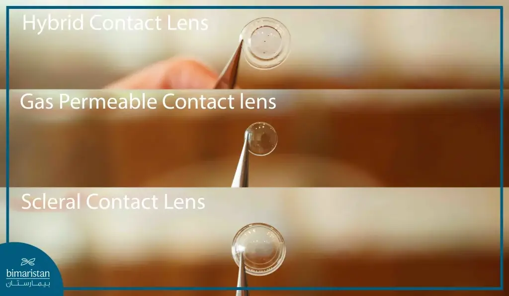
In the early stages of corneal thinning, vision can be corrected using prescription glasses or soft contact lenses. However, as the condition progresses, another type of contact lens such as rigid gas-permeable or scleral lenses may be needed due to irregular astigmatism.

Corneal Cross-Linking (CXL)
This procedure, also known as collagen cross-linking, treats keratoconus by strengthening the bonds between collagen fibers in the cornea and the surrounding proteins to maintain the corneal shape.
The patient receives local anesthesia with riboflavin-containing drops in the eye and then is exposed to UV light for 30 minutes.
INTACS Corneal Ring Segments
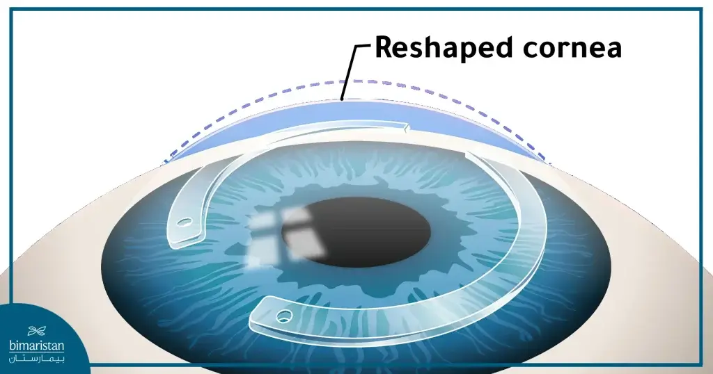
These are small crescent-shaped devices inserted into the cornea to improve vision by flattening the cornea and correcting its cone shape, thus allowing for better fitting of contact lenses.

Corneal Transplant
In advanced stages of the disease, such as scarring or severe thinning of the cornea, the doctor may opt for a corneal transplant, replacing the affected cornea partially or entirely with healthy tissue from a human donor.
Cost of Corneal Transplant in Turkey
The cost of a corneal transplant in Turkey starts from $5000, depending on the patient’s condition and the appropriate procedure for them. It is one of the best options compared to other countries, especially with the availability of the latest devices and highly competent specialists in all corneal transplant procedures.
Due to the vital role of the eyes, individuals should undergo regular examinations to detect eye disorders that may hinder daily life if left untreated, especially since they target middle-aged groups.
Therefore, do not hesitate to ask for answers to your inquiries or visit a medical center in Turkey to find the best treatment for keratoconus based on your condition.
The sources:
