Keratoconus affects the eye, causing vision problems that directly affect the patient’s life, as it is a disease that affects people at an early age and continues to develop.
Treatment for keratoconus is divided into conservative treatment and surgical treatment, and it is possible to combine them according to each patient’s need and the progression of the disease.
What is keratoconus?
Keratoconus is a condition that affects the eye and affects normal vision. The shape of the cornea slowly changes, turning from dome to cone, which prevents light rays entering the eye from being placed correctly on the retina, and thus vision becomes distorted and unclear.
Keratoconus is generally more aggressive when diagnosed in early adolescence as it can lead to significant vision loss in one or both eyes, while being diagnosed later in life is associated with a milder form of keratoconus and vision loss that is more easily controlled.
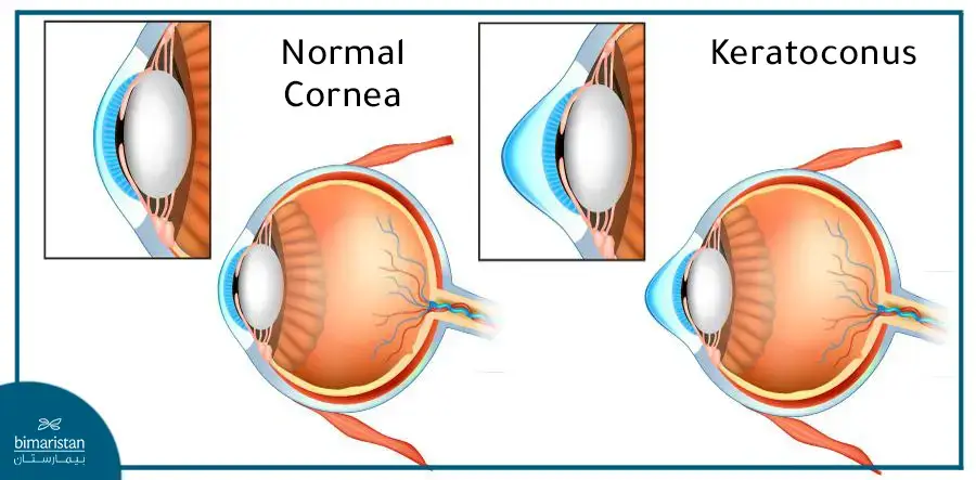
The causes of keratoconus are not completely known, but family history, some genetic conditions, and allergies that cause itchy eyes increase the risk of developing it.
The difference between keratoconus and normal cornea
In the normal state, the cornea of the eye (which is a transparent medium) has a round shape, which contributes to the refraction of light at a certain concentration so that it is positioned correctly on the retina, so that the person can see objects in their realistic and clear form.
When a person suffers from keratoconus, the cornea of the eye becomes thin, causing it to swell to become cone-shaped. As a result, slight blurring of vision occurs in the early stages, but as the condition progresses, the cornea swells more and the vision becomes blurred, and in the most serious cases, it causes a severe and sudden decline in the affected person’s vision.
Treatment for keratoconus
Unlike many diseases, keratoconus is an unusual condition in that it is a progressive disease in young people that tends to stabilize in middle age.
It also affects each eye differently, so it is not easy to choose the best treatment for keratoconus because of the way the disease develops.
There are many different treatment options available today for people diagnosed with keratoconus, ranging from wearing glasses in the early stages of the disease to a corneal transplant in more severe cases.
Treatment of keratoconus with corrective lenses
There are several types used in the treatment of keratoconus, differing in their designs and in the materials involved in their manufacture, to reflect characteristics that suit the different condition of each patient, including the following:
Custom thin contact lenses
Soft, thin lenses can be used to treat mild keratoconus and in athletes whose activity requires physical contact, but because they conform to the irregular shape of the cornea, they provide limited improvement in vision in cases of severe keratoconus.
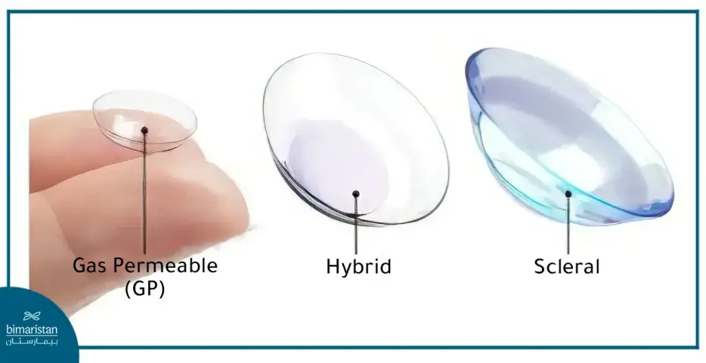
Rigid gas permeable contact lenses (RGP)
Scleral contact lenses provide the best long-term non-surgical solution for treating moderate to advanced keratoconus.
Although it may cause discomfort at first, most patients gradually get used to wearing it after 2-4 weeks of use.
Rigid lenses have a number of advantages compared to soft lenses, including:
- Made of silicone that is highly permeable to oxygen to deliver more oxygen to the front of the eye
- Clearer vision because it maintains the shape of the cornea
- It can last from one to two years with good care without tearing
Scleral contact lenses
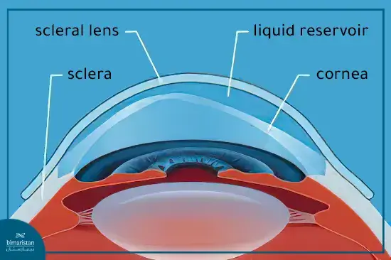
Scleral lenses have a wider diameter and, as the name suggests, rest on the sclera or the white of the eye, leaving a fluid chamber between the cornea and the lens itself that prevents corneal irritation and helps moisturize the eye.
The distinctive thing about scleral lenses used to treat keratoconus is that they provide optical correction in a severely irregular cornea and thus provide better clear vision.
Hybrid contact lenses
Hybrid lenses consist of a hard central part to improve vision connected to a soft periphery to increase comfort.
Although it is a reliable alternative, it is expensive and must be replaced every 6 to 12 months.
Piggybacking contact lenses
Tandem lenses or posterior lenses are a technique in which a soft contact lens is placed over the cornea and another hybrid lens is placed over the soft lens.
It was found that the dual lens system prevents the lens surface from irritating the sensitive cornea with the protective lens.
Intacs rings for the treatment of keratoconus
They are thin plastic semicircular rings that are inserted into the middle layer of the cornea.
It is used to treat keratoconus by flattening the shape of the cornea and helping the patient wear contact lenses more comfortably.
The procedure takes about 25 minutes, during which anesthetic drops are first placed in the eyes to comfort the patient and prevent blinking during the procedure.
After fixing the eye to ensure correct alignment, one small incision is made on the surface of the cornea and using a laser the middle layer is gently separated within a narrow circular field to allow the intacs ring to be clearly visible and the process is repeated to place the second ring.
It is recommended to rest after the operation for 1-2 days and frequent visits to monitor the healing process and evaluate the procedure.
Corneal cross-linking
Corneal cross-linking is a minimally invasive procedure and is the only treatment that can prevent keratoconus from getting worse and avoid a corneal transplant.
Preparing for the operation
On the day of the procedure do not apply cosmetics, shaving or perfume around the eyes.
In most cases, you can eat a light meal and drink fluids beforehand.
The patient needs a companion to drive him home after the operation, as vision may become blurry afterward.
During the process
The patient receives drops to numb the eyes and sedative medication if necessary.
The doctor places specially designed drops of riboflavin (vitamin B2), which allow the cornea to absorb light better.
It takes about 30 minutes for the drops to take their full effect.
Next, the patient lies on a chair and a type of ultraviolet ray is shined directly on the cornea.
This procedure causes new collagen cross-links to appear in the cornea.
These cross-links cause the thickness of the collagen layer to increase, making the cornea more solid and stronger.
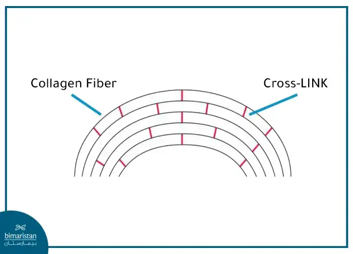
The procedure is performed in outpatient clinics and the entire procedure takes about 30-90 minutes, during which the patient does not feel pain.
After the operation, the doctor prescribes contact lenses that help speed up healing. It is also important not to rub the eyes for 5 days.
As is the case with most surgical operations, cross-linking may cause some symptoms, sometimes it is necessary to see a doctor as soon as they occur, such as:
- Sensitivity to light
- Blurred visio
- Pain or swelling in the eye
- Damage to the cornea or epithelium
- Eye infection
There are also patients for whom the crosslinking operation cannot be performed if:
- Having a very thin cornea (less than 350 microns)
- Having an active eye disease other than keratoconus
- Herpes keratitis
- Pregnant women
- The presence of active and uncontrolled eye allergies
- The presence of scarring in the cornea
Topographically guided conductive keratoplasty
Conductive keratoplasty uses radiofrequency energy to reshape the surface of the cornea by shrinking the collagen fibers in the cornea.
The procedure is performed after applying anesthetic and using a small probe, radio waves are applied to small areas of corneal tissue.
Vision may begin to improve the day after the procedure but may fluctuate for a few weeks to months.
Corneal transplant
Corneal transplantation, also known as corneal grafting or keratoplasty, involves removing the central portion of the diseased or scarred cornea with corneal tissue from a healthy donor using a femto laser or specialized tools in traditional microsurgery.
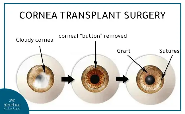
There are two types of keratoplasty used to treat keratoconus:
Total corneal transplant (conventional)
Penetating keratoplasty (PK) in which the entire front portion of the eye wall is replaced with a disc-shaped piece of donor corneal tissue is the most commonly used type of corneal transplant to treat advanced keratoconus.
Partial corneal transplant (DALK)
For many patients diagnosed with keratoconus, the layer of cells lining the back of the cornea and the thin supporting layers (Descemet’s membrane and above) remain normal.
In this case, the abnormal front part of the cornea is selectively replaced, This type is called: Deep Anterior Lamellar Keratoplasty (DALK).
This type has the advantage of reducing the risk of problems with transplant rejection and the need for a corneal transplant replacement in the future.
The recovery time after surgery is long, as the stitches remain in place for 12-18 months, and vision may not stabilize for two years, which affects the patient’s life, education, or work.
A corneal transplant is a life-changing procedure and no keratoconus patient should consider a corneal transplant until they have exhausted all contact lens options as only about 15% of people with keratoconus will need a transplant.
Can keratoconus be treated with laser?
Lasers are used as a procedure to correct vision in cases of refractive eye diseases such as myopia, farsightedness and astigmatism.
Keratoconus is a degenerative disease that leads to thinning of the cornea and causes an abnormal shape that cannot be corrected with laser surgery, as it may worsen the condition by making it thinner with unexpected changes in the shape of the cornea and thus loss of vision.
Stages of keratoconus treatmen
Depending on the severity of the condition and the aggravation of the disease, the stages of treatment of keratoconus can be divided into three stages:
- The first stage: the stage of slight changes in the corneal lining, during which the patient suffers from changes in vision, but vision remains good here if wearing glasses or contact lenses, which is considered a sufficient and effective option.
- The second stage: During which corneal swelling increases, the patient needs to undergo a ring implantation procedure to modify the surface of the cornea.
- The third stage: The disease worsens as the corneal tissue thins severely and the patient’s vision deteriorates, the doctor then resorts to corneal transplant surgery.
Duration of treatment of keratoconus
The duration of treatment for keratoconus varies depending on the severity of the disease and the nature of the procedures used. In early cases, when resorting to conservative treatment using contact lenses, the patient lives with the condition to prevent the worsening of keratoconus.
In the case of surgical treatment, such as a corneal transplant, it takes about two years for vision to stabilize almost completely.
Results and rate of treatment of keratoconus in Turkey
The success rates of eye surgery in Turkey are high due to the great progress it has made in attracting the best ophthalmologists.
The success rate of keratoconus surgery in Turkey is between 85% and 95% and depends on the condition of each individual patient.
The cost of treatment of keratoconus in Istanbul, Turkey
Prices for surgical treatment of keratoconus in Turkey vary depending on the treatment plan for each patient, but the best prices can be obtained in parallel with receiving excellent health care at the hands of the best experts and in health centers equipped with modern technologies.
The price of corneal stabilization with collagen ranges from about $1,500 per eye, while corneal transplant surgery in Turkey is approximately $7,000 for each eye separately, and this includes follow-up and optimal hospitalization for the patient.
It is necessary to periodically examine eye diseases in children and adults, as corneal keratoconus can be detected in its early stages and its development can be limited by selecting the appropriate treatment, especially with the development of surgeries and techniques in treatment of keratoconus.