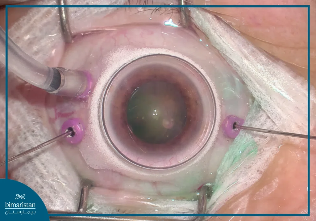
Vitrectomy is a surgical procedure designed to address cases where the vitreous material in the eye leads to vision issues or discomfort, such as blurriness, flashing lights, or unusual reflections. It is a widely performed operation in ophthalmology, particularly in Türkiye, where medical services are known for their high quality and affordability.
The procedure is generally recommended when vitreous-related visual disturbances persist or significantly affect eyesight. The procedure aims to enhance vision clarity and alleviate associated symptoms. However, as with any surgery, there are potential risks, including bleeding or fluid leakage. Therefore, consulting an experienced ophthalmologist is crucial for evaluating the condition and determining whether surgery is necessary. Understanding the risks and benefits is essential to achieving the best possible outcome.
What is vitrectomy?
Vitrectomy is a type of eye surgery to treat specific problems with the retina and vitreous humor. During the procedure, the surgeon removes the vitreous humor, a jelly-like substance that fills the back of the eye, and replaces it with a solution.

The role of the vitreous body in the eye
Vitrectomy involves removing the vitreous humor, a transparent, gel-like substance that fills about two-thirds of the eye’s volume. This substance is crucial for maintaining the eye’s shape, ensuring its structural stability, and supporting its spherical form. It also serves as a light-transmitting medium, allowing light to reach the retina, where it is converted into nerve signals sent to the brain. Additionally, the vitreous humor acts as a protective cushion, absorbing shocks that could damage internal eye tissues. Furthermore, it helps regulate oxygen and nutrient levels, ensuring the health of the retina and lens. Through this procedure, surgeons address issues related to the vitreous humor to improve vision and alleviate discomfort.

Indications for vitrectomy
Vitrectomy is a surgical procedure used to address various eye conditions, including:
- A macular hole, which involves damage to the central part of the retina
- Infections within the eye, such as severe inflammatory disorders
- Eye injuries, including cases where foreign objects enter the eye
- Complications from cataract surgery, such as the displacement or loss of an artificial intraocular lens
Treatment of vitreous hemorrhage and retinopathy
Vitrectomy is performed to address vitreous hemorrhage, a condition where blood seeps into the vitreous humor inside the eye, causing blurred or lost vision. It is also used in cases of retinopathy, which involves damage to the retinal blood vessels and is commonly linked to diabetes or high blood pressure.
When is vitrectomy the optimal treatment for vitreous hemorrhage and retinopathy?
- Acute vitreous hemorrhage: When blood fails to reabsorb naturally within a few months, vitrectomy may be required to clear the blood and restore vision.
- Advanced diabetic retinopathy: The formation of abnormal blood vessels in the retina can lead to repeated bleeding or retinal detachment, necessitating surgical intervention.
In cases of retinal detachment or tear
Vitrectomy is used to treat retinal detachment, a serious condition where the retina separates from the eye wall, potentially causing vision loss if not addressed promptly. It is also performed for retinal tears, which involve a break or hole in the retina that may progress to full detachment if untreated.
When is vitrectomy the optimal solution for treating retinal detachment or tear?
- Complicated retinal detachment: When bleeding inside the eye or retinal fibrosis occurs, the procedure is performed to clear obstructions and reattach the retina.
- Retinal tear with hemorrhage: If a retinal tear leads to bleeding within the vitreous, vitrectomy helps remove the blood and repair the retina using laser or gas treatment.
How is the vitrectomy performed, and what are its detailed steps?
Vitrectomy is a surgical procedure used to address retinal and vitreous body issues. It consists of several precise steps:
- Postoperative care: The patient requires adequate rest and regular medical monitoring to ensure a smooth recovery.
- Anesthesia: Depending on the patient’s condition, local or general anesthesia is administered.
- Eye incisions: Three small openings are created in the sclera to allow the insertion of surgical instruments.
- Vitreous removal: The gel-like vitreous humor is extracted and replaced with a suitable fluid, such as saline or silicone oil.
- Retinal repair: If a tear or detachment is present, specialized solutions like saline or silicone oil are used to aid in restoration.
- Incision closure: The small entry points are either sealed with fine sutures or left to heal naturally.
Surgical techniques used
The choice of vitrectomy technique depends on several factors, including the patient’s condition, the severity of the eye issue, and the complexity of the procedure. Different surgical approaches exist, each with its own benefits and limitations.
- Open vitrectomy: Used in cases requiring direct access to the eye for extensive treatment.
- Minimally invasive vitrectomy: Performed with small incisions, reducing recovery time and minimizing complications.
- Robotic-assisted vitrectomy: Enhancing precision and control, particularly for complex retinal surgeries.
Duration of the operation and when the patient is discharged
The duration of the vitrectomy procedure ranges from 20 to 30 minutes, but it may take up to two hours in more complex cases.
When is the patient discharged?
In most cases, patients undergoing vitrectomy can be discharged on the same day if no complications arise. However, if further procedures, such as retinal repair or silicone oil injection, are necessary, an overnight hospital stay may be required for monitoring and observation.

Post-Vitrectomy Surgery: Recovery and Follow-up
After vitrectomy, the patient requires a recovery period that lasts from several weeks to several months, depending on the condition and type of procedure performed.
Post-operative instructions and precautions
- Rest and minimize strain: Patients should avoid intense activities such as heavy lifting or prolonged bending.
- Use prescribed eye drops: Doctors typically recommend anti-inflammatory and antibiotic drops to prevent infection.
- Maintain head positioning: In certain cases, the patient may need to keep their head in a specific position to support retinal stability.
- Avoid air travel: If intraocular gas is used during surgery, changes in atmospheric pressure could affect the eye.
When do the results appear?
- Vision gradually improves over a period of weeks to months, depending on the condition and procedure.
- If intraocular gas is used, visual recovery may take longer until the gas is completely absorbed.
- In some instances, additional procedures, such as silicone oil injections, might be required, which could influence the recovery timeline.
When should you consult a doctor?
Seek immediate medical attention if any of the following symptoms occur:
- Persistent or worsening pain that does not respond to pain medication
- Severe redness or abnormal discharge from the eye
- Sudden vision deterioration, including black spots or flashes of light
- Unusual swelling around the eye or eyelids
- Increased eye pressure or a sensation of pressure within the eye
In conclusion, vitrectomy is an effective treatment option for conditions affecting the retina and vitreous, such as vitreous hemorrhage, retinopathy, retinal detachment, and retinal tears. The success of the surgery depends on early diagnosis, regular medical follow-up, and adherence to recovery instructions. Patients should monitor any changes in vision after surgery and contact their doctor as needed to ensure the best possible results. With proper care, vitrectomy can improve vision quality and restore patients to a normal life with greater confidence.
Sources:
- American Academy of Ophthalmology. (n.d.). What is vitrectomy?. Retrieved May 30, 2025
- National Eye Institute. (n.d.). Vitrectomy. Retrieved May 30, 2025
