
Pituitary tumors are relatively common among skull base tumors and account for a significant proportion of intracranial tumors. They are sometimes discovered incidentally during brain imaging, while in other cases they present due to hormonal disturbances or direct pressure on the optic nerves. Although many of these tumors are benign and slow-growing, the deep and sensitive location of the pituitary gland means that managing them requires a high degree of precision and a well-thought-out treatment plan.
In recent years, endoscopic transnasal pituitary tumor surgery has become one of the advanced surgical options for treating many of these tumors, especially when the tumor is large, affects vision, or causes hormonal hypersecretion. This technique is characterized by the ability to access the tumor through the nose and sphenoid sinus without opening the skull in most cases, which helps minimize surgical trauma and speed up recovery when the appropriate patient is selected.
What is endoscopic pituitary tumor surgery?
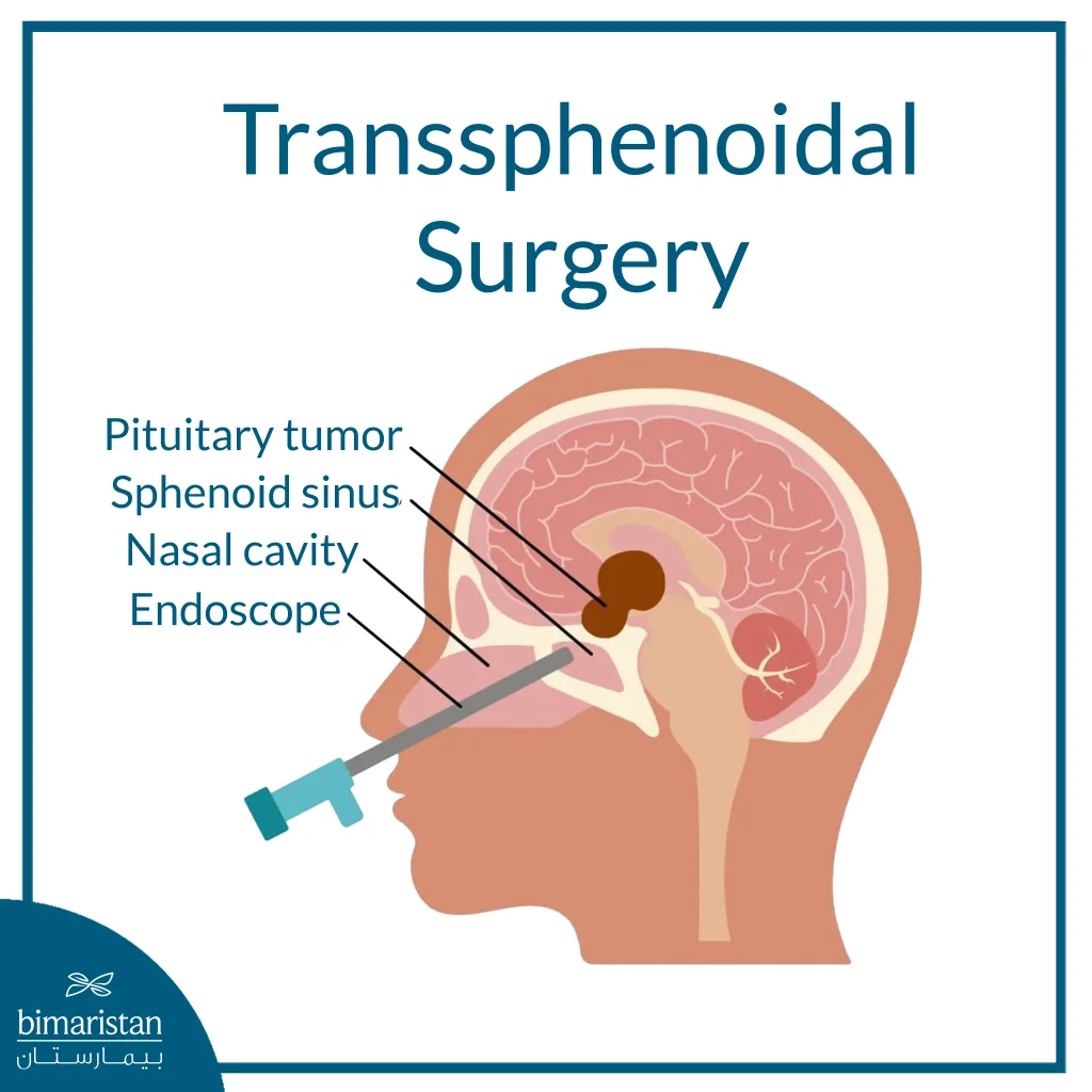
Endoscopic pituitary tumor surgery is a procedure typically performed through the nose to access the pituitary gland at the base of the brain and remove the tumor without the need to open the skull in most cases. During the procedure, the surgeon uses a thin endoscope with a high-resolution camera, inserted through the nostrils into the sphenoid sinus and then reaching the sella turcica, where the pituitary gland is located. This approach provides a magnified, clear view of the tumor and its surroundings, helping remove the tumor while preserving as much of the normal pituitary gland, optic nerves, and nearby blood vessels as possible.
This surgery is used primarily for pituitary tumors that put pressure on the optic nerves, disrupt hormone secretion, or show a significant increase in tumor size. It involves less surgical trauma than some conventional surgeries, often leaves no visible external scar, and may shorten hospital stay and recovery time when the patient is a good candidate. However, endoscopic surgery is not suitable for all cases; the decision depends on the tumor’s size, its extension into adjacent areas, its hormonal type, and the surgical team’s experience with skull base surgeries.
When does a patient need endoscopic pituitary tumor surgery?
A patient requires endoscopic pituitary tumor surgery when the tumor is not merely a small, stable mass that can be monitored, but rather causes noticeable symptoms or poses a threat to neurological and hormonal functions. Key indications include tumors that compress the optic nerves, causing visual impairment or a narrowed field of vision; tumors that gradually increase in size on MRI scans; or tumors that cause persistent headaches with signs of pressure on adjacent structures at the base of the brain.
Surgery is also performed for certain hormone-secreting tumors when medication is insufficient to control the hormonal imbalance, such as in some cases of growth hormone or adrenocorticotropic hormone (ACTH) excess. It may also be necessary if the tumor causes pituitary insufficiency, or if sudden symptoms appear, such as severe headache accompanied by vision loss due to bleeding within the tumor, a condition that requires urgent evaluation because it may threaten vision and pituitary function.
How are pituitary tumors diagnosed before surgery?
The preoperative diagnosis of pituitary tumors begins with an evaluation of the patient’s symptoms, particularly vision problems, headaches, and changes in weight, menstrual cycle, sexual function, or facial and hand features when a hormonal disorder is present. This is followed by an MRI with contrast, the most important test for determining the tumor’s size, location, and extent of invasion into the optic nerves, sinuses, or adjacent areas at the base of the brain, helping the surgeon develop a precise surgical plan.
Diagnosis cannot be made based on imaging alone; the patient requires comprehensive hormonal testing to determine whether the tumor is hormone-secreting or causing pituitary insufficiency. An eye exam and visual field test are also required if there is pressure near the optic chiasm. Additional tests may be performed depending on the case, such as CT scans or preoperative evaluation of the nose and sinuses, in order to select the most appropriate surgical approach and minimize risks during the procedure.
How is endoscopic pituitary tumor surgery performed?
Endoscopic pituitary tumor surgery is performed under general anesthesia, typically by a multidisciplinary team of a neurosurgeon and an otolaryngologist (ear, nose, and throat surgeon) to access the tumor with maximum precision. In most cases, the procedure does not require an external incision in the face or opening of the skull; instead, surgeons gain access through the natural passages of the nose to reach the base of the skull, then remove the tumor under magnified visualization using an endoscope and precision surgical instruments.
Access to the tumor through the nose and the sphenoid sinus
The surgeon begins by inserting the endoscope through the nose toward the sphenoid sinus, which is the air-filled sinus near the pituitary gland. Once this area is reached, a small section of bone at the base of the skull is opened to access the sella turcica, where the pituitary gland is located. Then, the layer covering the tumor is carefully opened to allow access to it without causing unnecessary damage to the surrounding tissues.
Tumor resection while preserving important tissues
After the tumor is exposed, the surgeon begins to remove it gradually using precision instruments, while attempting to separate the tumor tissue from the normal pituitary gland, the optic nerves, and nearby blood vessels. The goal is not only to remove as much of the tumor as possible but also to preserve hormonal and visual functions to the greatest extent possible. Therefore, the surgeon may leave a small portion of the tumor if it is adherent to sensitive structures and its complete removal would pose a risk to the patient.
Skull base closure and leak monitoring
After the tumor has been removed, the surgeon checks to ensure there is no significant bleeding or cerebrospinal fluid leak from the skull base. If the surgeon finds a leak or the risk is high, they seal the area with patches or other supportive materials. Tissue from inside the nose may be used to help repair the skull base. The patient is then monitored in the hospital to ensure that vision and hormone levels remain stable and that there are no signs of leakage or inflammation.
What are the advantages of endoscopic pituitary tumor surgery?
Endoscopic pituitary tumor surgery is characterized by the fact that it allows access to the tumor through the nose without a visible external incision or opening of the skull in most cases, which minimizes surgical trauma and helps make the recovery period easier compared to open surgery. The endoscope also provides the surgeon with a magnified, close-up view of the pituitary gland and the base of the skull, which allows for more precise tumor removal, especially when the tumor is located near the optic nerves or critical blood vessels.
Among its most important advantages are reduced hospital stay, alleviated postoperative pain, and a faster return to daily activities when the appropriate case is selected. This technique also helps relieve optic nerve pressure symptoms in many patients. It may also help regulate hormonal imbalances if the tumor is hormone-secreting, but postoperative follow-up is necessary to ensure vision and pituitary function remain stable.
What are the risks of endoscopic pituitary tumor surgery?
Although endoscopic pituitary tumor surgery is considered a precise and relatively safe procedure when performed by an experienced team, it may carry some risks due to the tumor’s proximity to the optic nerves, blood vessels, and the base of the brain. The most significant risks include bleeding, inflammation, or leakage of cerebrospinal fluid from the base of the skull, as well as temporary or, in rare cases, permanent vision loss if the optic nerves are affected during the procedure.
Hormonal disturbances may occur after surgery due to disruption of the normal pituitary gland, such as deficiencies in certain hormones or imbalances in fluid and electrolyte levels. Depending on the case, the patient may require temporary or permanent hormonal therapy. Additionally, part of the tumor may remain if it is adherent to sensitive areas, which requires follow-up imaging and laboratory tests, and sometimes subsequent medication or radiation therapy.
Recovery after endoscopic pituitary tumor surgery
Recovery after endoscopic pituitary tumor surgery begins in the hospital. During this time, healthcare providers monitor the patient’s vision, headaches, urine output, and blood electrolyte levels, as well as assess pituitary gland function to ensure no hormonal disturbances occur following the procedure. Typically, patients stay in the hospital for several days, depending on factors such as tumor size, overall condition, and whether there are complications like cerebrospinal fluid leakage or fluid and electrolyte imbalances.
After discharge, the patient needs to rest and avoid blowing their nose forcefully, lifting heavy objects, or bending over excessively during the first few weeks, as these activities may increase pressure in the nose and at the base of the skull. Follow-up visits with the neurosurgeon and endocrinologist will be scheduled, and an MRI and hormone tests may be ordered later to evaluate the outcome of the surgery and ensure that the pituitary gland is stable and the absence of tumor recurrence.
In conclusion, endoscopic pituitary tumor surgery is a minimally invasive surgical option that allows for tumor removal through the nose without opening the skull in most cases, while also relieving pressure on the optic nerves and improving certain hormonal disorders associated with the tumor. The procedure’s success depends on an accurate preoperative diagnosis, the medical team’s expertise in working in this sensitive area near the brain and blood vessels, and regular postoperative follow-up to monitor vision and pituitary gland function. At Bimaristan Medical Center, physicians specializing in pituitary and skull base surgery can coordinate treatment, helping patients safely and clearly choose the treatment plan best suited to their condition.
Sources: