
The medical field is witnessing continuous advancements in diagnostic and treatment methods, with a growing trend toward procedures that combine precision with minimal surgical intervention, This evolution is clearly reflected in the field of interventional radiology, which now encompasses a wide range of specialized procedures to treat and assist patients with various conditions.
Among the procedures performed in this field is the IVC filter placement, which is used in specific cases based on the physician’s assessment and the patient’s condition, This procedure is now available at many specialized medical centers, including advanced centers in Turkey that offer interventional radiology services.
What is the IVC filter placement procedure?
Inferior vena cava (IVC) filter placement is a minimally invasive medical procedure designed to place a small device inside the inferior vena cava, designed to help prevent large blood clots from traveling through the bloodstream and reaching the lungs, where they can cause pulmonary embolism. The filter acts as a barrier inside the vein, allowing blood to flow normally while trapping some of the clots carried by the bloodstream.
The procedure is typically performed by an interventional radiologist, who uses a catheter and imaging techniques to guide the filter and position it correctly within the vein after determining the appropriate type of filter based on the patient’s condition.
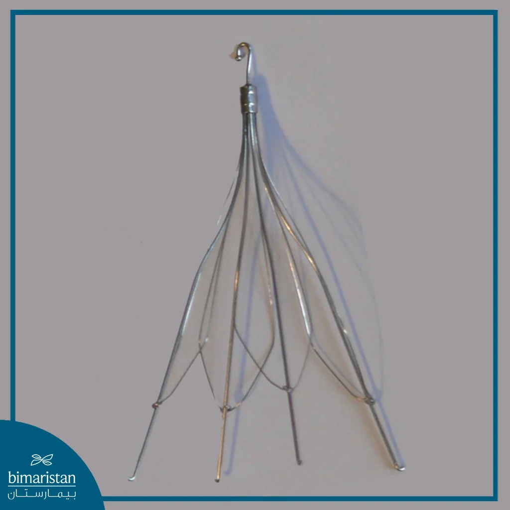
How is an inferior vena cava filter implanted?
The IVC filter placement is done using imaging techniques. The procedure begins with sterilizing the entry site and administering local anesthesia, followed by making a small incision to insert a thin catheter through one of the veins, most often the internal jugular vein or the femoral vein; the right internal jugular vein is preferred in many cases because of its relatively direct path to the inferior vena cava.
The doctor guides the catheter using imaging, and fluoroscopy is the most commonly used method during the procedure, although echocardiography or intravascular echocardiography may be used in some cases. Once the appropriate location has been identified, the filter is typically placed in the lower part of the inferior vena cava, below the junction of the renal veins.
The filter is then advanced through the catheter and deployed within the vein to expand and secure itself in place. In certain cases, such as the presence of a clot in the area where the filter is to be placed or during pregnancy, the filter may be placed in the portion located above the renal veins. After confirming its position and stability, the catheter is removed and the entry site is closed.
What are the types of inferior vena cava filters?
Inferior vena cava filters vary in design and how long they remain in the body, and can be broadly classified into:
- Permanent filters: Designed to remain permanently in the inferior vena cava; they are used when long-term protection against pulmonary embolism is needed or when removal of the filter is not appropriate.
- Retrievable filters: These filters can be removed once they are no longer needed; therefore, they are commonly used when anticoagulants cannot be used temporarily or when the risk of pulmonary embolism is high for a limited period of time.
- Convertible filters: These are designed so that their structure can be modified inside the vein once the filter is no longer needed, rather than being completely removed in some cases.
Which patients are candidates for inferior vena cava (IVC) filter placement?
Inferior vena cava (IVC) filter placement is primarily recommended for patients with or at risk of venous thromboembolism, particularly deep vein thrombosis or pulmonary embolism, when anticoagulants cannot be used or are inappropriate due to contraindications or a high risk of bleeding, Implantation of an inferior vena cava filter may also be considered in certain patients who remain at risk for pulmonary embolism despite appropriate anticoagulant therapy, or in selected cases where the risk of clot formation and embolism is high, such as during pregnancy, in immobile patients, and in those with coagulation disorders.
The decision to implant a filter is made after evaluating the patient’s condition, and weighing the risks of thrombosis and bleeding, bearing in mind that the filter is not a routine substitute for anticoagulant therapy when it is available.
What are the advantages and benefits of inferior vena cava (IVC) filter placement?
The main benefit of inferior vena cava (IVC) filter placement is to reduce the risk of blood clots traveling from the deep veins of the lower extremities to the lungs, thereby helping to prevent pulmonary embolism in patients who are suitable candidates for filter placement. Among its most notable benefits are:
- Minimally invasive procedure: The filter can be inserted through a vascular catheter via a small incision, without the need for open surgery in most cases.
- Prevention of Pulmonary Embolism: The filter acts as a barrier inside the inferior vena cava, helping to trap large blood clots before they reach the heart and lungs.
- This is an alternative treatment option when anticoagulants cannot be used: it may be an option for patients who cannot receive anticoagulants due to a risk of bleeding or a contraindication.
- Removal of Retrievable Filters: Depending on the patient’s condition and the doctor’s assessment, certain types of filters can be removed when they are no longer needed.
What are the disadvantages and risks of inferior vena cava (IVC) filter placement?
Although inferior vena cava filters offer benefits in reducing the risk of pulmonary embolism in appropriate patients, their placement may be associated with certain complications, either during the procedure or while the filter remains in the vein, Post-implantation medical follow-up is particularly important for the early detection of any potential complications, including:
- Bleeding or the formation of a hematoma at the catheter insertion site.
- Injury to a blood vessel during insertion or placement of the filter.
- The embolus moves from its original location and, in rare cases, may travel to another area within the blood vessels or to the heart.
- The position of the stent or its improper placement, which may affect its ability to trap clots.
- Blockage of the inferior vena cava or the formation of a clot around the filter, which may lead to increased swelling and pain in the lower extremities.
Removal of the Inferior Vena Cava Filter
The inferior vena cava filter is removed when it is no longer needed and the patient can be treated with anticoagulants, or when there is no longer a need to keep the filter in place. Removal is typically performed for retrievable filters, whereas permanent filters cannot be routinely removed. The doctor first assesses the patient’s condition to ensure that removal is appropriate, and may rely on medical imaging to locate the filter and ensure there are no clots or complications that would prevent its retrieval.
The procedure is performed using interventional radiology techniques, in which a catheter is inserted through a vein, usually the jugular or femoral vein, then precision instruments are used to grasp the filter and pull it out of the body. The procedure is usually performed under local anesthesia and, in most cases, does not require surgery.
It is preferable to remove the retrievable filter as soon as it is no longer medically necessary, because leaving it in place for a long time may increase the risk of certain complications, such as the filter becoming embedded in the vein wall, its tilting, or the formation of clots around it, which may make retrieval more difficult. However, it may be left in place if the attempt to remove it poses greater risks than benefits—such as when there is a large clot inside the filter or the filter has penetrated the vein wall.
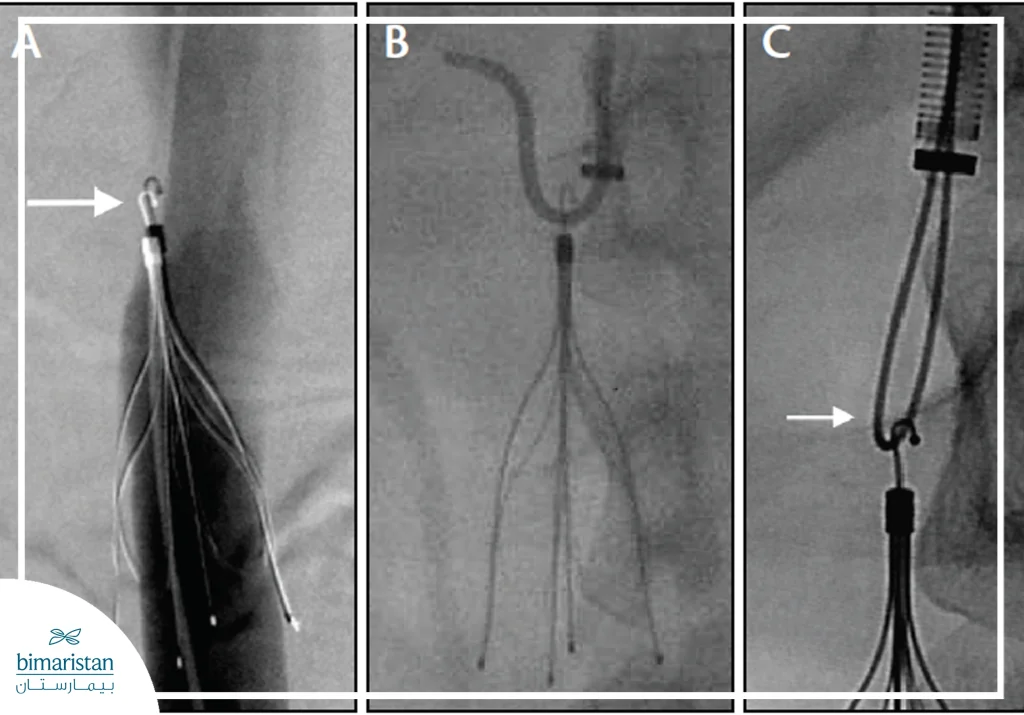
In conclusion, the inferior vena cava filter is an important option for preventing pulmonary embolism in patients whose condition warrants its use, however, the success of the treatment is not limited to the placement of the filter; it also includes careful follow-up and assessment of the need for its continued presence and timely removal, With the continuous advancement of interventional radiology techniques, the removal of retrievable filters has become more precise and safer, providing patients with effective treatment options involving the least possible surgical intervention, Choosing a specialist and an advanced medical center in this field is an essential step toward receiving comprehensive care—from initial assessment and selection of the appropriate filter to follow-up and removal when necessary.
Sources:
- Benedetti, R., Marino, S., Tangianu, F., & Imberti, D. (2024). Inferior vena cava filters: A clinical review and future perspectives. Journal of Clinical Medicine, 13(6), 1761
- Muneeb A, Dhamoon AS. Inferior Vena Cava Filter. . In: StatPearls . Treasure Island (FL): StatPearls Publishing; Jan. 2026-.
- RadiologyInfo.org. (September 23, 2024). IVC filters: Placement and removal.