
Some chest conditions affect the lungs’ ability to expand and function normally, and may cause shortness of breath, chest pain, or prolonged inflammation. The severity of these conditions varies by cause and stage, and treatment differs from patient to patient based on test and imaging results.
Medications or drainage procedures may be sufficient to treat some cases, while others may require surgical intervention if the problem persists or lung function does not improve. Thoracoscopic lung decortication is one surgical option that may be considered in appropriate cases after a thoracic surgeon evaluates the patient.
What is thoracoscopic lung decortication?
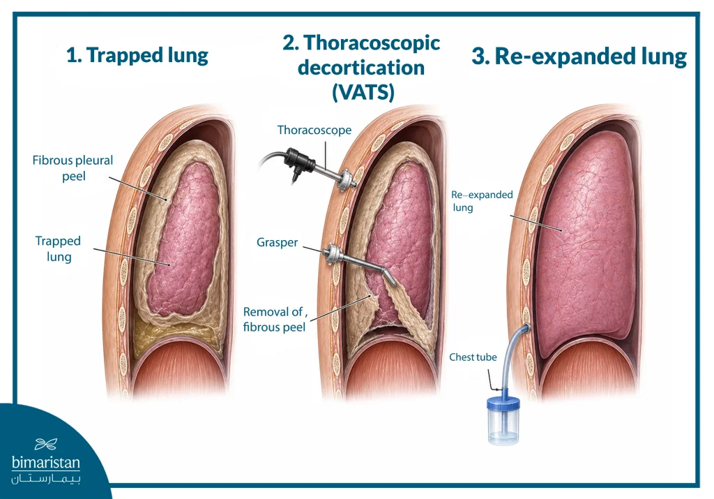
Thoracoscopic lung decortication is a surgical procedure that removes a thick, abnormal fibrous layer from the surface of the lung; it may also involve surrounding tissues such as the chest wall or diaphragm. The surgeon performs the procedure through small chest incisions, inserting a camera and precision surgical instruments to remove this layer and free the lung, allowing it to expand more fully during breathing.
Indications for thoracoscopic lung decortication
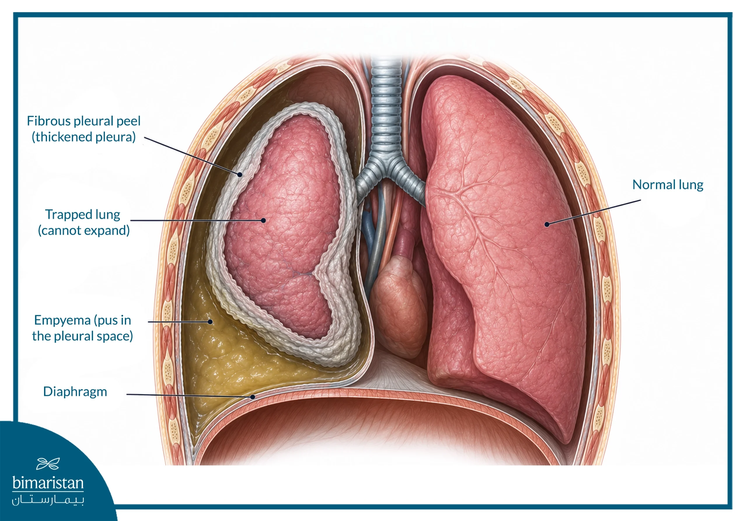
Thoracoscopic lung decortication is a surgical procedure performed when a thick fibrous peel forms around the lung, hindering normal expansion and causing significant respiratory symptoms. This condition often arises in cases of chronic pleural effusion, where there is an accumulation of pus in the pleural space surrounding the lung, chronic blood accumulation in this space, pleural fibrosis, or lung atelectasis. Additionally, this procedure may be indicated in certain situations involving pleural tumors or chronic infections that lead to the formation of adhesions and thickening of the tissues surrounding the lung.
A doctor usually recommends surgery when simpler procedures, such as draining fluid or pus with a needle or chest tube, are ineffective. Additionally, if X-rays reveal that the lung remains collapsed and cannot expand, surgery may be necessary. The decision to perform thoracoscopic surgery depends on the specific condition, the thickness of the fibrous peel, the patient’s overall health, and the thoracic surgeon’s assessment.
Tests and preparations before thoracoscopic lung decortication
Preparation for the surgery begins with a review of the patient’s medical history, including past or current smoking and exposure to radiation therapy or asbestos, followed by an assessment of the heart’s and lungs’ ability to tolerate the surgery. The doctor determines the necessary tests based on the patient’s condition and the nature of the problem; these may include:
- Chest X-ray and computed tomography (CT) scan to determine the thickness of the fibrous peel, the degree of lung atelectasis, and the condition of the lung tissue
- Pulmonary function tests to assess respiratory function
- Echocardiogram (echocardiography) or stress test to assess surgical tolerance
- Complete blood count, coagulation tests, liver and kidney function tests, and screening for infections
- Bronchoscopy to check the integrity of the airways when needed
- Magnetic resonance imaging (MRI) or thoracoscopy with tissue biopsy when a tumor is suspected
Compatible blood units may also be prepared before the procedure in anticipation of possible bleeding during the removal of the fibrous peel.
How is thoracoscopic lung decortication performed?
Thoracoscopic lung decortication is performed using Video-Assisted Thoracoscopy (VATS) under general anesthesia, typically through three small incisions in the chest wall; one is used to insert the camera, while surgical instruments are inserted through the other incisions. Sometimes the surgeon may use only one incision, or may need an additional port to improve visibility or to suction out blood and fluids.
Once the preparations are complete, the process is carried out according to the following steps:
- Inserting the thoracoscope into the pleural space, using CT imaging to determine the appropriate entry point
- Suctioning pus or fluids and cleaning the chest cavity of damaged tissue
- Breaking down adhesions and septations (partitions) within the pleural cavity
- Gradually removing the fibrous peel from the lung surface and pleura using precision instruments
- Repositioning the camera as needed to access various parts of the chest cavity
- Ensuring the lung is freed and capable of expanding, and addressing any air leaks if present
- Irrigating the chest cavity and placing two drainage tubes through the surgical incisions
- Removing the thoracoscope and closing the incisions
The procedure typically takes more than two hours, though the duration varies depending on the thickness of the fibrous peel and the extent of the adhesions. Thoracoscopic surgery may take longer in some cases, as the peel is removed in small fragments to allow for passage through the limited incisions.
Recovery after thoracoscopic lung decortication
After thoracoscopic lung decortication, the patient is closely monitored for the first few hours and may remain in the intensive care unit or observation unit for 12 to 24 hours, depending on their condition. Some patients may need a breathing tube for 24–48 hours, while most can breathe using an oxygen mask or nasal cannula.
Chest tubes are typically left in place for at least 48 hours to drain fluid and monitor for air leaks; they are removed when the fluid becomes clear and daily output drops below 50 mL. Postoperative care includes:
- Pain management using epidural anesthesia, nerve blocks, or analgesics
- Administration of antibiotics, fluids, and nutritional support as needed
- Chest X-rays to monitor lung expansion
- Monitoring oxygen levels and blood gases in certain cases
- Breathing exercises and chest physiotherapy
- Early mobilization to aid recovery and prevent complications
The patient can be discharged from the hospital 48–72 hours after the procedure if their condition is stable, the chest tubes have been removed, and they can eat. The first follow-up visit usually occurs 5–7 days after discharge to examine the incisions and use imaging to confirm the lung has fully expanded and that no air or fluid remains around it.
Results and benefits of thoracoscopic lung decortication
Thoracoscopic lung decortication helps restore lung expansion by removing the fibrous peel that restricts movement, improving lung compliance, ventilation efficiency, and gas exchange; it may also reduce shortness of breath and improve exercise tolerance. After the procedure, certain lung function parameters may also increase, such as forced vital capacity and forced expiratory volume in the first second, and the residual space around the lung may decrease, reducing areas where fluid may accumulate or infection may persist.
The degree of improvement depends on the integrity of the underlying lung tissue and disease severity; therefore, outcomes are typically better in patients with marked pleural fibrosis who have relatively preserved lung function.
Risks and complications of thoracoscopic lung decortication
Some complications may occur during or after thoracoscopic lung decortication, and their likelihood varies with disease severity, lung condition, and the surgical team’s experience. The most significant risks include:
- Persistent air leak from the lung; while this often resolves within a few days, a large leak may require suturing or further intervention
- Bleeding from the lung surface, pleura, or blood vessels during the removal of adhesions; in some cases, this may require a blood transfusion.
- Failure of the lung to fully re-expand or persistent collapse of a portion of the lung, particularly if the lung tissue is damaged
- Infection or a retained collection of pus within the pleural space, which, in severe cases, can lead to sepsis.
- Accumulation of fluid or air around the lung following the procedure
- Injury to adjacent tissues or organs, such as the diaphragm, esophagus, pericardium, or blood vessels
- Bronchopleural fistula, an abnormal connection between the airways and the pleural space, resulting in a significant and persistent air leak
- Respiratory complications and the need for prolonged respiratory support, especially in patients with pre-existing impaired lung function
Close monitoring of chest tubes, radiography, and postoperative respiratory therapy help detect and treat these complications early.
Thoracoscopic lung decortication compared to open surgery
The appropriate method of lung decortication varies by disease stage, fibrous peel thickness, and extent of adhesions. The following table outlines the main differences between thoracoscopic decortication and open surgery:
| Comparison element | Thoracoscopic lung decortication (VATS) | Open‑surgery lung decortication |
|---|---|---|
| Access method | Small incisions through which the camera and surgical instruments are inserted | A larger thoracic incision that provides direct access to the lung |
| Suitable cases | Often appropriate for less complex or carefully selected cases | Suitable for advanced cases, thick fibrous peel, and extensive adhesions |
| Postoperative pain | Usually less | Usually more due to the wider incision |
| Hospital stay & recovery | Shorter hospital stay and generally faster recovery | Relatively longer hospital stay and recovery period |
| Ability to remove the peel | Removing very thick peel may be difficult through small incisions | Provides a wider field for removing thick peel and complex adhesions |
| Outcomes | Effective in suitable patients and may achieve results comparable to open surgery | Particularly effective in complex and advanced cases |
In general, no single approach suits all patients; the thoracic surgeon determines the most appropriate surgical method after assessing lung condition and disease complexity, aiming for the best possible outcome with minimal intervention.
In conclusion, thoracoscopic lung decortication is a significant surgical option for freeing a restricted lung and restoring its ability to expand in suitable patients. Video-assisted thoracoscopic surgery (VATS) allows the procedure to be performed through limited incisions, rather than open surgery, potentially enabling faster recovery in appropriate cases. Treatment success depends on accurate diagnosis, selecting the optimal timing and surgical approach, and the thoracic surgeon’s expertise in managing each case.
Sources: