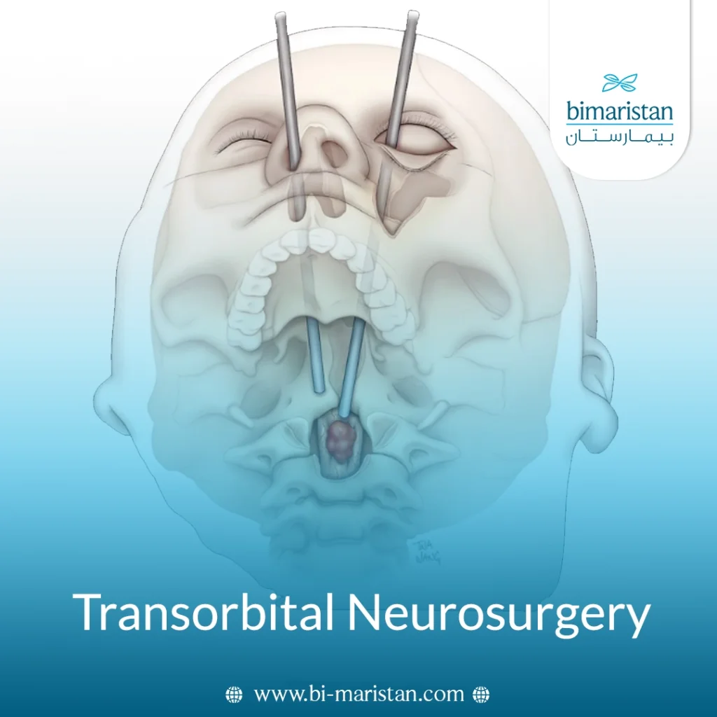
Transorbital neurosurgery is one of the modern minimally invasive techniques used to access certain deep-seated tumors and lesions at the base of the skull and in the brain. This surgery relies on a multidisciplinary team, typically a neurosurgeon, an otolaryngologist, and an ophthalmic plastic and reconstructive surgeon, to select a precise surgical route that minimizes the need for extensive craniotomy. For suitable patients, this technique can reduce incision size, limit exposure of brain tissue, and speed recovery.
What is transorbital neurosurgery?
Transorbital neurosurgery is a minimally invasive surgical procedure that uses the eye socket as a natural pathway to access tumors, lesions, and aneurysms located in the orbit, the base of the skull, or the anterior, middle, and lateral regions of the brain. The surgeon makes a small incision, often hidden within the natural folds around the eye, and then inserts an endoscope and precision instruments to reach the target area without removing the eye. The procedure aims to remove the tumor, treat the lesion, or close the aneurysm while preserving neural and visual functions as much as possible; it can be combined with transnasal endoscopic surgery when treating large or complex tumors.
Types of transorbital neurosurgery
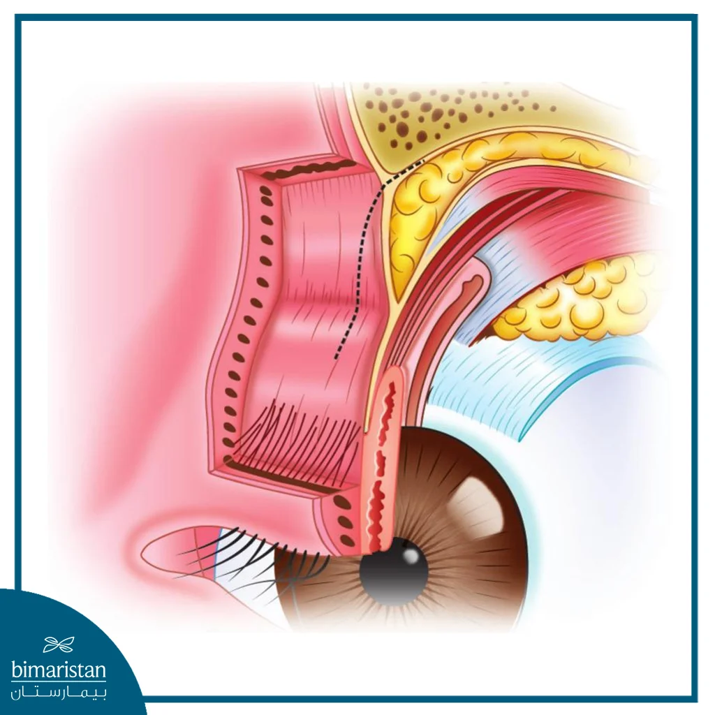
The surgeon selects the eyelid-based approach based on the tumor, lesion, or hemangioma location and its relationship to surrounding nerves and blood vessels. All approaches involve a small incision around the eye socket, which is often concealed within the natural creases of the eyelid or eyebrow to minimize visible scarring.
- Lateral orbital approach: The incision is made within the crease at the outer corner of the eye, near the “crow’s-feet” area, providing access to the lateral aspects of the orbit and the skull base.
- Supraorbital approach: The incision is placed within the eyebrow or the natural crease of the upper eyelid; it is used to access certain anterior and superior regions of the skull base.
- Transcaruncular approach: The incision is made in the small pink tissue at the inner corner of the eye, providing a medial route without leaving a visible skin scar.
- Transconjunctival approach: Access is gained through the inner surface of the eyelid, avoiding a visible skin incision, to reach the inferior or medial parts of the orbit.
Transorbital neurosurgery may be performed using one of these approaches, in which the surgeon inserts a thin endoscope that provides a magnified, high-resolution view of the skull base, the optic nerve, and adjacent structures. This allows for precise manipulation of instruments within a narrow surgical corridor, minimizing the need to resect brain tissue. Multiple approaches can also be combined, or the trans-orbital approach can be combined with transnasal endoscopic surgery when treating large or complex lesions.
When is transorbital neurosurgery used, and who are the candidates?
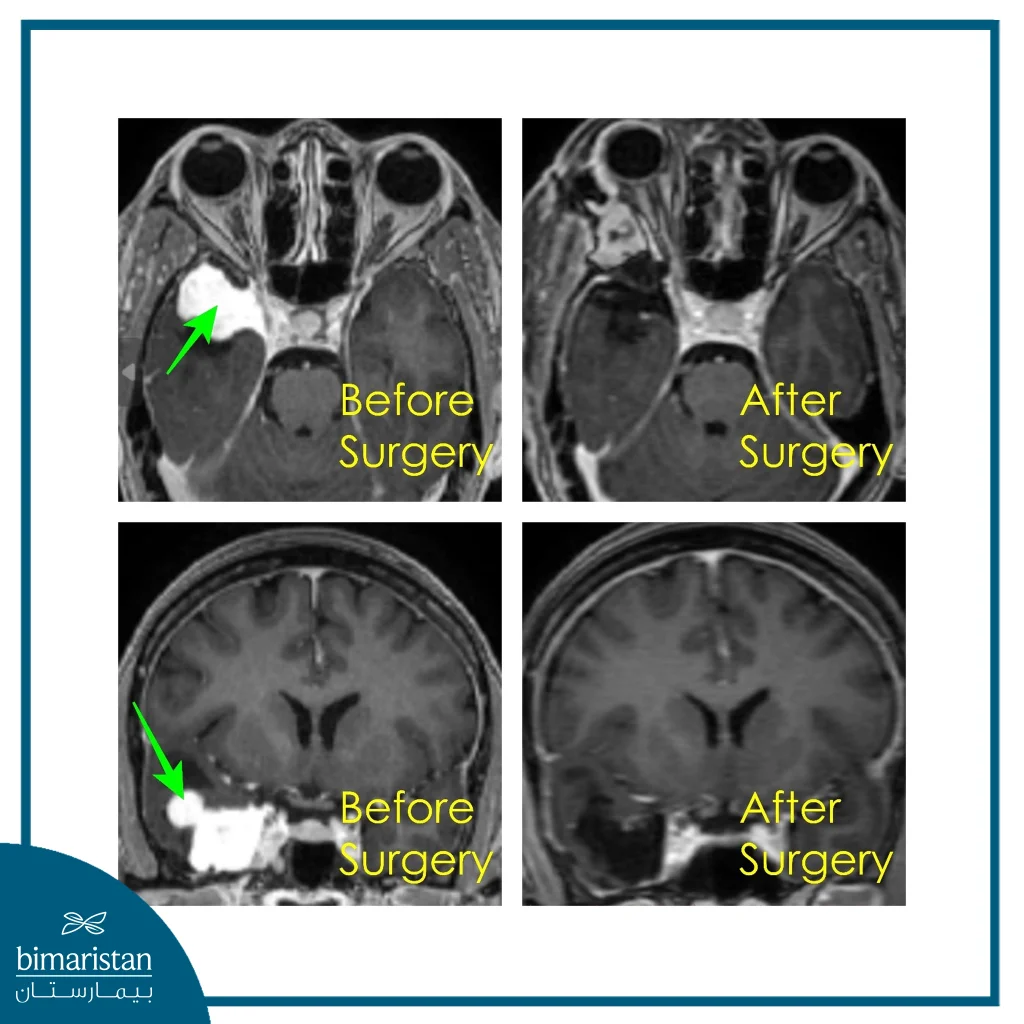
Transorbital neurosurgery is used to treat certain tumors and lesions within the orbit or at the skull base, when the orbit provides a direct and safe route that minimizes the need for brain tissue resection. Conditions that may be treated using this technique include:
- Spheno-orbital and sphenoid wing meningiomas
- Anterior clinoid process tumors
- Selected lesions of the lateral cavernous sinus and Meckel’s cave
- Schwannomas, including trigeminal nerve schwannomas
- Intraorbital vascular tumors and low-grade gliomas
- Arachnoid cysts and epidermoid tumors
- Osteomas
- Optic nerve compression
- Orbital inflammatory pseudotumors
- Selected inflammatory, cystic, and vascular lesions
- Fibrous dysplasia requiring decompression or bone remodeling
- Selected cerebral aneurysms
- Large, complex, or recurrent tumors (using a combined transorbital and other surgical approach)
A patient is considered a candidate for surgery when the lesion can be accessed via the orbit without placing visual or neurological functions at high risk; this is determined following a review of imaging studies and an evaluation by a multidisciplinary surgical team.
What is TONES (Transorbital Neuroendoscopic Surgery)?
Transorbital Neuroendoscopic Surgery (TONES) is a minimally invasive technique that provides access to the skull base and certain areas of the brain through the eye socket using a microendoscope. The procedure typically begins with a small incision near the inner corner of the eye or within one of its natural creases; the surgeon then inserts the endoscope and surgical instruments to obtain a magnified, high-resolution view of the target structures. This technique removes certain orbital tumors and lesions, relieves pressure on the optic nerve, and treats cerebrospinal fluid leaks while causing less damage to surrounding tissues than open cranial surgery. The choice of this procedure depends on the lesion’s size and location, the patient’s anatomy, and the surgical team’s expertise; therefore, it is not suitable for all cases.
How do you prepare for transorbital neurosurgery?
Preparation begins with a careful assessment of the tumor’s or lesion’s location, extent, and relationship to the optic nerve, blood vessels, and the base of the skull. The surgical team reviews MRI and CT scans, which may later be used in a surgical navigation system to precisely guide instruments during the procedure. The patient also undergoes a comprehensive neurological and ophthalmological examination, including an assessment of eye movement, visual acuity, and cranial nerves, as well as blood tests and an anesthesia evaluation. Patients are usually instructed to stop taking certain blood-thinning medications as directed by their doctor and to fast (abstain from food and drink) for a specified period before surgery.
How is transorbital neurosurgery performed?
Once preparations are complete, the procedure is carried out according to the following steps:
- Anesthesia and head stabilization: The patient undergoes general anesthesia, and the head is secured in a position that allows the surgical team safe access to the orbit and skull base.
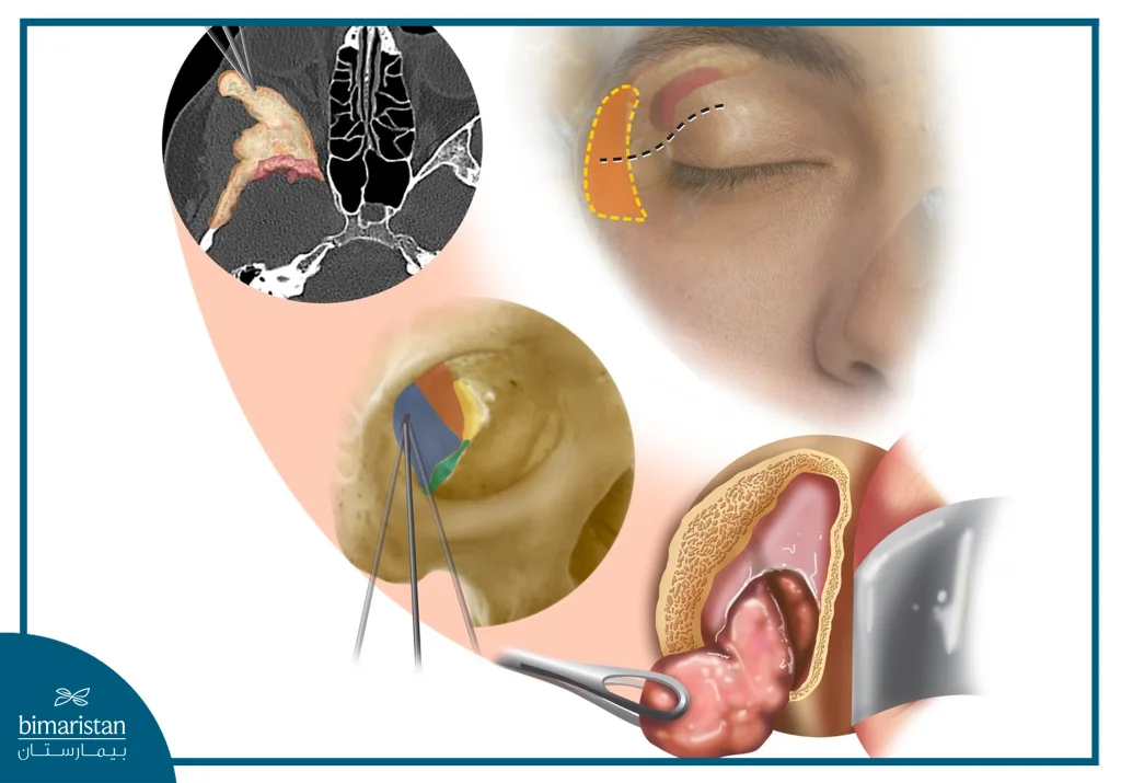
- Selection of entry point: The surgeon determines the optimal approach based on the lesion’s location; the incision may be made within the upper eyelid crease, the eyebrow, the inner corner of the eye, or from inside the eyelid.
- Insertion of the endoscope and instruments: The surgeon inserts a slender endoscope and precision surgical instruments through the incision; the endoscope provides a magnified, high-resolution view of deep tissues and structures.
- Accessing the skull base: The surgeon carefully dissects the tissues and may remove a limited portion of the orbital bone or skull base to widen the corridor and reach the target area.
- Resection of the tumor or lesion: The surgeon gradually removes pathological tissue while protecting the eye, optic nerve, cranial nerves, and adjacent blood vessels; surgical navigation may be used to ensure an accurate approach.
- Closing the surgical site: After ensuring hemostasis, the surgeon reconstructs bone or tissue if needed; a tissue patch or fat graft may be used to prevent cerebrospinal fluid leakage, and the incision is meticulously closed to achieve a good cosmetic outcome.
Benefits of transorbital neurosurgery
Transorbital neurosurgery provides a minimally invasive approach to reach certain deep lesions in the orbit and the base of the skull, which may reduce damage to surrounding tissues compared to conventional craniotomy. Its main advantages include:
- Faster recovery in suitable patients
- Avoid extensive craniotomy
- Reduced need for brain tissue removal
- Provides a direct route to tumors and deep lesions
- Minimizes manipulation of surrounding nerves and blood vessels
- Conceals the incision often within the folds of the eyelid or eyebrow
- Reduced visible scarring
- Shorter hospital stay in some cases
Risks and complications of transorbital neurosurgery
The procedure may cause temporary vision disturbances or double vision, as well as eyelid swelling or drooping and impairment of the eye muscles or the nerves responsible for eye movement. Less common complications include cerebrospinal fluid leakage, bleeding, infection, and injury to cranial nerves or blood vessels, while vision loss or stroke are considered rare complications. Careful planning and collaboration among the multidisciplinary surgical team help minimize these risks.
Recovery after transorbital neurosurgery
Recovery begins with hospital monitoring to ensure vision, eye movement, and nerve function are normal; the patient may be discharged within a day or a few days, depending on the extent of the surgery and the patient’s overall condition. Swelling of the eyelid, bruising, and mild or temporary pain around the eye are to be expected; these symptoms will gradually improve with medication, keeping the head elevated, and applying cold compresses as directed by the doctor.
Avoid bending, physical exertion, and heavy lifting during the initial recovery period, and attend scheduled neurological and ophthalmological follow-up appointments. The time it takes to resume normal activities varies depending on the type of lesion and the extent of the resection. Consult the surgical team immediately if you experience decreased vision, increased headaches, fever, or clear discharge from the nose or the surgical site.
Comparison of transorbital neurosurgery, craniotomy, and transnasal endoscopic surgery
The surgical approaches used to access the skull base vary depending on the lesion’s location, size, and extent, as well as its relationship to nerves and blood vessels. The following table outlines the main differences between the three approaches:
| Comparison aspect | Transorbital neurosurgery | Conventional craniotomy | Transnasal endoscopic surgery |
|---|---|---|---|
| Access route | Through a small incision around the orbit or eyelid folds | Through a scalp incision and removal of part of the skull bone | Through the nasal passages and sinuses |
| Best target areas | Lateral, anterior, and central skull base regions and orbital areas | Large lesions extending into multiple brain and skull base regions | Central skull base areas such as the pituitary region and clivus |
| Direction of approach | Lateral or anterolateral direct access | Varies depending on craniotomy location | Midline anterior access through the nose |
| Brain tissue resection | Usually minimal | May be significant depending on tumor size and location | Minimal in suitable cases |
| Incision size | Small and often hidden within eyelid folds | Larger and more visible | No external skin incision |
| Working field | Good for lateral and deep lesions but limited by narrow corridor | Wide field with strong control for complex lesions | Good visualization of midline areas; limited lateral access |
| Cosmetic impact | Minimal or barely visible scar | May leave a noticeable scalp scar | No external visible scar |
| Recovery duration | Shorter in selected cases | Usually longer due to extensive surgery | Relatively short in uncomplicated cases |
| Key advantages | Direct route, minimal brain tissue resection, excellent cosmetic results | Ability to manage large and complex tumors with wide control | Direct midline access without opening the skull |
| Main limitations | Not suitable for all tumors; may affect orbital nerves | More invasive with longer recovery | Limited access to lateral lesions behind carotid artery |
| Combination options | Can combine with transnasal endoscopic surgery for multi‑portal access | May combine with endoscopic routes in complex cases | Can combine with transorbital approach for expanded lateral access |
None of these approaches is considered the best in all cases; craniotomy may be necessary for large or extensive tumors, while the transnasal approach is suitable for lesions located in the middle of the skull base. Transorbital neurosurgery offers a particular advantage when targeting lateral or anterolateral lesions, and it can be combined with the transnasal approach to treat certain complex tumors from multiple directions.
Transorbital neurosurgery is a modern, minimally invasive option for accessing certain tumors and lesions of the facial region and the base of the skull, as it provides a direct pathway to deep areas while minimizing the need to open the skull and resect brain tissue. The safety and effectiveness of this technique depend on careful patient selection, multidisciplinary team expertise, and a thorough assessment of the lesion’s location and its relationship to the optic nerve, cranial nerves, and blood vessels. Transorbital neurosurgery may be used alone or combined with transnasal endoscopic surgery to access large and complex tumors from multiple directions.
Sources:
- Vural, A., Carobbio, A. L. C., Ferrari, M., Rampinelli, V., Schreiber, A., Mattavelli, D., Doglietto, F., Buffoli, B., Rodella, L. F., Taboni, S., Tomasoni, M., Gualtieri, T., Deganello, A., Hirtler, L., & Nicolai, P. (2021). Transorbital endoscopic approaches to the skull base: A systematic literature review and anatomical description. Neurosurgical Review, 44(5), 2857–2878.
- Ramakrishna, R., Kim, L. J., Bly, R. A., Moe, K., & Ferreira, M., Jr. (2016). Transorbital neuroendoscopic surgery for the treatment of skull base lesions. Journal of Clinical Neuroscience, 24, 99–104.
- Ramakrishna R, Kim LJ, Bly RA, Moe K, Ferreira M Jr. Transorbital neuroendoscopic surgery for the treatment of skull base lesions. J Clin Neurosci. Feb 2016;24:99-104. doi: 10.1016/j.jocn.2015.07.021. Epub 2015 Nov 10. PMID: 26563603; PMCID: PMC5955706.